The Impact of Big Gradients on the Future of MRI
Susie Huang1
1Massachusetts General Hospital, United States
1Massachusetts General Hospital, United States
Synopsis
Keywords: Physics & Engineering: Hardware, Contrast mechanisms: Diffusion
The engineering advances required to achieve strong gradient amplitudes and fast slew rates have directly benefitted the radiological sciences and clinical imaging by encouraging all the major scanner vendors to incorporate stronger and faster gradients into their commercially available products. A new generation of ultra-high gradients are now being adopted, such that hundreds of mT/m will be more readily accessible and routinely available for imaging patients. This lecture will highlight key clinical and research applications that will benefit most from such powerful gradients and advance our limits of detecting, understanding, and managing disease in patients across a range of pathologies. The gradient system is a key component
of the MRI machine, being responsible for the spatial encoding in image
generation and integral to controlling a range of physiological imaging
contrasts, most notably diffusion-weighted MRI. The design and performance of
the gradient system has substantial influence on the overall quality of the
acquired images and has been the focus of intense engineering efforts over the
last 3 decades in the quest for better image quality and ever-faster imaging
speed.
Gradient performance is parameterized by the maximum gradient amplitude, which is measured in mT/m, and the slew rate, which describes how fast a gradient can attain a desired amplitude within a given amount of time and is measured in T/m/s. Since the inception of MRI, gradient amplitudes and slew rates have increased by orders of magnitude, roughly doubling every 10 years since the 1990’s. In parallel, the push for stronger and faster gradients has been spurred by research applications, particularly in the brain. A seminal breakthrough in whole-body gradient design was achieved for the Human Connectome Project (HCP), culminating in the installation of the first Connectom MRI scanner at the MGH Martinos Center in 2011, which featured a whole-body gradient with a peak gradient performance of 300 mT/m at a slew rate of 200 T/m/s. More recently, we have developed the next-generation Connectom scanner (Connectom 2.0) with the goal of comprehensive multi-scale mapping of structure and connectivity across the entire living human brain.
While advances in gradient technology have improved our understanding of the human brain through large-scale research efforts like the HCP and NIH BRAIN Initiative, the engineering advances required to achieve such strong gradient amplitudes and fast slew rates have directly informed and benefitted the radiological sciences and clinical imaging by encouraging all the major scanner vendors to incorporate stronger and faster gradients into their commercially available products. The latest commercial scanners now feature integrated whole-body gradient systems with a maximum gradient amplitude of at least 80 mT/m, and in many cases, greater than 100 mT/m, with maximum slew rates of at least 200 T/m/s.
Where does the future lie with such powerful technology, and how can we best leverage such advances to make a difference for our patients? Based on our collective experience with the original MGH Connectom scanner, we believe we are at the cusp of a new generation of ultra-high gradients for whole-body imaging, where hundreds of mT/m will be more readily accessible and routinely available for imaging patients. In this talk, I will highlight a few of the key clinical and research applications that will benefit most from such powerful gradients, listed in order of those that are closest to our current clinical practice to those that will advance our limits of detecting, understanding, and managing disease in patients across a range of pathologies.
Gradient performance is parameterized by the maximum gradient amplitude, which is measured in mT/m, and the slew rate, which describes how fast a gradient can attain a desired amplitude within a given amount of time and is measured in T/m/s. Since the inception of MRI, gradient amplitudes and slew rates have increased by orders of magnitude, roughly doubling every 10 years since the 1990’s. In parallel, the push for stronger and faster gradients has been spurred by research applications, particularly in the brain. A seminal breakthrough in whole-body gradient design was achieved for the Human Connectome Project (HCP), culminating in the installation of the first Connectom MRI scanner at the MGH Martinos Center in 2011, which featured a whole-body gradient with a peak gradient performance of 300 mT/m at a slew rate of 200 T/m/s. More recently, we have developed the next-generation Connectom scanner (Connectom 2.0) with the goal of comprehensive multi-scale mapping of structure and connectivity across the entire living human brain.
While advances in gradient technology have improved our understanding of the human brain through large-scale research efforts like the HCP and NIH BRAIN Initiative, the engineering advances required to achieve such strong gradient amplitudes and fast slew rates have directly informed and benefitted the radiological sciences and clinical imaging by encouraging all the major scanner vendors to incorporate stronger and faster gradients into their commercially available products. The latest commercial scanners now feature integrated whole-body gradient systems with a maximum gradient amplitude of at least 80 mT/m, and in many cases, greater than 100 mT/m, with maximum slew rates of at least 200 T/m/s.
Where does the future lie with such powerful technology, and how can we best leverage such advances to make a difference for our patients? Based on our collective experience with the original MGH Connectom scanner, we believe we are at the cusp of a new generation of ultra-high gradients for whole-body imaging, where hundreds of mT/m will be more readily accessible and routinely available for imaging patients. In this talk, I will highlight a few of the key clinical and research applications that will benefit most from such powerful gradients, listed in order of those that are closest to our current clinical practice to those that will advance our limits of detecting, understanding, and managing disease in patients across a range of pathologies.
Acknowledgements
No acknowledgement found.References
No reference found.Figures
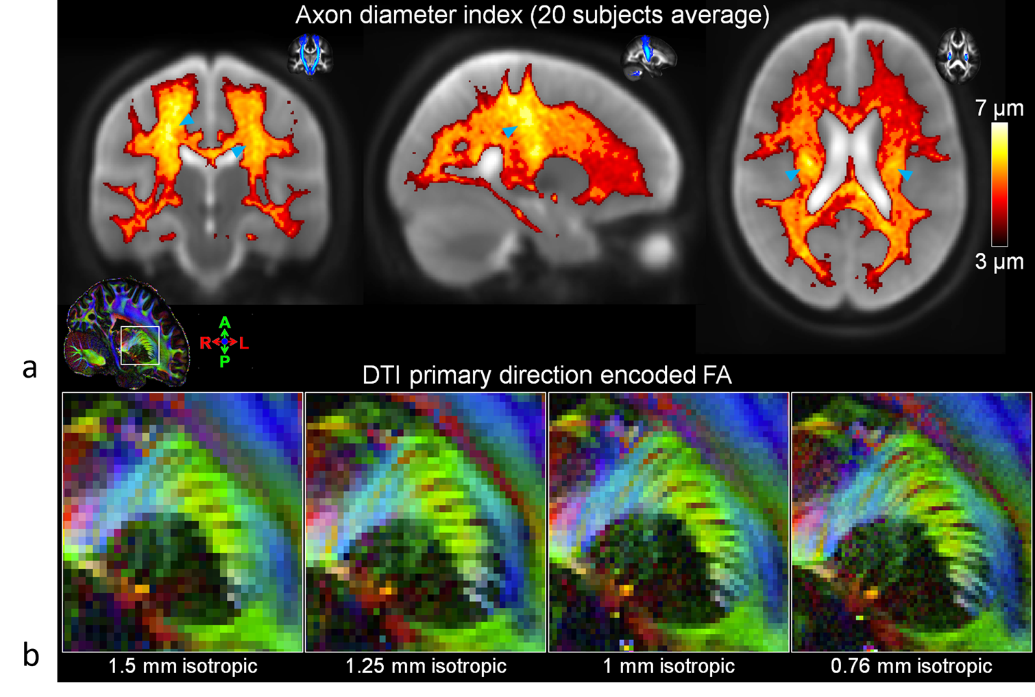
Benefits of strong
gradients for diffusion MRI. (a) In vivo axon diameter index maps enabled by high b-values using Gmax~200-300
mT/m. Average axon diameter across 20 healthy subjects (1). (b) Fractional anisotropy (FA) maps of
internal capsule at different spatial resolutions down to sub-millimeter 0.76
mm3 spatial resolution (2).