Perfusion Across Field Strengths
1University of Southern California (USC), Los Angeles, CA, United States
Synopsis
Keywords: Contrast mechanisms: Perfusion, Physics & Engineering: High-Field MRI, Physics & Engineering: Low-field MRI
Motivation: Ultrahigh field (UHF>=7T) and high performance low field (HPLF<1T) MR systems have greatly expanded the field strengths and associated applications for perfusion MRI.
Goal(s): To summarize recent advances of perfusion MRI across field strengths.
Approach: UHF offers dual benefits for ASL with increased SNR and blood T1, and increased sensitivity for dynamic contrast based perfusion MRI. HPLF systems provide improved field homogeneity and prolonged T2* and is ideal for cardio-pulmonary perfusion imaging.
Results: Perfusion MRI across a wide range of field strengths represent specific opportunities and challenges for specific clinical applications to be further exploited in near future.
Impact: Perfusion MRI across a wide range of field strengths represent specific opportunities and challenges for specific clinical applications to be further exploited in near future.
Introduction
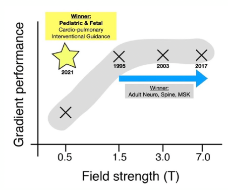
Recent developments of ultrahigh field (UHF >= 7T) and high performance low field (HPLF < 1T) MR systems have greatly expanded the field strengths and associated applications for perfusion MRI. UHF provides increased SNR that is proportional to or has superlinear relationship with field strength, allowing (sub)millimeter resolution and high sensitivity for perfusion imaging for CNS and MSK applications. However, B1/B0 field inhomogeneity and SAR limit are major challenges. HPLF systems were developed during the past 5 years that take advantage of the high performance gradient system while lowering the main magnetic field to be less than 1 Tesla. HPLF systems are ideally suited for pediatric and fetal imaging, cardio-pulmonary imaging and interventional guidance due to their low sensitivity to susceptibility artifacts (Figure 1).Methods and Results
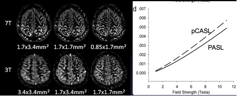
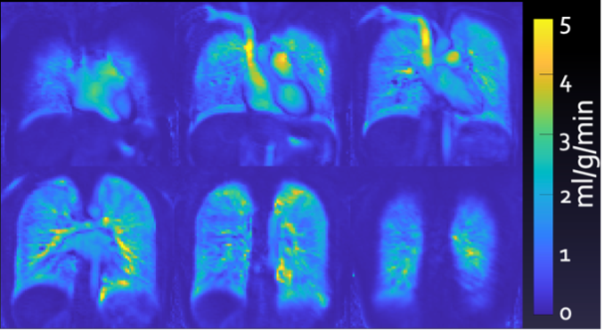
UHF offers dual benefits for ASL with increased SNR and blood T1 [1, 2](Figure 2). Both pulsed and pseudo-continuous ASL (pCASL) have been optimized for 7T, which showed increased SNR compared to corresponding techniques at 3T [3-5]. Laminar perfusion MRI with zoomed 3D GRASE pCASL and isotropic 1mm resolution has been recently developed [6]. Increased tissue and blood T1 values at UHF also benefit dynamic contrast-based perfusion MRI techniques, allowing increased contrast agent detection sensitivity and/or reduced contrast dose for mapping perfusion and blood-brain barrier (BBB) permeability [7].HPLF systems provide improved field homogeneity and prolonged T2* and is ideal for cardio-pulmonary perfusion imaging [8]. Feasibility studies have been performed at 0.55T showing excellent image quality with minimal susceptibility artifacts for cardio-pulmonary perfusion MRI [9] (Figure 3).Summary and Discussion
Recent developments of perfusion MRI at UHF (>= 7T) and HPLF (< 1T) have greatly expanded the field strengths and associated clinical applications for perfusion MRI. Perfusion MRI across a wide range of field strengths represent specific opportunities and challenges for specific clinical applications to be further exploited in near future.Acknowledgements
NIH grants UF1-NS100614 and R01-EB032169References
1. Zuo, Z., et al., Turbo-FLASH Based Arterial Spin Labeled Perfusion MRI at 7 T. Plos One, 2013. 8(6).
2. Wang, J.J., et al., Comparison of quantitative perfusion imaging using arterial spin labeling at 1.5 and 4.0 tesla.Magnetic Resonance in Medicine, 2002. 48(2): p. 242-254.
3. Zhao, C., et al., Whole-Cerebrum distortion-free three-dimensional pseudo-continuous arterial spin labeling at 7T.Neuroimage, 2023. 277: p. 120251.
4. Wang, K., et al., Optimization of adiabatic pulses for pulsed arterial spin labeling at 7 tesla: Comparison with pseudo-continuous arterial spin labeling. Magn Reson Med, 2021. 85(6): p. 3227-3240.
5. Wang, K., et al., Optimization of pseudo-continuous arterial spin labeling at 7T with parallel transmission B1 shimming. Magn Reson Med, 2022. 87(1): p. 249-262.
6. Shao, X., et al., Laminar perfusion imaging with zoomed arterial spin labeling at 7 Tesla. Neuroimage, 2021. 245: p. 118724.
7. Knutsson, L., et al., Dynamic Susceptibility Contrast MRI at 7 T: Tail-Scaling Analysis and Inferences About Field Strength Dependence. Tomography, 2017. 3(2): p. 74-78.
8. Campbell-Washburn, A.E., et al., Opportunities in Interventional and Diagnostic Imaging by Using High-Performance Low-Field-Strength MRI. Radiology, 2019. 293(2): p. 384-393.
9. Campbell-Washburn, A.E., et al., Cardiac MRI at Low Field Strengths. J Magn Reson Imaging, 2023.
Figures