Spectral Encoding: Fundamentals & Challenges
1Center for Stroke Researech Berlin (CSB), Charite Universitätsmedizin Berlin, Berlin, Germany
Synopsis
Keywords: Contrast mechanisms: Spectroscopy, Contrast mechanisms: Spectroscopic Imaging (MRSI), Neuro: Brain
Spectral encoding provides a means to obtain additional and often complementary information in comparison to magnetic resonance imaging (MRI). Using the chemical shift σ in MR spectroscopy (MRS) metabolic information can be extracted from resulting spectra acquired as single voxel or multi-voxel MR spectroscopic imaging (MRSI) data. The composition of MR spectra for sample molecules is outlined. In addition, typical acquisition protocols and more advanced MRS methodology with a particular importance of required adjustments and including fast MRSI schemes are presented. Metabolite quantification is briefly addressed. Finally, remaining challenges including the relatively low sensitivity and possible amendments are discussed.Syllabus
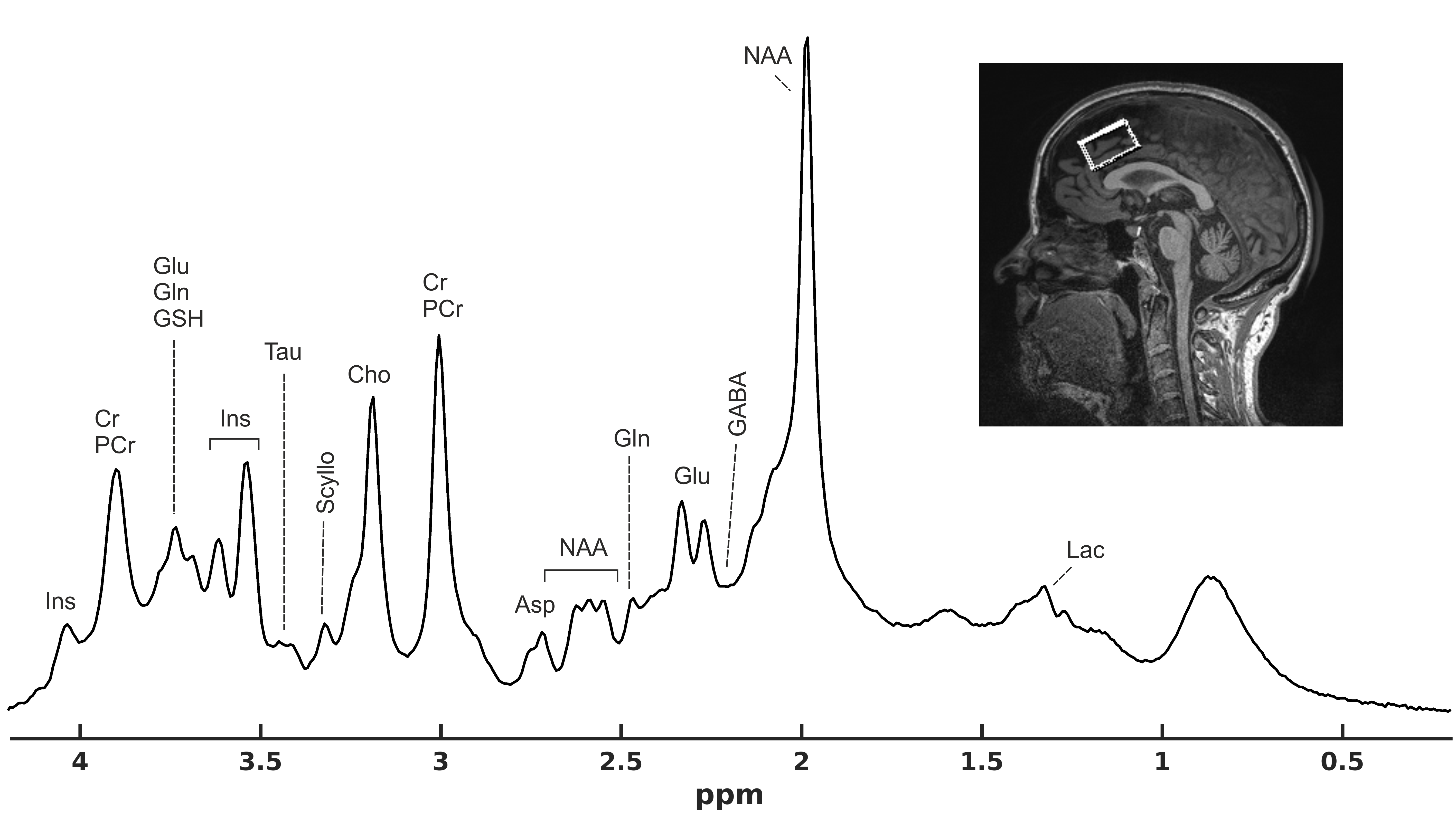
Spectral encoding provides a means to obtain additional information in comparison to magnetic resonance imaging (MRI). Metabolic information can be assessed using MR spectroscopy (MRS), where the chemical shift σ of different molecules (metabolites) yields specific resonance patterns in frequency space. Some basics with respect to the composition and acquisition of MR spectra with the focus on 1H MRS will be discussed. Typically, MRS data are measured from a localized volume as single voxel data or, if phase encoding is applied in addition, from multiple voxels in MR spectroscopic imaging (MRSI). Some specific MRS acquisition schemes will be described starting with most widely available sequences protocols, but also considering more advanced methodology with the particular importance of adjustments, such as radiofrequency (RF) calibration, B0 shimming, water suppression (WS), and outer volume saturation (OVS). For MRSI, in addition to classical phase encoding methods, some approaches to acceleration, including echo-planar based methods (EPSI), non-cartesian k-space trajectories, undersampling, and ultrashort TR acquisitions will be described. Analysis of MR spectra to yield metabolite concentrations using linear combination model (LCM) fitting will be outlined. The interpretation of metabolic information obtained from MRS will be presented for the example of 1H MRS in the brain. One principal challenge of MRS is the relative low signal-to-noise ratio (SNR) due to the low abundance of metabolites often resulting in long scan times and limited coverage of regions of interest. In this context, MRS at high B0 fields (≥ 3T) is advocated, where MRS has a twofold benefit - enhanced sensitivity and increased spectral resolution. When adequately executed, MRS can be used to non-invasively study metabolic function and psychopathological conditions or even for treatment monitoring. Lastly, as an alternative method of spectral encoding, chemical exchange saturation transfer (CEST) imaging will be very briefly referenced.Syllabus - Key Points
- Spectral encoding provides a means to obtain additional information in comparison to magnetic resonance imaging (MRI)
- Metabolic information can be assessed using MR spectroscopy (MRS), where the chemical shift σ of different molecules (metabolites) yields specific resonance patterns in frequency space
- Basic composition and acquisition of MR spectra will be discussed for single voxel and MR spectroscopic imaging data with the focus on 1H MRS
- Challenge for MRS is relatively low sensitivity due to the low abundance of metabolites that can be addressed by advanced MRS methodology including careful adjustments and measurements at high B0 fields
- Using advanced methodology, MRS can be used to non-invasively study metabolic function and psychopathological conditions or even for treatment monitoring
- This lecture should provide an introduction to the basic principles of spectral encoding as exemplified for MR spectroscopy (MRS) including single voxel and MR spectroscopic imaging (MRSI) approaches. Remaining challenges for MRS are discussed as well
- A second type of spectral encoding is chemical saturation transfer imaging (CEST), where the water signal is used to detect changes in small pools of protons (solute) via chemical exchange that reside at different resonance frequencies via selective RF irradiation
Acknowledgements
The author wishes to thank all colleagues and collaborators, who provided input and feedback to this lecture.References
Books and (Review) Papers MR Spectroscopy (MRS)
1. de Graaf RA. In Vivo NMR Spectroscopy: Principles and Techniques, 3rd ed. Hoboken, NJ: John Wiley & Sons, Ltd, 2019.
2. Graves MJ, McRobbie DW, Moore EA, Prince MR. MRI from Picture to Proton, 3 ed. Cambridge: Cambridge University Press, 2017.
3. Prost RW. Magnetic resonance spectroscopy. Med Phys 2008;35:4530-4544.
4. Buonocore MH, Maddock RJ. Magnetic resonance spectroscopy of the brain: a review of physical principles and technical methods. Rev Neurosci 2015;26:609-632.
5. Hajek M, Dezortova M. Introduction to clinical in vivo MR spectroscopy. Eur J Radiol 2008;67:185-193.
6. Barker PB, Lin DDM. In vivo proton MR spectroscopy of the human brain. Progress in Nuclear Magnetic Resonance Spectroscopy 2006;49:99-128.
7. Xin L, Tkáč I. A practical guide to in vivo proton magnetic resonance spectroscopy at high magnetic fields. Anal Biochem 2017;529:30-39.
8. Kreis R. Issues of spectral quality in clinical 1H-magnetic resonance spectroscopy and a gallery of artifacts. NMR Biomed 2004;17:361-381.
9. Govindaraju V, Young K, Maudsley AA. Proton NMR chemical shifts and coupling constants for brain metabolites. NMR Biomed 2000;13:129-153.
10. Oz G, Alger JR, Barker PB, et al. Clinical proton MR spectroscopy in central nervous system disorders. Radiology 2014;270:658-679.
11. Wilson M, Andronesi O, Barker PB, et al. Methodological consensus on clinical proton MRS of the brain: Review and recommendations. Magn Reson Med 2019;82:527-550.
12. Kreis R, Boer V, Choi IY, et al. Terminology and concepts for the characterization of in vivo MR spectroscopy methods and MR spectra: Background and experts' consensus recommendations. NMR Biomed 2020;34:e4347.
13. Lin A, Andronesi O, Bogner W, et al. Minimum Reporting Standards for in vivo Magnetic Resonance Spectroscopy (MRSinMRS): Experts' consensus recommendations. NMR Biomed 2021;34:e4484.
14. Öz G, Deelchand DK, Wijnen JP, et al. Advanced single voxel (1) H magnetic resonance spectroscopy techniques in humans: Experts' consensus recommendations. NMR Biomed 2020:e4236.
15. Maudsley AA, Andronesi OC, Barker PB, et al. Advanced magnetic resonance spectroscopic neuroimaging: Experts' consensus recommendations. NMR Biomed 2021;34:e4309.
16. Juchem C, Cudalbu C, de Graaf RA, et al. B(0) shimming for in vivo magnetic resonance spectroscopy: Experts' consensus recommendations. NMR Biomed 2021;34:e4350.
17. Near J, Harris AD, Juchem C, et al. Preprocessing, analysis and quantification in single-voxel magnetic resonance spectroscopy: experts' consensus recommendations. NMR Biomed 2021;34:e4257.
18. Choi IY, Andronesi OC, Barker P, et al. Spectral editing in (1) H magnetic resonance spectroscopy: Experts' consensus recommendations. NMR Biomed 2021;34:e4411.
19. Bogner W, Otazo R, Henning A. Accelerated MR spectroscopic imaging-a review of current and emerging techniques. NMR Biomed 2021;34:e4314.
20. Henning A. Proton and multinuclear magnetic resonance spectroscopy in the human brain at ultra-high field strength: A review. Neuroimage 2018;168:181-198.
Books and (Review) Papers Chemical Exchange Saturation Transfer (CEST)
21. Michael T. McMahon AAG, Jeff W.M. Bulte, Peter C.M. van Zijl, ed. Chemical Exchange Saturation Transfer Imaging Advances and Applications. Singapore: Pan Stanford Publishing Pte. Ltd., 2017.
22. Ward KM, Aletras AH, Balaban RS. A new class of contrast agents for MRI based on proton chemical exchange dependent saturation transfer (CEST). J Magn Reson 2000;143:79-87.
23. Wu B, Warnock G, Zaiss M, et al. An overview of CEST MRI for non-MR physicists. EJNMMI Phys 2016;3:19.
24. van Zijl PC, Yadav NN. Chemical exchange saturation transfer (CEST): what is in a name and what isn't? Magn Reson Med 2011;65:927-948.
25. Vinogradov E, Sherry AD, Lenkinski RE. CEST: from basic principles to applications, challenges and opportunities. J Magn Reson 2013;229:155-172.
Figures