How To Measure Brain Temperature Using MR
Kagayaki Kuroda1
1Tokai University, Japan
1Tokai University, Japan
Synopsis
Keywords: Contrast mechanisms: Thermometry, Physics & Engineering: Interventional, Neuro: Brain
Different levels of techniques and accuracy should be adopted for MR thermometry. Proton resonance frequency (PRF) shift with phase mapping technique is suitable for monitoring brain thermal therapy using laser or high intensity focused ultrasound. For monitoring hypothermia therapy or for detecting ischemia and traumatic brain injury, PRF shift detected spectroscopically with internal reference such as NAA, chorine and creatine may help. However, careful consideration regarding variation of bulk susceptibility, distributions of electrolyte and macromolecules as well as status of pH should be made. The purpose of this lecture is to discuss thermometry techniques and the factors influencing temperature quantification.Introduction
Most proton (1H) magnetic resonance (MR) parameters are temperature dependent(1). These include the proton resonance frequency (PRF) or chemical shift (CS) of hydroxyl or other groups with hydrogen bonding, spin-lattice relaxation time, T1 (2-5); spin-spin relaxation time, T2 (3, 6, 7); proton density or thermal equilibrium magnetization, M0 (8, 9); and the diffusion coefficient, D (10, 11). Among these parameters, PRF can be measured independently from the other parameters, which are determined using signal amplitudes and are thus affected by each other within a limited measurement time. Moreover, the thermal coefficient of PRF in water molecules is almost tissue insensitive. Thus, water PRF is thought to be the most reasonable and practical indicator for temperature measurement. There are two major approaches, MR spectroscopy (MRS) (12-17) and phase mapping (18, 19) to measure the water PRF. Although the phase mapping techniques have been widely accepted (20-22) for monitoring thermal therapies (23-26), they yield only relative temperature change with reference to a baseline temperature. On the other hand, MRS techniques have potential to be used for monitoring hypothermia therapy or for detecting ischemia and traumatic brain injury(27) as shown in Figure 1. To make MRS technique to be practical for those applications, some factors affecting on temperature quantifications has to be considered.Principle of temperature dependence of water PRF
Hydrogen bonds between the protons in the hydroxyl groups and other more electronegative atoms are primarily responsible for inducing the temperature dependence of the chemical shift of protons(28). Briefly, the electrical currents from the electronic orbitals surrounding the MR nuclei produce a weak magnetic field that opposes the external applied field (B0) producing a shielding effect(29). Consequently, the resonance frequency of the water proton decreases with the rate around -0.01ppm/oC(30). A schematic diagram of this temperature dependence including effect of volume susceptibility is depicted in Figure 2(31).Factors for temperature dependence of water PRF
ElectrolytesWe can also observe the downward shift of the MR frequency in water when univalent electrolytes are added. This phenomenon is referred to as “bond-breaking processes”(32) . When sodium or potassium ions are dissolved in water, their positive charges attract oxygen atoms in the hydroxyl group, which increases the likelihood of breaking the hydrogen bonds between other water molecules(32). Interestingly, the temperature coefficient of water PRF slightly increase with ionic concentrations up to 1 M applied(33).
Bulk susceptibility
When a group of nuclei are in a isotropic region at temperature T, the nuclei experience a local magnetic field, ,described elsewhere (34, 35). The rate of volume susceptibility change with temperature in an aqueous medium is negative, and is several-fold smaller (~ -0.002 ppm/oC)(34, 35) than that of the screening constant of water protons. However, it is also known that the blood oxygenation dependent susceptibility during functional activation reaches as large as 0.02 ppm(36), which corresponds to the PRF shift equivalent to 2 oC temperature change. Moreover, the susceptibility difference between completely deoxygenated and completely oxygenated red blood cells can be 0.264 ppm(37).
pH
The effect of the pH on the PRF of an aqueous hydroxyl group is presently unresolved. Some of the published results are summarized in Table 1 (15, 17, 38, 39). If the pH is constant during the period temperature is being monitored and only relative temperature change is of interest, the effect of pH can generally be neglected. Meanwhile, we do need to consider pH effects when attempting absolute temperature measurements.
Macromolecules
Generally, water molecules tend to cluster around large protein molecules due to hydrogen bonding, and there is exchange between these molecules and freer water in the cytosol. Averaged over the time period of observation, the hydrogen bonding causes a small increase in the water PRF as compared to that of pure water. Some representative results of temperature dependence of the proton chemical shifts in tissues obtained with internal references are summarized in Table 1 based on the previous studies (15, 16, 38-43). The possible factors described above are depicted in Figure 3 together with variation of the chemical shift between water and the methyl resonance of N-acetylaspartate (NAA) in a normal volunteer.
Acquisition Strategies for brain temperature measurement
MRS techniques with internal reference are focused here. Figure 4 shows the basic idea of using MRS technique for temperature measurement. By using an internal reference signal, temperature change of bulk susceptibility can be cancelled, and thus only the change in the shielding constant is detected. Single voxel spectroscopy using conventional Point Resolved Spectroscopy (PRESS) has been extensively explored the temperature dependence of the water PRF, and to measure temperature at various spatially localized sites in tissue.(15, 42, 44, 45) As an internal reference in brain, a temperature-insensitive reference metabolite such as NAA (~1.98 ppm) has been preferred because of its prominence and relatively isolated chemical shift position in cerebral spectra(15, 16, 38-41, 45). The resonances from choline-containing metabolites (Cho; ~3.2 ppm) and total creatine (Cr; ~3.0 ppm) have also been used(41, 42).Signal averaging is essential for detecting reference signals from relatively low concentration (~10 mM) of these metabolites. Multivariate analysis in conjunction with spectral processing techniques is one of the key techniques for improving both inter-subject reproducibility, resilience, and the quantification of the thermal change of water-metabolite chemical shifts(41).
2D- and 3D- Magnetic Resonance Spectroscopic Imaging (MRSI) have been explored for imaging temperature.(43, 46-48). Recent technique with adiabatic selective refocusing pulse such as sLASER (semi localized adiabatic selective refocusing)(49) in conjunction with sophisticated shimming techniques such as FASTESTMAP and GRESHIM(50) has greatly improved the spectral quality good for thermometry(27).
To overcome the low temporal resolution of the phase-encoding-based SI approach, echo-planar spectroscopic imaging (EPSI) (51, 52) and line-scan EPSI (LSEPSI) (40, 48, 53) have been introduced for thermometry. The latter is a combination of the echo-planar technique with column scanning that accelerates multi-voxel MRS, while avoiding spectral degradation due to eddy currents from the phase-encoding process. It should be noted that the EPSI and LSEPSI approaches are substantially similar to the multiple gradient-echo phase mapping technique(54).
Other possible means
Other than the PRF approach, there has been a few attempts to use diffusion coefficient of cerebrospinal fluid (CSF) to measure brain temperature(55). Indeed, there is clear temperature dependence in diffusion coefficient in pure water(56), we cannot deny the effect of macromolecules or other factors in the diffusivity of the water molecules in CSF.Some nuclei other than protons such as 13C (57), 31P (58, 59), or 59Co (60) also demonstrate properties that may be suitable for thermometry. However, it is difficult to justify their use in the practical consequences of their lower abundance and sensitivity, and the hardware/software limitations of MR scanners.
Hyperpolarized 129Xe in a cryptophane-A cage has a temperature dependent chemical shift of 0.29 ppm/oC (61). Although this temperature coefficient is more than 10 times larger than that of water protons and this phenomenon is interesting as basic science, the hyperpolarization process is laborious, and clinical translation has not yet to be seen. Refer to table 2 for reviewing some non-PRF or non-proton parameters for temperature measurement.
Acknowledgements
The author thanks all his colleagues for their collaboration and contribution.
References
1. Kuroda K, Tsutsumi S,. Chapter C: NMR tomography II: Temperature imaging by proton chemical shift. In: Miyakawa M BJ, editor. Non-invasive thermometry of the human body. Boca Raton: CRC Press Inc; 1996.2. Lewa CzJ, A B. Proton magnetic relaxation in dehydrated normal and neoplasmic animal tissue. Bull Cancer. 1977;64:37.
3. Bottomley PA, Foster TH, Argersinger RE, Pfeifer LM. A review of normal tissue hydrogen NMR relaxation times and relaxation mechanisms from 1 - 100 MHz: Dependence on tissue type, NMR frequency, temperature, species, excision, and age. Med Phys. 1984;11:425.
4. Parker DL. Applications of NMR imaging in hyperthermia. IEEE Trans Biomed Eng. 1984;BME-31:161.
5. Dickinson RJ, Hall AS, Hind AJ, Young IR. Measurement of changes in tissue temperature using MR imaging. J Compt Assist Tomogr. 1986;10:468.
6. Nelson TR, Tung SM. Temperature dependence of proton relaxation times in vitro. Magn Reson Imag. 1987;5:189.
7. Kuroda K, Tsutsumi S, A S. Non-invasive temperature distribution measurement by simultaneous use of multiple NMR parameters. Trans IEICE. 1990;J73-A(8):1431.
8. Kamimura Y. Nagoya University, PhD Thesis. 1985.
9. Chen J, Daniel BL, Pauly KB. Investigation of proton density for measuring tissue temperature. J Magn Reson Imaging. 2006;23(3):430-4.
10. Hall AS, Prior MV, Hand JW, Young IR, Dickinson RJ. Observation by MR imaging of in vivo temperature changes induced by radio frequency hyperthermia
. J Compt Assist Tomogr. 1990;14:430.
11. Le Bihan D, Delannoy J, Levin RL. Temperature mapping with MR imaging of molecular diffusion imaging: Application to hyperthermia. Radiology. 1989;171:853.
12. Hall LD, Talagala Sl. Mapping of pH and temperature distribution using chemical-shift-resolved tomography. J Magn Reson,. 1985;65:501.
13. Kuroda K, Abe K, Tsutsumi S, Ishihara Y, Suzuki Y, Satoh K. Water proton magnetic resonance spectroscopic imaging. Biomed Thermol 1994;13:43-62.
14. Kuroda K, Suzuki Y, Ishihara Y, Okamoto K, Suzuki Y. Temperature mapping by water proton chemical shift obtained with 3D-MRSI - Feasibility in vivo -. Magn Reson Med. 1996;35:20-9.
15. Corbett RJT, Laptook AR, Tollefsbol G, Kim B. Validation of a noninvasive method to measure temperature in vivo using 1H NMR spectroscopy. Neurochem J. 1995;64:1224-30.
16. Corbett RJT, Laptook AR, Weatherall P. Noninvasive measurements of human brain temperature using volume-localized proton magnetic resonance spectroscopy. J Cereb Blood Flow Metab. 1997;17:363-9.
17. Cady EB, D'souga PC, Penrice J, Ann L. The estimation of local brain temperature by in vivo 1H magnetic resonance spectroscopy. Magn Reson Med. 1995;33:862-7.
18. Ishihara Y, Calderon A, Watanabe H, Okamoto K, Suzuki Y, Kuroda K, et al. A precise and fast temperature mapping using water proton chemical shift. Magn Reson Med. 1995;34:814-23.
19. Poorter JD, Wagter CD, Deene YD, Thomsen C, Stahlberg F, Achten E. Non-invasive MRI thermometry with the proton resonance frequency shift (PRF) method: in vivo results in human muscle. Magn Reson Med. 1995;33:74-81.
20. Rieke V, Vigen KK, Sommer G, Daniel BL, J.M. P, Butts K. Referenceless PRF Shift Thermometry. Magn Reson Med. 2004;51:1223-31.
21. Cline HE, Hynynen K, Schneider E, Hardy CJ, Maier SE, Watkins RD, et al. Simultaneous magnetic resonance phase and magnitude temperature maps in muscle. Magn Reson Med. 1996;35:309-15.
22. Peters RD, Hinks RS, Henkelman RM. Ex vivo tissue-type independence in proton-resonance frequency shift MR thermometry. Magn Reson Med. 1998;40:454-9.
23. Hynynen K, Freund WR, Cline HE, Chung AH, Watkins RD, Vetro JP, et al. A clinical, noninvasive, MR imaging-monitored ultrasound surgery method. Radiographics. 1996;16(1):185-95.
24. Moriaty JA, Chen JC, Purcell CM, Ang LC, Hinks RS, Peter RD, et al. MRI monitoring of interstitial microwave-induced heating and thermal lesions in rabbit brain in vivo. J Magn Reson Imag. 1998;8:128-35.
25. Kettenbach J, Silverman SG, Hata N, Kuroda K, Saiviroonporn P, Zientara GP, et al. Monitoring and visualization technique for MR-guided laser ablation in an open MR system. J Magn Reson Imag. 1998;8:933-43.
26. Kahn T, Harth T, Kiwit JCW, Schawarzmaier H-J, Wald C, Mödder U. In vivo MRI thermometry using a phase-sensitive sequence: preliminary experience during MRI-guided laser-induced interstitial thermotherapy of brain tumors. J Magn Reson Imag. 1998;8:160-4.
27. Dehkharghani S, Qiu D. MR Thermometry in Cerebrovascular Disease: Physiologic Basis, Hemodynamic Dependence, and a New Frontier in Stroke Imaging. AJNR Am J Neuroradiol. 2020;41(4):555-65.
28. Kuroda K. Non-Invasive MR thermography using water proton chemical shift. Int J Hyperthermia 2005;21(6):547-60.
29. Muller N, Reiter R. Temperature dependence of chemical shifts of protons in hydrogen bonds. J Chem Phys. 1965;42(9):3265-9.
30. Hindman J. Proton resonance shift of water in gas and liquid states. J Chem Phys. 1966; 44:4582.
31. Kuroda K. Temperature Monitoring Using Chemical Shift. In: Bottomley PA, Griffiths JR, editors. Handbook of Magnetic Resonance Spectroscopy In Vivo: MRS Theory, Practice and Applications. Chichester, UK: John Wiley & Sons Ltd; 2016. p. pp 401-14.
32. Hindman J. Nuclear magnetic resonance effects in aqueous solutions of 1-1 elecrolytes. J Chem Phy. 1962;36(4):1000-15.
33. Kuroda K, Itoh Y, Chung A, Hynynen K, Jolesz F, editors. Temperature dependence of water proton chemical shift in tissue. 4 th Meeting ISMRM; 1996; New York.
34. Poorter J. Non-invasive MRI thermometry with the proton resonance frequency method: study of susceptibility effects. Magn Reson Med. 1995;34:359-67.
35. Sprinkhuizen S, Konings M, van der Bom M, Viergever M, Bakker C, Bartels L. Temperature-induced tissue susceptibility changes lead to significant temperature errors in PRFS-based MR thermometry during thermal interventions. Magn Reson Med. 2010;64(5):360-72.
36. Kennan RP, Scanley BE, Innis RB, Gore JC. Physiological basis for BOLD MR signal changes due to neuronal stimulation: separation of blood volume and magnetic susceptibility effects. Magn Reson Med. 1998;40(6):840-6.
37. Spees WM, Yablonskiy DA, Oswood MC, Ackerman JJ. Water proton MR properties of human blood at 1.5 Tesla: magnetic susceptibility, T(1), T(2), T*(2), and non-Lorentzian signal behavior. Magn Reson Med. 2001;45(4):533-42.
38. Arus C, Chang YC, Baramy M. N-Acetylaspartate as an intrinsic thermometer for 1H NMR of brain slices. J Magn Reson, . 1985;63:376.
39. Lutz KW, Kuesel AC, Hull WE. A 1H-NMR method for determining temperature in cell culture perfusion systems. Magn Reson Med 1993;29:113-8.
40. Kuroda K, Takei N, Mulkern RV, Oshio K, Nakai T, Okada T, et al. Feasibility of Internally-Referenced Temperature Imaging Using Metabolite Signals. Magn Reson Med Sci. 2003;2(1):17-22.
41. Cady EB, Penrice J, Robertson NJ. Improved reproducibility of MRS regional brain thermometry by 'amplitude-weighted combination'. NMR in biomedicine. 2011;24(7):865-72.
42. Zhu M, Bashir A, Ackerman JJ, Yablonskiy DA. Improved calibration technique for in vivo proton MRS thermometry for brain temperature measurement. Magn Reson Med. 2008;60(3):536-41.
43. Weis J, Covaciu L, Rubertsson S, Allers M, Lunderquist A, Ortiz-Nieto F, et al. Phase-difference and spectroscopic imaging for monitoring of human brain temperature during cooling. Magn Reson Imaging. 2009;30(10):1505-11.
44. Corbett R, Laptook A, Weatherall P. Noninvasive measurements of human brain temperature using volume-localized proton magnetic resonance spectroscopy. J Cereb Blood Flow Metab. 1997;17:363-9.
45. Corbett RJT, Purdy PD, Laptook AR, Chaney C, Garcia D. Noninvasive measurement of brain temperature after stroke. Am J Neuroradiol. 1999;20:1851-7.
46. Hall L, Talagala S. Mapping of pH and temperature distribution using chemical-shift-resolved tomography. J Magn Reson. 1985;65:501.
47. Kuroda K, Suzuki Y, Ishihara Y, Okamoto K, Suzuk iY. Temperature mapping by water proton chemical shift obtained with 3D-MRSI - Feasibility in vivo -. Magn Reson Med. 1996;35:20-9.
48. Mulkern R, Chung A, Jolesz F, Hynynen K. Temperature monitoring of ultrasonically heated muscle with RARE chemical shift imaging. Med Phys. 1997;24:1899-906.
49. Scheenen TW, Klomp DW, Wijnen JP, Heerschap A. Short echo time 1H-MRSI of the human brain at 3T with minimal chemical shift displacement errors using adiabatic refocusing pulses. Magn Reson Med. 2008;59(1):1-6.
50. Zhong X, Lyubich YM, DeVito T, Shah S, Knight-Scott J, editors. Improving In Vivo 1H-MRS with Robust Automated Shim Techniques: A Comparison Study of FASTESTMAP and GRESHIM. Proc 20th Int Soc Magn Reson Med; 2012; Melbourme, Australia.
51. Posse S, DeCarli C, LeBihan D. Three-dimensional echo-planar MR spectroscopic imaging at short echo times in the human brain. Radiology. 1994;192:733-8.
52. Kuroda K, Oshio K, Mulkern R, Panych L, Nakai T, Moriya T, et al. Temperature Mapping Using Water Proton Thermal Shift: Self-Referenced Method with Echo Planar Spectroscopic Imaging. Magn Reson Med 2000;43:220-5.
53. Oshio K, Kyriakos W, Mulkern R. Line scan echo planar spectroscopic imaging. Magn Reson Med. 2000;44(4):521-4.
54. Mulkern R, Panych L, McDannold N, Jolesz F, Hynynen K. Tissue temperature monitoring with multiple gradient echo imaging sequences. J Magn Reson Imag 1998;8:493 - 502.
55. Sparacia G, Sakai K. Temperature Measurement by Diffusion-Weighted Imaging. Magnetic resonance imaging clinics of North America. 2021;29(2):253-61.
56. Mills R. Self-diffusion in normal and heavy water in the range 1-45.deg. The Journal of Physical Chemistry. 1973;77(5):685-8.
57. Bemsk iG, Leon V, Manzo F. Temperature dependence of 13C chemical shifts in myoglobin. Acta cientifica venezolana. 1980;31(2):125-7.
58. Temussi P, Guidoni L, Ramoni C, Podo F. Temperature dependence of 31P-NMR spectra of chicken erythrocyte nucleosomes. Physiological chemistry and physics. 1979;11(5):445-52.
59. Gorenstein D, Findlay J, Momii R, Luxon B, Kar D. Temperature dependence of the 31P chemical shifts of nucleic acids. A prode of phosphate ester torsional conformations. Biochemistry. 1976;15(17):3796-803.
60. Smith N, Webb A, Ellis D, Wilmes L, Obrien W. Experimental-Verification of Theoretical in-Vivo Ultrasound Heating Using Cobalt Detected Magnetic-Resonance. Ieee T Ultrason Ferr. 1995;42(4):489-91.
61. Schilling F, Schröder L, Palaniappan K, Zapf S, Wemmer D, Pines A. MRI Thermometry Based on Encapsulated Hyperpolarized Xenon. Chem Phys Chem. 2010;11(6):3529-33.
Figures
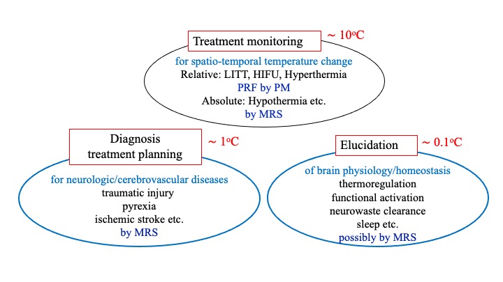
Figure
1 Needs for brain temperature measurement
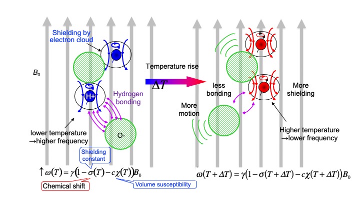
Figure 2 Schematic diagram of temperature dependence of water proton resonance frequency (PRF)
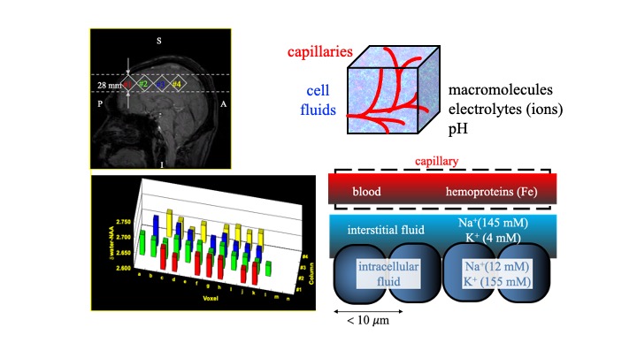
Figure 3 Distribution of absolute values of chemical shift between water and NAA in a normal volunteer measured by LSEPSI, and a schematic diagram of the possible factors affecting on the chemical shift in a voxel.
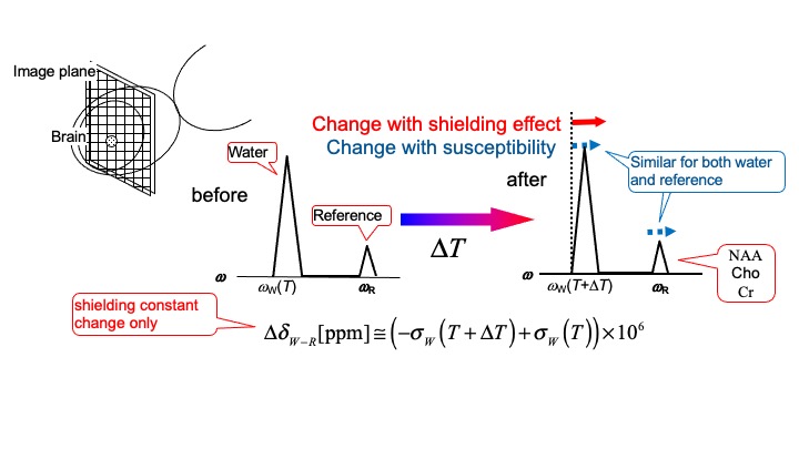
Figure 4 Basic concept of using MRS technique for thermometry
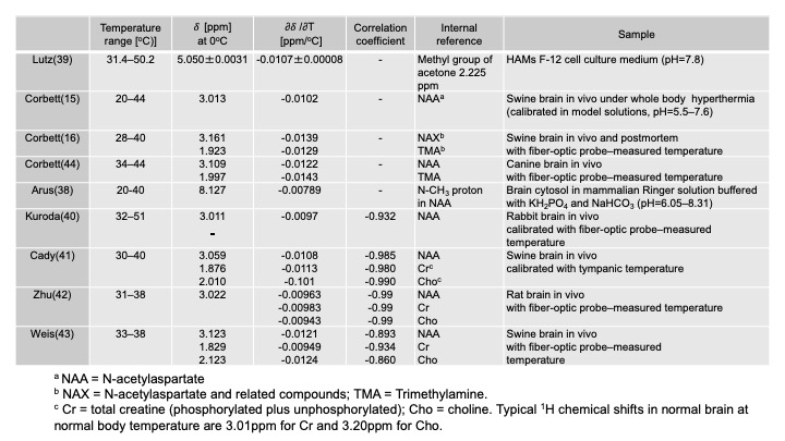
Table 1 temperature dependence of the proton chemical shifts in tissues obtained with internal references
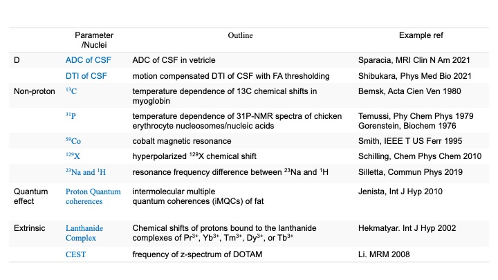
Table 1 Examples of other possible techniques