What Lies Beyond 7T on the Horizon
1NeuroSpin/CEA, Gif sur Yvette, France, 2Irfu/CEA, Gif sur Yvette, France, 3Siemens France, Courbevoie, France, 4INSERM-CEA Unicog, CNRS, Gif sur Yvette, France, 5Jacob, CEA, Gif sur Yvette, France
Synopsis
Keywords: Physics & Engineering: High-Field MRI, Neuro: Brain
The supralinear gains of signal-to-noise and contrast-to-noise ratios have been a driving force for ultra-high field MRI. Many exciting projects worldwide have emerged to leverage this gain and boost the spatiotemporal resolution of brain images, gain sensitivity in fMRI and increase the spectral peak separation. In this context, the highest magnetic field used to date in vivo on humans is 11.7T at CEA, Saclay France. This work presents the latest achievements at this unprecedented field strength, including the first images ever acquired in vivo, as well as a few lessons learnt on the way.Introduction
Understanding the human brain is one of the greatest scientific quests of the 21st century. Along with many important technological developments enabling its observation both at the microscopic and macroscopic scale, MRI may be the most promising approach to explore the human brain in vivo and understand its structure and organization at the mesoscopic scale (few thousands of neurons). In this context, important hardware developments have been carried out, e.g. with receive arrays and gradient coils [1,2], to fetch the NMR signal. The magnetization, however, essentially comes from the polarization of the nuclear spins arising from the interaction between their magnetic moments and the external magnetic field. The signal boost therefore has been the driving force to design and build more powerful magnets for MRI. Experimental data in vivo [3] and on phantoms [4] have confirmed supralinear increases of SNR with field strength. Leveraging the potential of ultra-high field (UHF) machines yet requires overcoming technological and physiological obstacles. The highest magnetic field to date to do MRI on humans is 11.7T at NeuroSpin-CEA, Saclay-France. The whole-body magnet was delivered to NeuroSpin in 2017 while the 11.7T field strength was reached in 2019 for the first time. After many gradient-magnet interaction tests to secure the magnet, first images were acquired on a pumpkin in 2021. Approval from the regulatory body and ethics committee to perform first tests in vivo on adult volunteers was obtained in February 2023. This first protocol above all consisted of performing safety tests to confirm the absence of adverse effects, as well as performing first imaging tests. This presentation reveals the few lessons learnt as well as the first in vivo images.Challenges
The nature of the challenges associated to UHF and beyond 7T MRI are technological, methodological and physiological. Technological challenges for instance incorporate meeting MRI specifications such as field homogeneity, temporal stability and magnet safety. The Lorentz force being proportional to current and magnetic field B0, vibrations of conducting structures in the magnet cryostat induce eddy-currents leading to Joule effect increasing with field strength, thereby rising the temperature of the He bath [5,6]. Care is usually taken to avoid exciting directly the most problematic frequencies. Exciting indirectly mechanical resonances yet can hardly be avoided and with more powerful gradient coils [1,2] and magnets, appropriate safety margins should be determined, and possibly anticipated at the design stage, prior to MR operation to avoid hardware damage (e.g. magnet quench). The heating of the magnet, of the iron shims and of the gradient coil by the same token can perturb the stability of the magnetic field which can then deteriorate data quality. Finally, even when the equipment is not at risk, increased gradient-magnet interactions can lead to more field perturbations [6], causing more difficulties for MR exploitation. Experience to date at 11.7T reveals field monitoring [7,8] as a powerful approach to control the good functionning of the MR scanner and troubleshoot problems.The physiological aspects are clearly uncharted territory and caution is advised whenever a new record magnetic field becomes available to do MRI on humans. In [9], chronic exposures of mice at 16.4T suggested possible long term impairment of the inner ear, while no similar results were observed at 10.5T. Other tests and measurements were performed at 10.5T on adult volunteers and no deleterious effects were reported [10]. To seek approval to perform first in vivo experiments on volunteers at 11.7T, given the results above it remained necessary to investigate potential effects on the vestibular system at that field strength. Chronic exposures thereby were repeated on anaesthetized mice at 11.7T and 17T. Motor, balance and swimming tests were performed as well as audio brainstem response measurements [11]. No significant difference with the control group was found. The differences with the CMRR results (17T versus 16.4T) could be due to the absence of anaesthesia in their case and the fact that mice could move inside the magnet. Impairment of the vestibular system yet still remains to date the most plausible effect that should be monitored as field strengths continue increasing. In addition, the vibrations induced by gradient activity lead to increased sound pressure levels with B0. Acoustic noise thus becomes an increasing concern and should not be underestimated. Stronger gradient coils to compensate for the shortening of the T2 in tissues with B0 or decreasing the distortions in fMRI while increasing resolutions otherwise may not reach their full potential [1]. Efforts to mitigate acoustic noise at the source, possibly also at the design stage, therefore should be undertaken to fully leverage the potential of UHF scanners [12]. Finally, although significant reductions of PNS thresholds could be achieved thanks to simulation tools [13], likewise PNS shall become an important obstacle if optimal operation of higher field MR scanners heavily depends on more powerful gradient coils.
Methodological challenges involve elaborating the methods to tackle the fundamental pillar problems such as RF field inhomogeneity, static field inhomogeneity, SAR and motion. Parallel transmission is the most promising approach to tackle the former problem and many demonstrations of its potential can be found in the literature [14]. But despite many efforts led by industrials and academia, efficient use of the technology is still reserved to a handful of experts and it remains to be determined whether plug and play approaches so far demonstrated at 7T [15] and 9.4T [16] could be sufficient at higher fields. If subject-tailored RF pulses are warranted, efforts to facilitate the workflow should be undertaken while human resources having the pTx expertise would be unavoidable. RF pulse design however should be carried out jointly with RF coil developments [17,18]. Clearly, the number of transmit channels can also be an asset to tackle the RF field inhomogeneity problem, yet with consequences on RF pulse design complexity and SAR supervision. SAR furthermore tends to increase moderately with field strength [19], but may still require some work to reduce some of the pTx safety margins, or alternatively to rely on most relevant safety metrics such as temperature [20,21]. Static field homogeneity is another major obstacle where the dispersion of the B0 field distribution for a same head anatomy grows linearly with field strength. Physics imposes practical limits on the homogeneity that can be achieved with shim coils [22], while odd-order shim coils can lead to undesired coupling with the gradient terms [6]. At this point, shim-arrays [23-25] constitute a promising approach to tackle the problem. Finally, motion correction certainly becomes an important challenge not only because of the increased resolution targets but also because associated field perturbations are amplified [26]. Naturally, performing motion correction goes hand in hand with faster sequences [27] possibly employing also non cartesian k-space trajectories [28,29].
First in vivo results at 11.7T
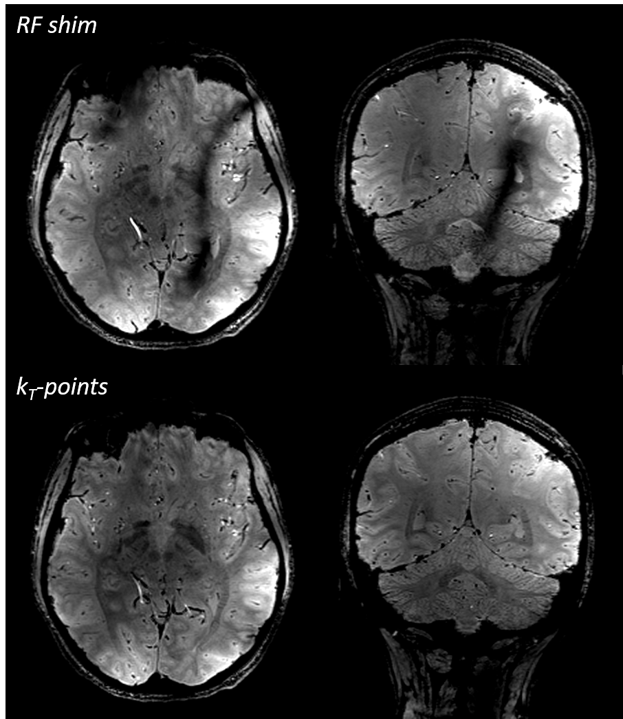
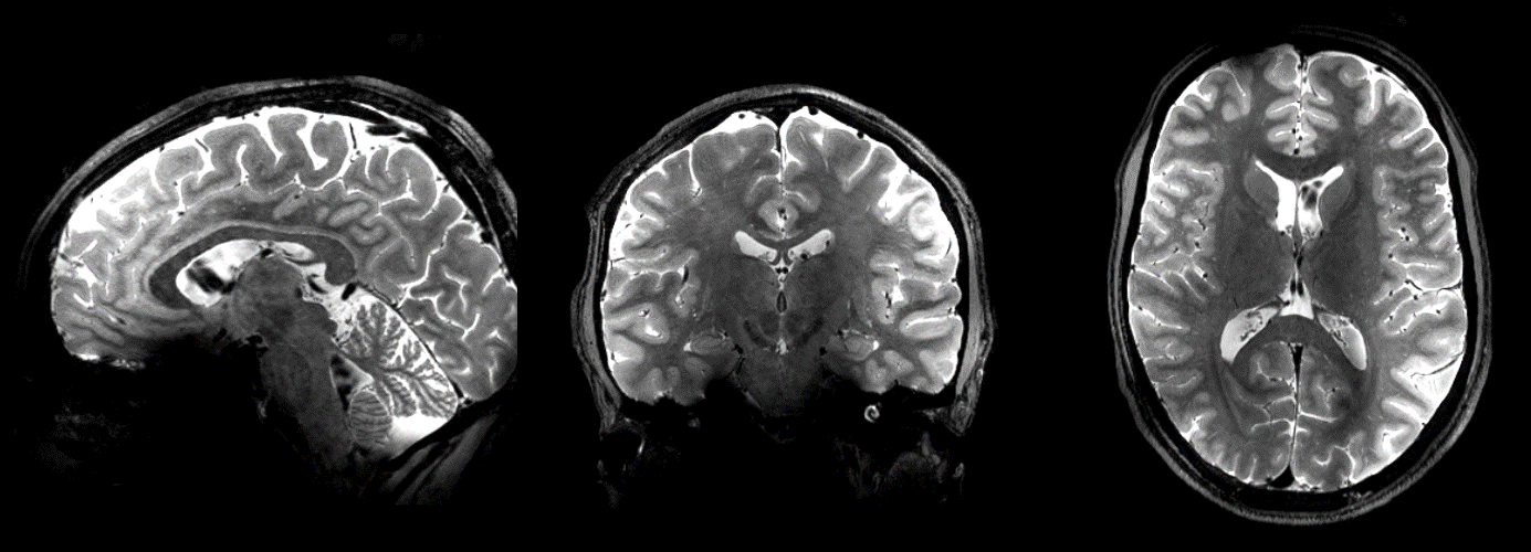
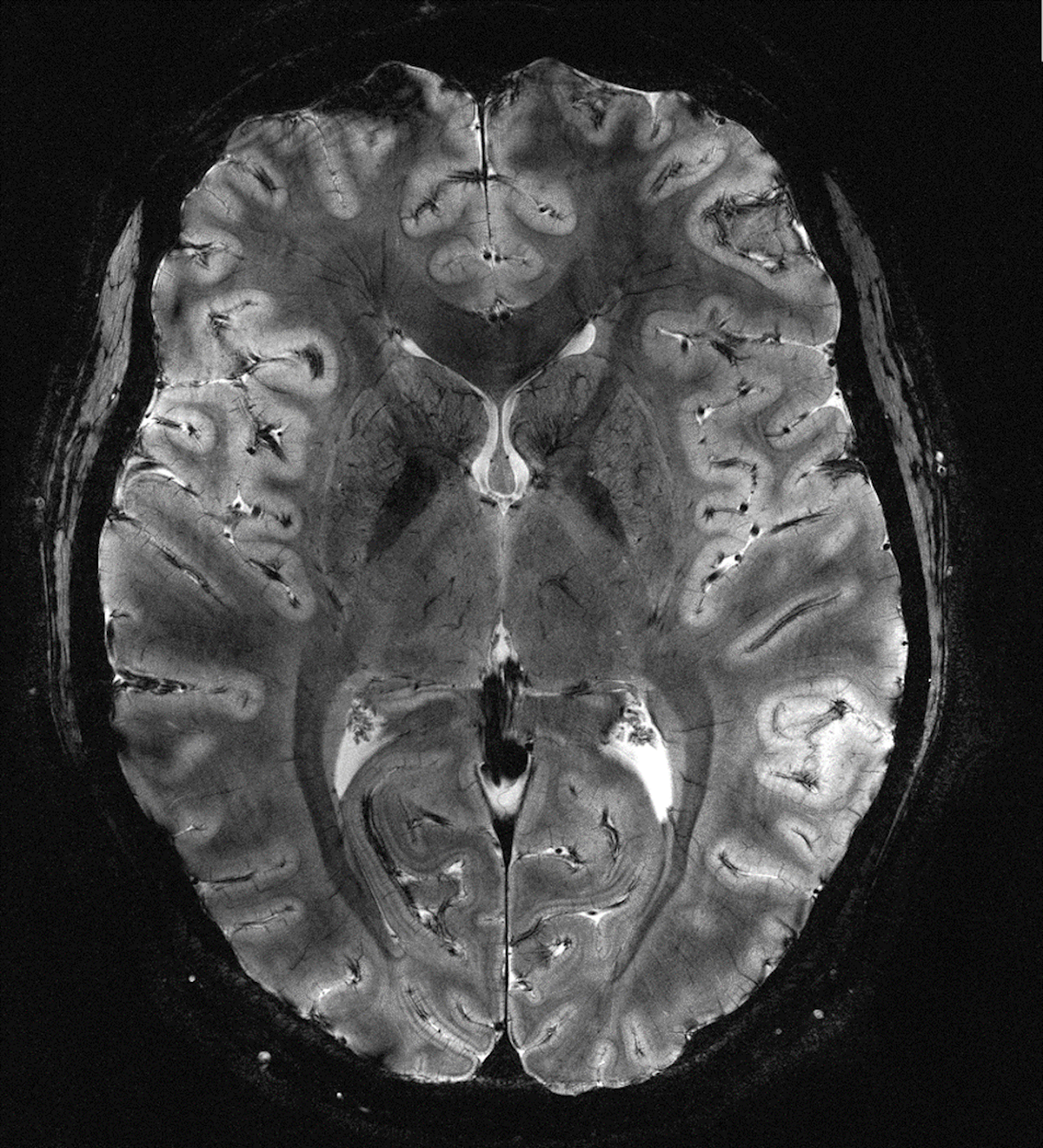
Twenty healthy volunteers were scanned at 11.7T between July 2023 and February 2024. The study was approved by the French national regulatory body (ANSM) and a national ethics committee while written informed consent was obtained from each participant. After passing an anxiety test, the participants underwent cognitive (before, during and after the MRI exam) and vestibular (before and after) tests. Blood samples were drawn before and after the exams to conduct a genotoxic [30] analysis subcontracted to an external and certified laboratory. Arterial pressure and cardiac pulsation were characterized before, during and after the MR exam. Another set of twenty volunteers was exposed to a 0T, nocebo, field and underwent the same tests (except genotoxicity). The environment was identical, i.e. the 11.7T scanner, for the latter group but with the magnetic field ramped down. The volunteers in that case were not informed of the absence of the field and received the same instructions as the 11.7T group. The sound of MR sequences for the 0T group was mimicked by recordings played by a loudspeaker hidden at the back of the magnet. For all tests performed throughout this study, no significant differences between the two groups was identified. Follow up phone calls by the NeuroSpin nurses up to 1 week after exposure at 11.7T did not indicate any abnormality.The MR sessions were 90 min long and targeted anatomical T1, T2 and T2* contrasts. The experiments were iteratively consolidated by the physics team at NeuroSpin in terms of workflow, sequence parameters and RF pulse design. A home-made pTx coil with dedicated VOPs was used [31]. Increased power losses at 500 MHz combined with the pairing strategy of 16 transmit elements to be fed by 8 RF power amplifiers made inversion pulses particularly difficult to design, with very localized B1+ field artefacts. The results underlined the necessity to have high power RF amplifiers (here 2 kW per channel) and the importance of low cable losses, transmit efficiency and possibly higher transmit channels count. The team at NeuroSpin was able to mitigate the RF field inhomogeneity problem at 11.7T in 3D T2*-weighted GRE (illustration in figure 1) in small flip angle excitation pulses (NRMSE≈8%) and in T2-weighted variable flip angle refocusing pulses (NRMSE≈11%) (figure 2) with kT-point [32] and GRAPE universal pulses [33] respectively. All acquisitions were performed with up to second order shimming using a home-made optimization and a computed brain mask. Comparisons obtained with similar data acquired at 3T and 7T revealed less than linear B0 field dispersions over the 3D brain, indicating more than satisfying results in terms of effective field homogeneity.
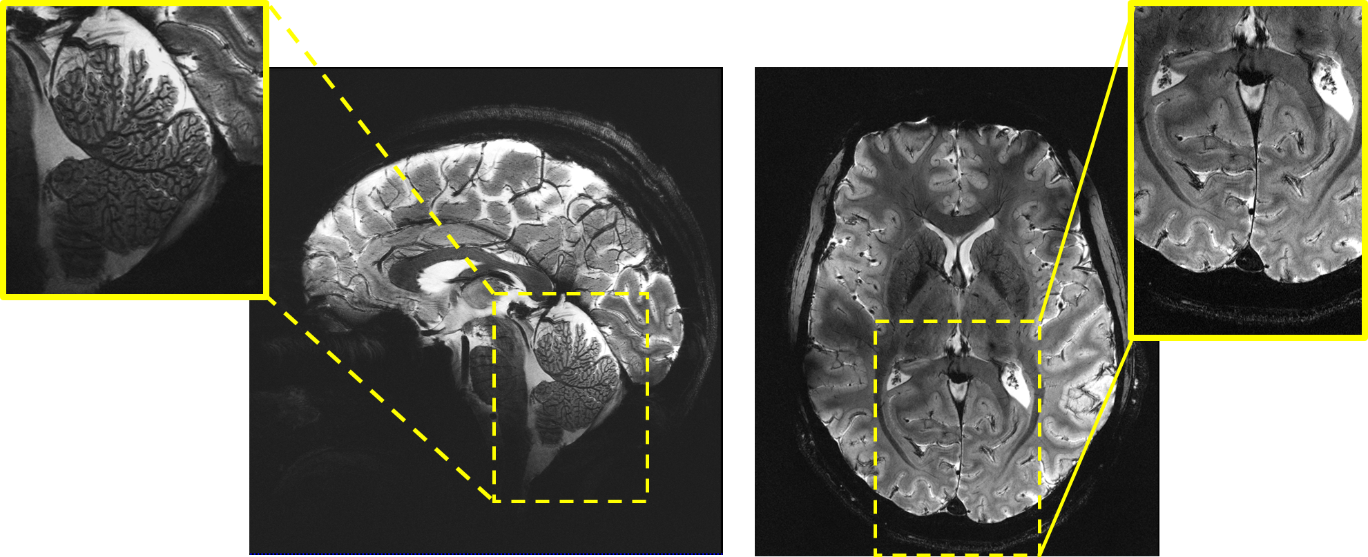
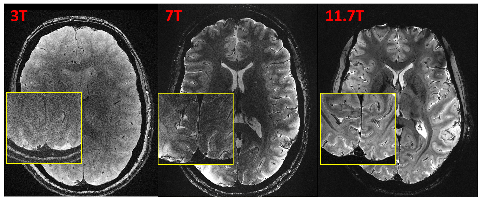
Figure 3 presents 0.2 × 0.2 × 1 mm3 2D T2*-weighted GRE acquisitions using selective spoke pulses [34,35]. Similar scans were performed at 3T and 7T on different subjects to visualize the gains brought by field strength (figure 4). A larger TE at 3T and 7T could increase the contrast to noise ratio but at the expense of less SNR. The signal and contrast were relatively robust with respect to flip angle, given the relatively small variation of T1 versus field strength. Careful analysis of the sequences aided with field monitoring and sequence adjustments were performed to minimize field perturbations during the acquisitions [6]. Finally, figure 5 reports the highest resolution result achieved in this first 11.7T in vivo protocol, i.e. with a T2*-weighted 2D GRE with resolution = 0.19 × 0.19 × 1 mm3. The SNR is still high enough to provide interesting details at this ultra-high resolution.
Discussion and conclusion
The first in vivo images acquired at 11.7T follow nearly 20 years of research and development. About 6 years of commissioning were dedicated to tests aimed at not only securing the magnet but also characterizing the behavior of the system. Needless to say, complexity in the MR operation increases on all fronts and NeuroSpin has benefitted from its great experience throughout the years at 7T. CMRR also has been a pioneering ally who performed safety tests and studies which inspired some of the safety investigations at 11.7T. When aiming at the highest resolution images in this protocol, success rate was low (~1/5) because of head motion and emphasizes the necessity to develop and implement motion correction tools [36,37] together with highly accelerated sequences [27]. Deployment of more efficient RF coils [17] and more receive channels count also is underway. These efforts fit in the context of a European effort gathering several expert partners (https://aroma-h2020.com/). Such collaborations should be encouraged to face the broad challenges and maximize the benefits to do neuroscience in a reasonable timeframe. Complexity but also larger financial investments therefore lie beyond the 7T horizon. With the first images ever acquired at 11.7T revealing superb brain details, but also with the safety tests confirming the absence of adverse effects, UHF MRI beyond 7T constitutes a unique platform to learn new physics, develop and merge new technologies and open new horizons to explore the human brain in vivo.Acknowledgements
AROMA H2020 FET-Open (885876). ANR-21-ESRE-0006 (“Investissements d'avenir"). The authors are immensely grateful to the Irfu department at CEA for designing and commissioning the Iseult magnet, and to the NeuroSpin platform personnel without whom the experiments would not have been possible. The authors also thank Siemens Healthineers for valuable support.References
[1] D. A Feinberg, A. JS Beckett, A.T Vu, J. Stockmann, L. Huber, S. Ma, S. Ahn, K. Setsompop, X. Cao, S. Park, C. Liu, L.L Wald, J.R Polimeni, A. Mareyam, B. Gruber, R. Stirnberg, C. Liao, E. Yacoub, M. Davids, P. Bell, E. Rummert, M. Koehler, A. Potthast, I. Gonzalez-Insua, S. Stocker, S. Gunamony, P. Dietz. Next-generation MRI scanner designed for ultra-high-resolution human brain imaging at 7 Tesla. Nat Methods 2023. https:// doi. org/ 10. 1038/s41592- 023- 02068-7.
[2] T. K F Foo, T.T Ek , M.E Vermilyea, Y. Hua, E. Fiveland, J.E Piel, K. Park, J. Ricci, P.S Thompson, D. Graziani, G. Conte, A. Kagan, Y. Bai, C. Vasil, M. Tarasek, D.TB Yeo, F. Snell, D. Lee, A. Dean , J.K De Marco, R.Y Shih, M.N Hood, H. Chae, V.B Ho. Highly efficient head-only magnetic field insert gradient coil for achieving simultaneous high gradient amplitude and slew rate at 3.0T (MAGNUS) for brain microstructure imaging. Magn Reson Med 2020 ;83:2356–2369.
[3] R. Pohmann, O. Speck, K. Scheffler. Signal-to-noise ratio and MR tissue parameters in human brain imaging at 3, 7, and 9.4 tesla using current receive coil arrays. Magn Reson Med 2016 ;75:801–809.
[4] C. Le Ster, A. Grant, P-F. Van de Moortele, A. Monreal-Madrigal, G. Adriany, A. Vignaud, F. Mauconduit, C. Rabrait-Lerman, B. Poser, K. Ugurbil, N. Boulant. Magnetic field strength dependent SNR gain at the center of a spherical phantom and up to 11.7T. Magn Reson Med 2022 ; 88:2131–2138.
[5] N. Boulant, L. Quettier, the Iseult consortium (2023) Commissioning of the Iseult CEA 11.7T whole-body MRI: current status, gradient–magnet interaction tests and first imaging experience. Magn Reson Mater Phys. https:// doi. org/ 10. 1007/ s10334- 023- 01063-5.
[6] N. Boulant, C. Le Ster, A. Amadon, G. Aubert, A. Beckett, J. Belorgey, C. Bonnelye, D. Bosch, D. O Brunner, G. Dilasser, O. Dubois, P. Ehses, D. Feinberg, S. Feizollah, V. Gras, S. Gross, Q. Guihard, H. Lannou, D. Le Bihan, F. Mauconduit, F. Molinié, F. Nunio, K. Pruessmann, L. Quettier, K. Scheffler, T. Stöcker, C. Tardif, K. Ugurbil, A. Vignaud, A. Vu, X. Wu. The possible influence of third-order shim coils on gradient–magnet interactions: an inter-field and inter-site study. Magn Reson Mater Phys. 2024 ; https://doi.org/10.1007/s10334-023-01138-3.
[7] C. Barmet, N. De Zanche, K.P Pruessmann. Spatiotemporal Magnetic field monitoring for MR. Magn Reson Med 2008 ; 60:187–197.
[8] S.J Vannesjo, M. Haeberlin, L. Kasper, M. Pavan, B.J Wilm, C. Barmet, K.P Pruessmann. Gradient system characterization by impulse response measurements with a dynamic field camera. Magn Reson Med 2013 ; 69:583–593.
[9] I. Tkáč, M. A. Benneyworth, T. Nichols-Meade, E. L. Steuer, S. N. Larson, G. J. Metzger, K. Uğurbil. Long-term behavioral effects observed in mice chronically exposed to static ultra-high magnetic fields. Magn Reson Med 2021; 86:1544-1559.
[10] A. Grant, G. J. Metzger, P-F. Van de Moortele, G. Adriany, C. Olman, L. Zhang, J. Koopermeiners, Y. Eryaman, M. Koeritzer, M. E. Adams, T. R Henry, K. Uğurbil. 10.5 T MRI static field effects on human cognitive, vestibular, and physiological function. MRI 2020 ; 73 :163-176.
[11] C. Le Ster, E. Selingue, R. Poirier, J-M. Edeline, S. Mériaux, N. Boulant. Behavioral and functional assessment of mice inner ear after chronic exposure to an ultrahigh B0 field of 11.7 T or 17.2 T. Magn Reson Med 2023 ; 90:699-707.
[12] S. A. Winkler, A. Alejski, T. Wade, C. A. McKenzie, B. K. Rutt. On the accurate analysis of vibroacoustics in head insert gradient coils. Magn Reson Med 2017 ; 78 :1635-1645.
[13] M. Davids, P. Dietz, G. Ruyters, M. Roesler, V. Klein, A. J.S. Beckett, A. T. Vu, B. Guérin, D. A. Feinberg, L. L. Wald. Peripheral nerve stimulation informed design of a high-performance asymmetric head gradient coil. Magn Reson Med 2023 ;90 :784-801.
[14] V. Gras, X. Wu, N. Boulant. Ultra-high field Neuro MRI. Elsevier Academic press. Editors : K. Markenroth Bloch, M. Guye, B. A. Poser.
[15] V. Gras, A. Vignaud, A. Amadon, D. Le Bihan, N. Boulant. Universal Pulses: a new concept for calibration-free parallel transmission. Magn Reson Med 2017; 77:635-643.
[16] O. Geldschläger, D. Bosch, S. Glaser, A. Henning. Local excitation universal parallel transmit pulses at 9.4T. Magn Reson Med 2021 ;86 :2589-2603.
[17] S. Chu, V. Gras, F. Mauconduit, A. Massire, N. Boulant, S. Gunamony. Electromagnetic and RF pulse design simulation based optimization of an eight-channel loop array for 11.7T brain imaging. Magn Reson Med 2023; 90:770-783.
[18] M. A. Ertürk, X. Wu, Y. Eryaman, P-F. Van de Moortele, E. J. Auerbach, R. L. Lagore, L. DelaBarre, J. T. Vaughan, K. Uğurbil, G. Adriany, G. J. Metzger. Toward imaging the body at 10.5 tesla 2017;77:434-443.
[19] Z. Cao, J. Park, Z-H Cho, C. Collins. Numerical evaluation of image homogeneity, signal-to-noise ratio, and specific absorption rate for human brain imaging at 1.5, 3, 7, 10.5, and 14T in an 8-channel transmit/receive array. Journal of magnetic resonance imaging. 2015:41(5):1432-9.
[20] C. Le Ster, F. Mauconduit, C. Mirkes, A. Vignaud, N. Boulant. Measuring radiofrequency field-induced temperature variations in brain MRI exams with motion compensated MR thermometry and field monitoring. Magn Reson Med 2022;87:1390-1400.
[21] N. Boulant, X. Wu, G. Adriany, S. Schmitter, K. Ugurbil, P-F Van de Moortele. Direct control of the temperature rise in parallel transmission: simulations at 10.5 T. Magn Reson Med 2016; 75:249-256.
[22] B. Pinho Meneses, A. Amadon. Physical limits to human brain B0 shimming with spherical harmonics, engineering implications thereof. Magnetic Resonance Materials in Physics, Biology and Medicine 35 (6), 923-941.
[23] C. Juchem, T.W Nixon, S. McIntyre, V.O Boer, D.L Rothman, R.A.d Graaf. Dynamic multi-coil shimming of the human brain at 7T. J Magn Reson 2011 ; 212 : 280–288. doi:10.1016/j.jmr.2011.07.005.
[24] J.P Stockmann, N.S Arango, T. Witzel, A. Mareyam, C. Sappo, J. Zhou, L. Jenkins, L. Craven-Brightman, E. Milshteyn, M. Davids, W.S Hoge, M. Sliwiak, S. Nasr, B. Keil, E. Adalsteinsson, B. Guerin, J.K White, K. Setsompop, J.R Polimeni, L.L Wald. A 31-channel integrated “AC/DC” B0 shim and radiofrequency receive array coil for improved 7T MRI. Magn Reson Med 2022 ;87 :1074-1092.
[25] B. Pinho Meneses, J-P Stockmann, N. Arango, P-F Gapais, E. Giacomini, F. Mauconduit, V. Gras, N. Boulant, A. Vignaud, M. Luong, A. Amadon. Shim coils tailored for correcting B0 inhomogeneity in the human brain (SCOTCH): Design methodology and 48-channel prototype assessment in 7-Tesla MRI. NeuroImage 2022 ; 261, 119498.
[26] A. Aranovitch, M. Haeberlin, S. Gross, B. E. Dietrich, B. J. Wilm, D. O. Brunner, T. Schmid, R. Luechinger, K. P. Pruessmann. Prospective motion correction with NMR markers using only native sequence elements. Magn Reson Med 2018 ;79 :2046-2056.
[27] R. Stirnberg, T. Stöcker. Segmented K-space blipped-controlled aliasing in parallel imaging for high spatiotemporal resolution EPI. Magn Reson Med 2021 ;85 :1540-1551.
[28] M. Engel, L. Kasper, C. Barmet, T. Schmid, L. Vionnet, B. Wilm, K. P. Pruessmann. Single-shot spiral imaging at 7 T. Magn Reson Med 2018 ;80 :1836-1846.
[29] C. Lazarus, P. Weiss, N. Chauffert, F. Mauconduit, L. El Gueddari, C. Destrieux, I. Zemmoura, A. Vignaud, P. Ciuciu. SPARKLING: variable-density k-space filling curves for accelerated T2*-weighted MRI. Magn Reson Med 2019 ;81 :3643-3661.
[30] C. Herate, P. Brochard, F. De Vathaire, M. Ricoul, B. Martins, L. Laurier, J-R Deverre, B. Thirion, L. Hertz-Pannier, L. Sabatier. The effects of repeated brain MRI on chromosomal damage. Eur Radiol Exp 6, 12 (2022). https://doi.org/10.1186/s41747-022-00264-2.
[31] M. Luong, G. Ferrand, E. Chazel, P-F Gapais, V. Gras, N. Boulant, A. Amadon. A Compact 16Tx-32Rx Geometrically Decoupled Phased Array for 11.7T MRI. Proceedings of the annual meeting of the ISMRM, 2023, Toronto, Canada, p 0707.
[32] M.A Cloos, N. Boulant, M. Luong, G. Ferrand, E. Giacomini, D. Le Bihan, A. Amadon. kT-points: short three dimensional tailored RF pulses for flip angle homogenization over an extended volume. Magn Reson Med 2012 ; 67:72–80.
[33] L. Van Damme, F. Mauconduit, T. Chambrion, N. Boulant, V. Gras. Universal nonselective excitation and refocusing pulses with improved robustness to off-resonance for Magnetic Resonance Imaging at 7 Tesla with parallel transmission. Magn Reson Med 2021 ;85 : 678-693.
[34] F. Mauconduit, V. Gras, A. Vignaud, N. Boulant. MetaPulse2D: methodology to enable universal slice specic spokes pulses in parallel transmission. Proceedings of the annual meeting of the ISMRM, 2022, London, UK, p 0454.
[35] S. Orzada, S. Maderwald, B. A. Poser, A. K. Bitz, H. H. Quick, M. E. Ladd. RF excitation using time interleaved acquisition of modes (TIAMO) to address B1 inhomogeneity in high-field MRI. Magn Reson Med 2010 ;64 :327-333.
[36] M. Riedel, T. Ulrich, K. Pruessmann. Real-time correction of rigid motion and 1st-order shims using rapid 3D orbital navigators. Proceedings of the annual meeting of the ISMRM, 2023, Toronto, Canada, p 1017.
[37] M. Serger, R. Stirnberg, P. Ehses, T. Stoecker. Combining FID navigators with field probe monitoring for improved head motion tracking and prospective correction at 7T. Proceedings of the annual meeting of the ISMRM, 2023, Toronto, Canada, p 1834.
Figures