MRI for Active Surveillance of Renal Masses
Kye Jin Park1
1Asan Medical Centre, Korea, Republic of
1Asan Medical Centre, Korea, Republic of
Synopsis
Keywords: Body: Kidney, Cross-organ: Cancer
Synopsis:- Introduction: Active surveillance of renal mass is defined as serial imaging that monitors serial changes regarding growth and morphology.
- Patient selection: According to several published guidelines and white papers, small renal masses especially < 2 cm with significant comorbidities and/or limited life expectancy are eligible for active surveillance.
- Active surveillance of solid renal masses: The role of MRI will be discussed with the use of the clear cell likelihood ratio as a supportive tool.
- Active surveillance of cystic renal masses: Serial changes in morphology is important in cystic renal masses.
MRI Surveillance for Small Renal Masses
Approximately 20% of small (≤ 4 cm) renal masses are benign neoplasms, and the rest of tumors are malignant. Small solid masses are frequently indolent with low rates of disease progression or distant metastasis, although they are malignant. Therefore, surveillance using imaging for renal masses can be justified in elderly patients or those with comorbidities. Several guidelines and white papers determine the criteria and protocols of active surveillance for renal masses. Recommendations for follow-up intervals vary among the guidelines and white papers. However, tumor growth exceeding 0.4-0.5 cm per year or significant morphologic changes prompt consideration of surgical or ablative treatment. MRI enables improved characterization of small renal masses by detecting macroscopic/microscopic fat, fibrosis, hemorrhage, and evaluating enhancement characteristics. Clear cell likelihood ratio for solid renal mass and Bosniak classification for cystic renal masses can be utilized as MRI reporting tool for monitoring lesion characteristics or morphology.Acknowledgements
NoneReferences
1. Shieda N, Krishna S, Pedrosa I, et al. Active Surveillance of Renal Masses: The Role of Radiology. Radiology 2022; 302:11–24.
2. Shetty AS, Fraum TJ, Ballard DH, et al. Renal Mass Imaging with MRI Clear Cell LikelihoodScore: A User’s Guide. RadioGraphics 2023; 43(7):e220209.
Figures
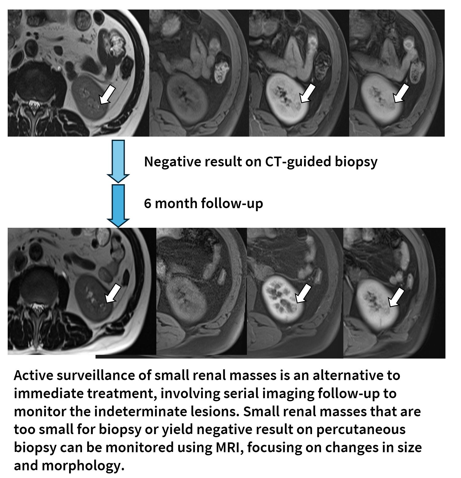
A
63-year-old man who underwent radical right nephrectomy for clear cell renal
cell carcinoma. On postoperative follow-up imaging, a new 9-mm T2 isointense
nodule with moderate contrast enhancement was observed (above). CT-guided
percutaneous biopsy was attempted, but yielded negative results because of targeting failure. The patient is
on active surveillance for the small renal mass at 6 month intervals.