Uterine Cancer: T2W or Texture?
1Department of Diagnostic Imaging and Nuclear Medicine, Kyoto Univesity Hospital, Kyoto, Japan
Synopsis
Keywords: Body: Urogenital
Motivation: Conventional MRI is crucial for uterine cancer in clinical settings. Radiomics, utilizing quantitative imaging features, has rapidly evolved in research. A brief review provides insights for addressing current clinical challenges and guiding future developments.
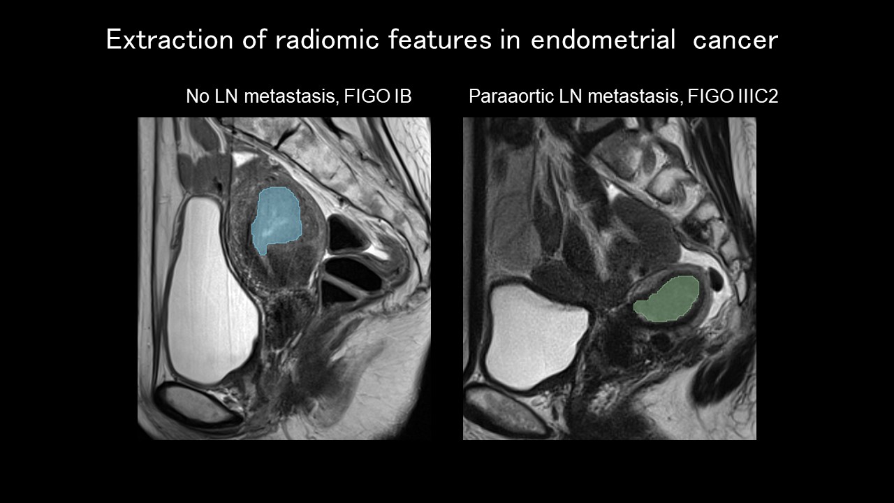
Goal(s): The goals are to grasp the roles of conventional MRI, achievements in radiomic studies for uterine cancer (particularly endometrial cancer), and the limitations and improvements in imaging quality.
Approach: Reviewing the latest research on MRI and radiomics in uterine cancer, with a primary focus on endometrial cancer.
Results: Despite limitations, radiomics continues to advance. Improvements in conventional MRI quality for gynecologic imaging are clinically significant and enhance radiomics.
Impact: Grasping current improvements in MRI quality and radiomics, along with identifying challenges, offers insights for addressing clinically relevant issues in uterine cancer.
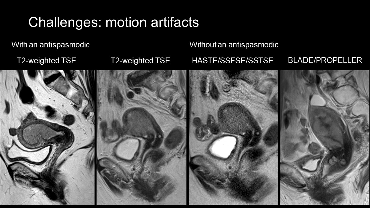
Commonly cited drawbacks of radiomics include issues related to generalizability and reproducibility. While clinical implementation may take time, steady progress is being made in overcoming these challenges [2, 3]. In considering MRI radiomics in uterine cancer, vulnerability to artifacts is identified as a specific issue: motion artifacts from respiratory movements or intestines; susceptibility artifacts by rectal air often affecting DWI and its apparent diffusion coefficient (ADC) map. Motion artifacts by intestines is particularly noteworthy when performed without the administration of antispasmodics. Improving the robustness of T2WI and DWI to artifact is essential to ensure the generalizability of radiomics.
Several promising techniques have been reported for improving image quality. As a representative, deep learning (DL) reconstruction is gaining attention [4]. DL reconstruction enhances spatial resolution, reduces noise, and shortens acquisition time. The reduction of acquisition time, while maintaining image quality, directly leads to artifact resistance. Obtaining high-quality T2WI and DWI, even without antispasmodics, not only improves conventional imaging diagnostics but also benefits image analysis techniques, including radiomics. Furthermore, exploration of user-friendly clinical tools based on advanced computation techniques is crucial in future imaging diagnostics, extending beyond radiomics.
Acknowledgements
No acknowledgement found.References
1. Kido, A., et al., Preoperative Imaging Evaluation of Endometrial Cancer in FIGO 2023. J Magn Reson Imaging, 2023.
2. Lefebvre, T.L., et al., Development and Validation of Multiparametric MRI-based Radiomics Models for Preoperative Risk Stratification of Endometrial Cancer. Radiology, 2022. 305(2): p. 375-386.
3. Zwanenburg, A., et al., The Image Biomarker Standardization Initiative: Standardized Quantitative Radiomics for High-Throughput Image-based Phenotyping. Radiology, 2020. 295(2): p. 328-338.
4. Tsuboyama, T., et al., Impact of Deep Learning Reconstruction Combined With a Sharpening Filter on Single-Shot Fast Spin-Echo T2-Weighted Magnetic Resonance Imaging of the Uterus. Invest Radiol, 2022. 57(6): p. 379-386.
Figures