Al in Hepatobiliary MRI: Landing on the Moon or Journey to Mars
Takeshi Nakaura1 and Toshinori Hirai2
1Kumamoto University, Honjo 1-1-1, Kumamoto, Japan, Kumamoto, Japan, 2Kumamoto University, Kumamoto, Japan
1Kumamoto University, Honjo 1-1-1, Kumamoto, Japan, Kumamoto, Japan, 2Kumamoto University, Kumamoto, Japan
Synopsis
Keywords: Education Committee: Clinical MRI
Deep Learning Reconstruction (DLR) evolutionizes MRI with noise reduction and super-resolution. This advancement positions DLR as the likely future standard for MRI examinations. Concurrently, the performance of large language models (LLM) has seen remarkable improvements, epitomized by the development of ChatGPT. The application of these models in the medical arena, particularly in the hepatobiliary sector, marks one of today's most promising AI frontiers. This presentation delves into the fundamentals of Deep Learning, elucidating the underlying mechanisms of DLR and LLM, and showcases their clinical applications with an emphasis on the hepatobiliary and pancreatic regions.Introduction
The integration of Artificial Intelligence (AI) into radiology has catalyzed a paradigm shift in diagnostic methodologies, particularly in Magnetic Resonance Imaging (MRI). Among the numerous AI advancements, Deep Learning Reconstruction (DLR) stands out for its revolutionary approach to image processing. It is a technology that has already reached practical levels and is widely used in clinical settings, offering significant improvements in image quality and diagnostic accuracy without the trade-offs traditionally associated with enhanced imaging techniques. Simultaneously, the evolution of large language models (LLM), exemplified by developments like ChatGPT, has introduced unprecedented capabilities in understanding and generating human-like text. While not yet widely adopted, these advancements hold great potential for revolutionizing medical diagnostics in the near future, including applications in the hepatobiliary field. This abstract explores the dual innovations of DLR and LLM, focusing on their fundamentals, technological advancements, and clinical applications, with a particular emphasis on their impact on hepatobiliary and pancreatic imaging.Advancements in Deep Learning Reconstruction (DLR)
DLR represents a major leap forward in MRI technology, utilizing deep learning algorithms to reconstruct images in a way that significantly reduces noise while preserving, if not enhancing, spatial resolution. This is achieved through the application of neural networks that have been trained on vast datasets of high-quality MRI scans, allowing the algorithms to "learn" how to optimize image reconstruction for clarity and detail. The implications of this technology are profound, offering the potential to reduce scan times, improve patient comfort, and enhance the detection and characterization of lesions, particularly in the complex and often challenging areas of the hepatobiliary and pancreatic regions. This section will also discuss our recent studies (1) on the application of DLR including super-resolution DLR for the hepatobiliary and pancreatic fields.Breakthroughs in Large Language Models (LLM)
The introduction of the Transformer architecture and subsequent development of LLMs like the Generative Pre-trained Transformer (GPT) series have further revolutionized AI. LLMs, exemplified by ChatGPT, have shown potential in automating tasks such as report generation in radiology, saving time and improving diagnostic efficiency and accuracy. However, despite their utility, there's a lack of comprehensive reviews on the development of LLMs for clinical radiologists. This session aims to provide a concise history and overview of LLMs' application in radiology for clinical radiologists. Additionally, this section will also discuss our recent studies (2) on the application of LLMs to automate the generation of radiological reports.Conclusion
The advent of AI in medical imaging, particularly through the advancements in Deep Learning Reconstruction and Large Language Models, represents a transformative era in the diagnosis and treatment of hepatobiliary and pancreatic diseases. As these technologies continue to evolve and mature, their integration into clinical practice is likely to become the standard, offering unprecedented precision and efficiency in medical diagnostics.Acknowledgements
We thank Ms. Tae Hamakawa from Department of Diagnostic Radiology, Kumamoto University, Japan, for her help with the measuring in the quantitative analysis.References
1. Deep learning-based reconstruction and 3D hybrid profile order technique for MRCP at 3T: evaluation of image quality and acquisition time. Kaori Shiraishi, Takeshi Nakaura et al. Eur Radiol. 2023 Nov;33(11):7585-7594.
2. Preliminary assessment of automated radiology report generation with generative pre-trained transformers: comparing results to radiologist-generated reports. Takeshi Nakaura, Naofumi Yoshida et al. Jpn J Radiol. 2024 Feb;42(2):190-200.
Figures
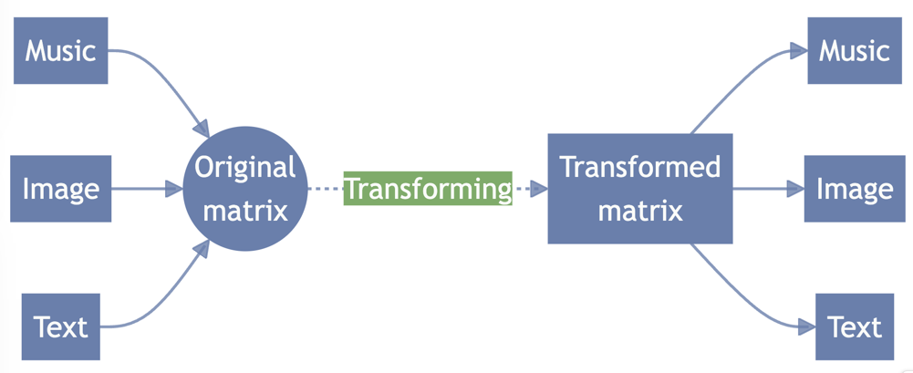
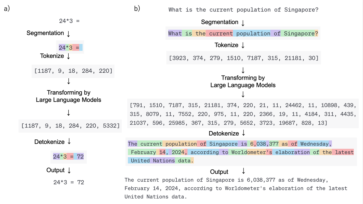
This schema represents how deep learning handles different types of data. Various input data are converted to matrices, which are then converted to other matrices and then back to various data types. Deep learning training is designed to improve the accuracy of this transformation.
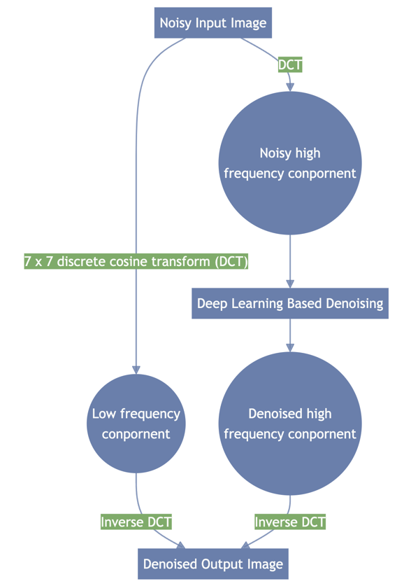
A typical denoising schema based on deep learning (AiCE; Canon Medical Systems). Noisy input image is separated into high-frequency and low-frequency components; high-frequency components are denoised by deep learning to remove noise and preserve complex structures. The denoised high-frequency and low-frequency components are combined after the denoising process.
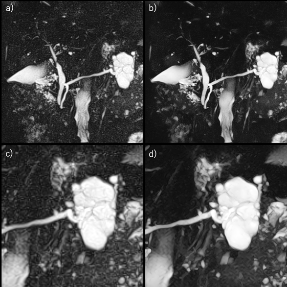
Figure shows 3D-MRCPs within a single breathhold images of without SR-DLR (a and c) and MRCP with SR-DLR (b and d). Compared with MRCP with SR-DLR, MRCP without DLR shows lower
contrast, more noise, and lower overall image quality. DLR, deep learning reconstruction; MRCP, MR cholangiopancreatography.
Examples of a) computation and b) conversation by Large Language Model. All of these different language processing tasks can be accomplished using the same process: converting input data into a matrix using a tokenizer, transforming it into another matrix using a Large Language Model, and then converting it back into output data.