Diffusion-Weighted MRS: Insights into the Brain Microstructure from Metabolites
André Döring1
1CIBM - Center for Biomedical Imaging, Lausanne, Switzerland
1CIBM - Center for Biomedical Imaging, Lausanne, Switzerland
Synopsis
Keywords: Contrast mechanisms: Diffusion-Weighted MR Spectroscopy, Contrast mechanisms: Diffusion
Magnetic resonance spectroscopy (MRS) allows quantifying concentrations of cell-type specific metabolites. This enabled to monitor metabolic pathways. However, alongside metabolic changes, alterations in cellular microstructure represent pivotal pathomechanistic shifts in the onset and progression of neurodegenerative-diseases (e.g., glial morphology in neuroinflammation or neuronal integrity in cerebral atrophy). By integrating diffusion-weighting into MRS (dMRS), we can measure the diffusion properties of individual, celltype-specific metabolites that can serve as probes for cellular microstructure. In combination with simulation techniques, enabling an absolute quantification of cellular-features, dMRS holds immense promise in offering a new class of biomarkers for identifying cell-type specific (patho)morphological alterations.Overview
Diffusion-weighted MRS (dMRS) has witnessed remarkable advancements in recent years. This lecture is a gateway to the understanding of the intricacies of dMRS and tailored for enthusiasts and those eager to explore its potential.1. Demystifying dMRS: Discover how dMRS works to unlock cellular secrets. From fundamental techniques like pulsed gradient [PG] encoding, to sophisticated methods such as oscillating [OG] and free waveform [FWF] gradients, to double-diffusion-encoding [DDE] and the concept of diffusion-time [TD].
2. Selecting the Right Sequence: Navigate the maze of dMRS sequences tailored to human and animal applications. Uncover the optimal sequence choices for your study considering microstructural sensitivity, gradient performance, SAR, and PNS limitations.
3. Crafting a dMRS Protocol: Master the art of setting up a meticulous dMRS acquisition protocol. Learn essential steps of shimming, water reference scanning, and gradient-polarity inversion, to ensure high-quality dMRS spectra.
4. Processing and Fitting Pipeline: Harness the power of open-source tools for processing and fitting dMRS data and learn about common signal distortions, pitfalls and how to avoid them.
5. Unveiling Cellular Insights: Explore new studies where dMRS sheds light on (patho)morphological alterations and gain an understanding where dMRS can help unraveling the mysteries of the brain microstructure.
Acknowledgements
No acknowledgement found.References
No reference found.Figures
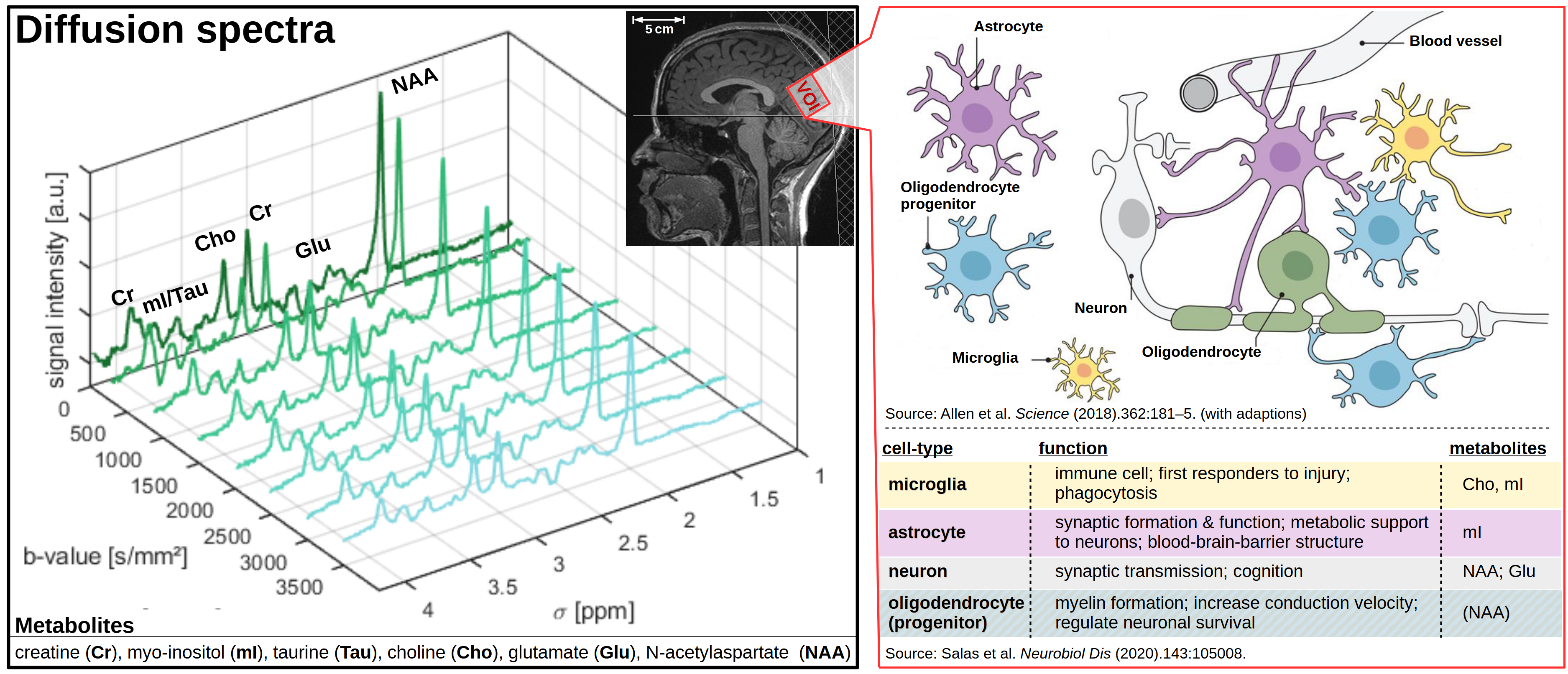
Diffusion-weighted spectra and cell-specific metabolites.