Clinical Applications of MRS in the Body
1University of Newcastle, Callaghan, Australia
Synopsis
Keywords: Body: Body, Contrast mechanisms: Spectroscopy, Education Committee: Clinical MRI
Motivation:MR Spectroscopy (MRS) offers a unique perspective into the body's biochemistry, providing insights into various diseases beyond what conventional imaging techniques can reveal.Goal(s):To elucidate the clinical applications of MRS, highlighting its role in diagnosing and monitoring diseases.
Approach:To explore MRS techniques, examining its integration with MRI, and discussing specific applications in various pathologies through case studies and recent research.
Results:MRS's effectiveness in precise diagnosis and treatment monitoring,revealing its potential in clinical scenarios from cancer to metabolic disorders.
Impact:MRS's capabilities are highlighted, prompting further research into its diagnostic precision. This enables clinicians to transform how they care for their patients.
Introduction to MR Spectroscopy
- Explain the concept of Magnetic Resonance, focusing on its non-invasive nature.
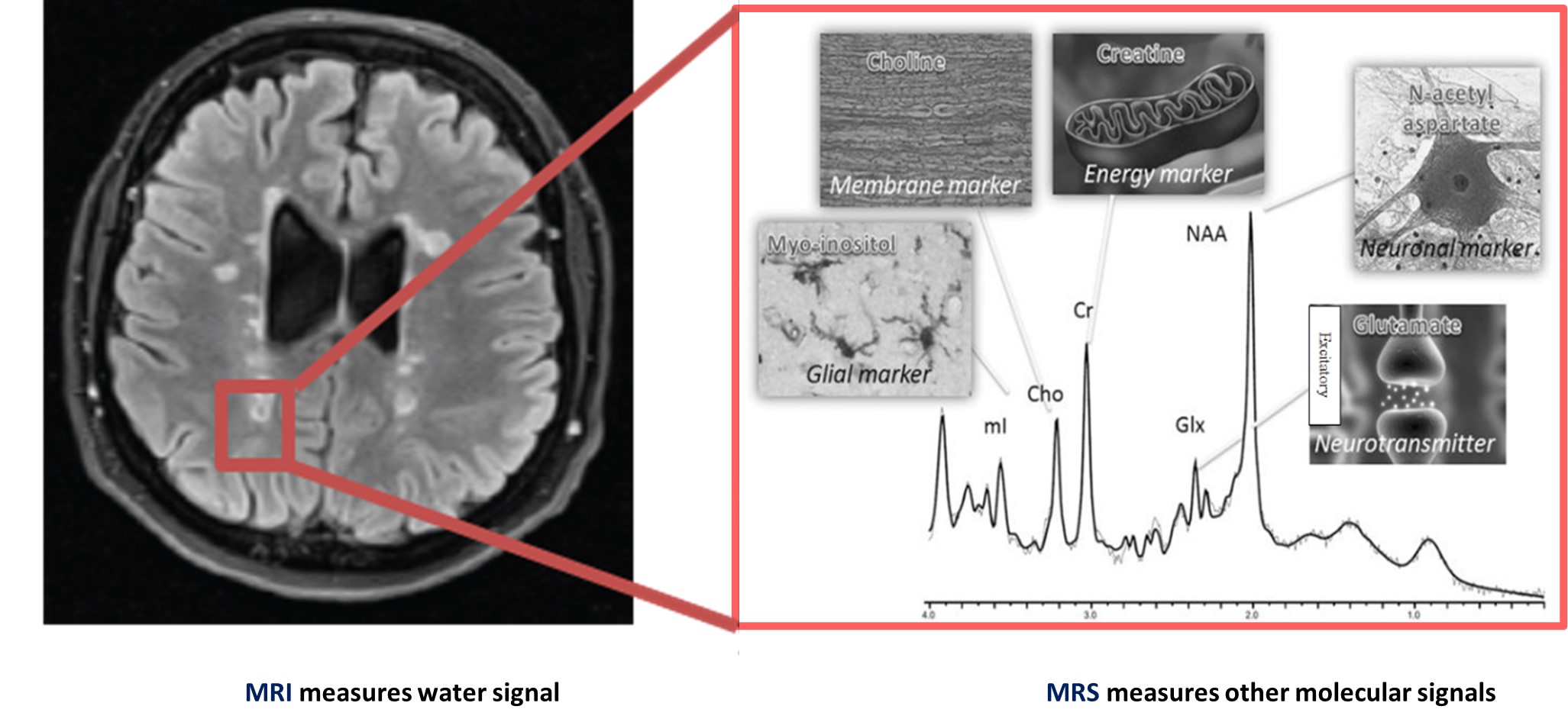
- Compare MRI and MRS: While MRI visualizes anatomical structures, MRS provides metabolic information1 (Figure 1).
- Describe how it’s acquired and how it measures the chemical composition of tissues.
Key Nuclei in MRS
- Elaborate on the significance of different nuclei:
- Proton (1H) is commonly used due to its abundance in biological tissues and sensitivity.
MRS and MRI: A Synergistic Approach
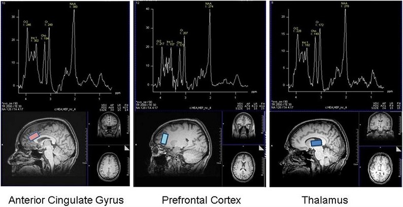
- Discuss how MRI aids 1HMRS by offering anatomical context for spectral data.
- Example: In brain studies, MRI helps localize regions for spectroscopic analysis, enhancing diagnostic accuracy2(Figure 2).
Clinical Application: Neurological Diseases
- Discuss 1HMRS application in neurology, particularly in identifying neurotransmitter imbalances3.
- Highlight the use of 1HMRS in detecting changes in brain chemistry related to multiple sclerosis4, brain tumors and lesions5.
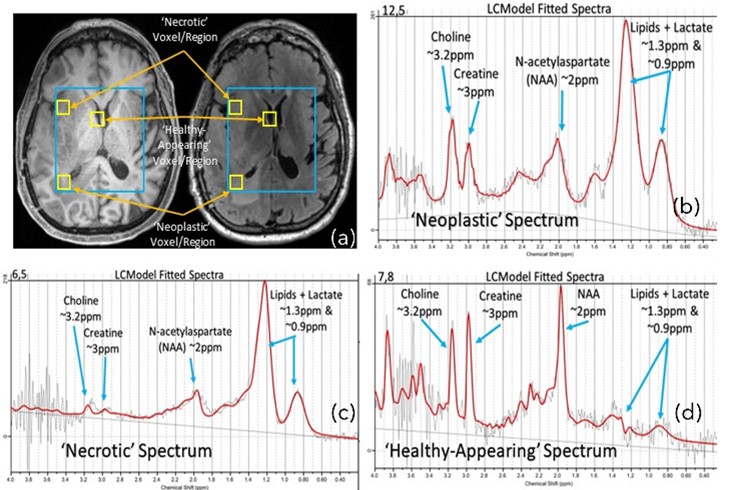
- Case example: Differentiating tumor types based on metabolic profiles observed in MRS(Figure 3).
MRS in Cancer Diagnosis
- Discuss how MRS identifies unique metabolic fingerprints of different tumor types.
- Differentiating benign from malignant tumors based on the presence or absence of certain metabolites.
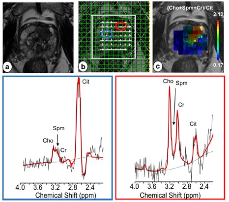
- Example: In prostate cancer, MRS reveals decreased levels of citrate and increased levels of choline and creatine, compared to normal prostate tissue6(Figure 4).
MRS in Dementia and Parkinson’s Disease
- Explain how MRS helps in understanding the biochemical changes in the brain associated with dementia and Parkinson's disease7.
- Mention studies showing altered metabolite levels in these conditions.
MRS in Liver Diseases
- Highlight the role of MRS in metabolic diseases, such as measuring liver fat content in diabetes or assessing intramuscular lipid levels8.
- Discuss how MRS can assess liver function by detecting metabolic changes associated with conditions like cirrhosis or fatty liver disease.
MRS in Breast Assessment
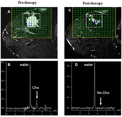
- Discuss the enhanced Detection role of MRS for clearer identification of key metabolites related to breast cancer, improving diagnostic accuracy9 (Figure 5).
- Discuss roles of 1D/2D allowing for more precise analysis of breast tissue composition.
- Discuss Clinical Utility for early cancer detection and monitoring treatment effects.
MRS in Musculoskeletal Disorders
- Discuss the use of MRS in evaluating muscle disorders by measuring metabolites like creatine and lactate, indicating muscle metabolism and health10 .
MRS in Pediatric Disorders
- Discuss unique applications in pediatrics, like assessing brain development in neonates or metabolic disorders in children11.
Future Directions in MRS
- Discuss potential future applications, such as personalized medicine or real-time monitoring of treatment responses.
Challenges and Limitations of MRS
- Address limitations such as lower spatial resolution compared to MRI, sensitivity to motion, and the need for expert interpretation12.
- Discuss technical challenges, including signal interference from water and lipids, and the need for precise localization and water suppression techniques.
Conclusion
- Summarize the key roles of MRS in clinical diagnosis and research, emphasizing its non-invasive nature and potential for future advancements.
Acknowledgements
The authors acknowledge the facilities and scientific and technical assistance of the National Imaging Facility, a National Collaborative Research Infrastructure Strategy (NCRIS) capability, at the Hunter Medical Research Institute Imaging Center, University of Newcastle.References
1. Al-Iedani O, Lechner-Scott J, Ribbons K, Ramadan S. Fast magnetic resonance spectroscopic imaging techniques in human brain- applications in multiple sclerosis. J Biomed Sci 2017;24:17.
2. Mountford CE, Stanwell P, Lin A, Ramadan S, Ross B. Neurospectroscopy: the past, present and future. Chemical reviews 2010;110:3060-3086.
3. Arm J, Oeltzschner G, Al-Iedani O, Lea R, Lechner-Scott J, Ramadan S. Altered in vivo brain GABA and Glutamate levels are associated with multiple sclerosis central fatigue. European journal of radiology 2021:109610.
4. Al-Iedani O, Lea R, Ribbons K, Ramadan S, Lechner-Scott J. Neurometabolic changes in multiple sclerosis: Fingolimod versus beta interferon or glatiramer acetate therapy. Journal of neuroimaging : official journal of the American Society of Neuroimaging 2022;32:1109-1120.
5. Horská A, Barker PB. Imaging of brain tumors: MR spectroscopy and metabolic imaging. Neuroimaging clinics of North America 2010;20:293-310.
6. Gholizadeh N, Greer PB, Simpson J, Fu C, Al‐iedani O, Lau P, Heerschap A, Ramadan S. Supervised risk predictor of central gland lesions in prostate cancer using 1H MR spectroscopic imaging with gradient offset‐independent adiabaticity pulses. Journal of Magnetic Resonance Imaging 2019;50:1926- 1936.
7. Firbank MJ, Harrison RM, O'Brien JT. A comprehensive review of proton magnetic resonance spectroscopy studies in dementia and Parkinson's disease. Dement Geriatr Cogn Disord 2002;14:64- 76.
8. Lăpădat AM, Florescu LM, Manea NC, Gheonea DI, Pirici D, Tudoraşcu DR, Ene R, Gheonea IA. MR spectroscopy of the liver - a reliable non-invasive alternative for evaluating non-alcoholic fatty liver disease. Rom J Morphol Embryol 2020;61:73-80.
9. Sharma U, Baek HM, Su MY, Jagannathan NR. In vivo 1H MRS in the assessment of the therapeutic response of breast cancer patients. NMR in biomedicine 2011;24:700-711.
10. Deshmukh S, Subhawong T, Carrino JA, Fayad L. Role of MR spectroscopy in musculoskeletal imaging. The Indian journal of radiology & imaging 2014;24:210-216.
11. Blüml S, Saunders A, Tamrazi B. Proton MR Spectroscopy of Pediatric Brain Disorders. Diagnostics (Basel) 2022;12.
12. Lee P, Adany P, Choi IY. Imaging based magnetic resonance spectroscopy (MRS) localization for quantitative neurochemical analysis and cerebral metabolism studies. Anal Biochem 2017;529:40-47.
Figures