Mid- & Low-Field MRI Imaging of Trauma: Is There a Role?
1Radiology, Mayo Clinic, Rochester, MN, United States
Synopsis
Keywords: Physics & Engineering: Low-Field MRI, Image acquisition: Visualization, Education Committee: Clinical MRI
Traumatic injuries impose a significant global health burden, resulting in substantial morbidity and mortality. Therefore, their timely and accurate diagnosis is crucial to ensure appropriate management and improve patient outcomes. Magnetic Resonance Imaging (MRI) has revolutionized diagnostic imaging, offering excellent soft tissue contrast and multiplanar capabilities. While high-field MRI scanners have been the cornerstone of trauma imaging, the emerging interest in mid- and low-field MRI systems has raised debates regarding their role in trauma assessment. This review aims to critically evaluate the potential of mid- and low-field MRI in trauma imaging, considering their advantages, limitations, and current evidence.Learning objectives:
- Understand trauma definition in the context of physical injury and learn about trauma classification.
- Learn about different techniques of choice for the diagnosis of varying trauma types.
- Explore the advantages and limitations of CT versus MRI as a diagnostic tool in trauma diagnosis.
- Understand the differences between high-field, mid-field, and low-field MRI systems.
- Explore the advantages and limitations of mid- and low-field MRI in imaging of traumatic injuries.
- Discuss the potential clinical applications of mid- and low-field MRI in trauma assessment and management.
- Analyze case studies or examples showcasing the utility of mid- and low-field MRI in specific types of traumatic injuries.
- Identify challenges and considerations when implementing mid- and low-field MRI in trauma imaging protocols.
- Discuss strategies for optimizing mid- and low-field MRI use in trauma care, considering imaging quality, cost-effectiveness, and accessibility.
Background:
Traumatic injuries place a considerable burden on global health, leading to significant levels of illness and death. Thus, prompt and precise diagnosis is essential for appropriate treatment and enhanced patient outcomes. One of the most widespread diagnostic tools for trauma imaging is computed tomography (CT), which has strengths and limitations, such as ionizing radiation exposure and limited soft tissue characterization. In contrast, MRI systems offer promising alternatives with improved soft tissue contrast and reduced radiation risks. Currently, most modern clinical MRI scanners operate at field strengths of 1.5T or 3T, enabling rapid and high-resolution imaging for various clinical indications. However, these high-end systems can be expensive to purchase and operate due to the significant expenses encompassing the MRI systems, necessary supplementary equipment, and costs related to their placement, setup, and maintenance1. Recently, lower-field MRI scanners have emerged, boasting improved image post-processing algorithms and more economical hardware, offering promise for cost reduction compared to conventional higher-field systems2,3. Hence, it is crucial to assess the practicality of employing these systems to gauge their suitability for routine clinical application, especially in the context of trauma patients.Purpose:
Low- to mid-field MRI scanners viability in clinical settings, particularly for trauma, garners medical attention. Innovations narrow their performance gaps with high-field scanners, enabling more comprehensive applications. This review examines technical strides, emphasizing their potential for balanced imaging quality, affordability, and portability.Methods:
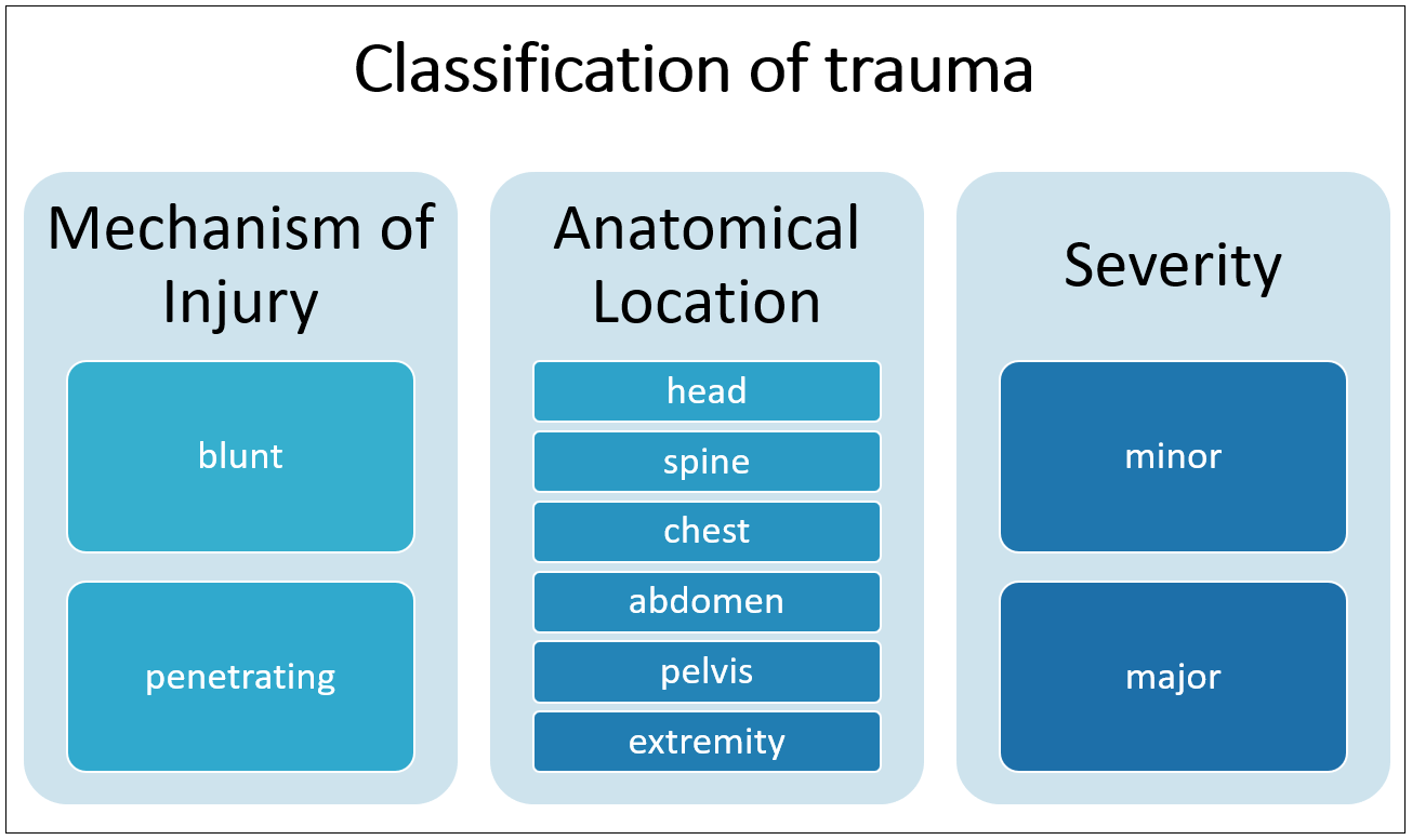
Firstly, the review explains the concept of trauma in the context of physical injury and expounds upon its various classifications (Figure 1). Secondly, it provides insights into the array of diagnostic techniques available for trauma diagnosis, including the advantages and limitations of each modality. A comparative analysis between computed tomography (CT) and MRI is particularly emphasized to discern their roles in trauma diagnostics. Furthermore, the distinctions among high-field, mid-field, and low-field MRI systems are provided.The discussion further explores the feasibility and diagnostic accuracy of mid- and low-field MRI in trauma assessment, particularly in detecting brain4 (Figure 2), spine5, and musculoskeletal injuries6 (Figure 3). Challenges such as image quality and protocol standardization are addressed, emphasizing ongoing research efforts to optimize imaging protocols and integrate advanced reconstruction algorithms. Interdisciplinary collaboration between radiologists, physicists, and clinicians is deemed crucial in tailoring mid- and low-field MRI protocols to the unique requirements of trauma imaging. Despite existing challenges, mid- and low-field MRI holds promise as a complementary modality in trauma assessment, offering advantages such as non-ionizing radiation and potential cost-effectiveness. In conclusion, while further research is warranted to validate its clinical utility, mid- and low-field MRI can enhance diagnostic capabilities and broaden access to advanced imaging in trauma care, ultimately improving patient outcomes and quality of life.
Acknowledgements
I want to thank the authors of the published articles for using figures in this work. I acknowledge that the reproduction of these figures is for academic purposes, and I have made efforts to ensure appropriate citation and attribution.References
1. Runge VM, Heverhagen JT. Advocating the Development of Next-Generation, Advanced-Design Low-Field Magnetic Resonance Systems. Investigative Radiology. 2020;55:747-753.
2. Vosshenrich J, Breit HC, Bach M, Merkle EM. [Economic aspects of low-field magnetic resonance imaging: Acquisition, installation, and maintenance costs of 0.55 T systems]. Der Radiologe. 2022 May;62(5):400-404.
3. Sarracanie M, Salameh N. Low-Field MRI: How Low Can We Go? A Fresh View on an Old Debate. Frontiers in Physics. 2020;8.
4. Sheth KN, Mazurek MH, Yuen MM, et al. Assessment of Brain Injury Using Portable, Low-Field Magnetic Resonance Imaging at the Bedside of Critically Ill Patients. JAMA Neurology. 2021;78(1):41.
5. Breit HC, Vosshenrich J, Hofmann V, et al. Image Quality of Lumbar Spine Imaging at 0.55T Low-Field MRI is Comparable to Conventional 1.5T MRI – Initial Observations in Healthy Volunteers. Academic Radiology. Published online February 2023.
6. Arnold TC, Freeman CW, Litt B, Stein JM. Low‐field MRI: Clinical Promise and Challenges. Journal of Magnetic Resonance Imaging. 2022;57(1).
Figures
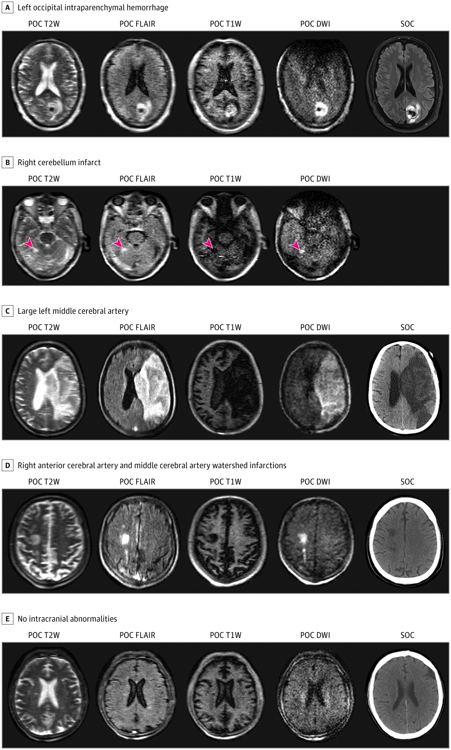
From: Assessment of Brain Injury Using Portable, Low-Field Magnetic Resonance Imaging at the Bedside of Critically Ill Patients. JAMA Neurol. 2021;78(1):41-47. doi:10.1001/jamaneurol.2020.3263
Figure 2. Examples of Point-of-Care (POC) MRI vs Standard-of-Care (SOC) Imaging in 5 Patients: A - left occipital intraparenchymal hemorrhage. B - right cerebellum infarct (arrowheads). C - large left middle cerebral artery infarct with hemorrhagic transformation. D - right anterior/middle cerebral arteries watershed infarction. E - no intracranial abnormalities.
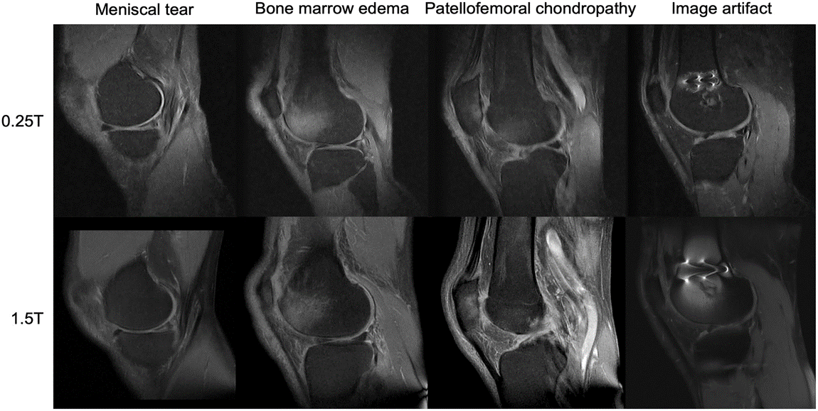
From: Low‐field MRI: Clinical promise and challenges. Magnetic Resonance Imaging, Volume: 57, Issue: 1, Pages: 25-44, First published: 19 September 2022, DOI: (10.1002/jmri.28408)
Figure 3. Examples of musculoskeletal imaging at 0.25T (top) and 1.5T (bottom). [Images for the article were provided courtesy of Dr. Riccardo Monti, Dr. Frederico Bruno, Prof. Antonio Barile, and Prof. Carlo Masciocchi, University of L'Aquila, Italy].