5194
The role of magnetic resonance in the diagnosis of intracranial berry aneurysm: A case report1MEDICAL IMAGING TECHNOLOGY, University for Development Studies ( UDS), KUMASI, Ghana, 2MEDICAL IMAGING TECHNOLOGY, University for Development Studies ( UDS), Tamale, Ghana, 3Medical Imaging, University for Development Studies, Wa, Upper West Region, Ghana
Synopsis
Keywords:
Motivation: Headaches associated with intracranial aneurysms are often misdiagnosed and poorly understood in the Ghanaian population.
Goal(s): Our goal was to use gadolinium-enhanced MR angiography to radiologically describe features of intracranial berry aneurysm (IBA) in a clinically asymptomatic patient presenting with right-sided headaches.
Approach: Axial, sagittal and coronal brain MR angiography was conducted with 50 mg/ml gadolinium contrast and radiologically evaluated.
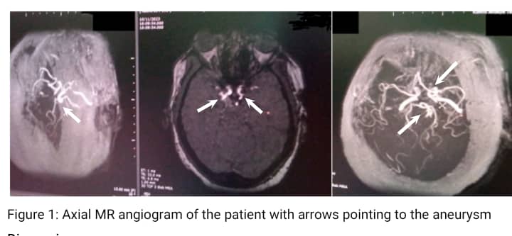
Results: A right posterior communicating artery aneurysm was found, suggesting compression of the oculomotor nerve which is characteristic of IBA.
Impact: The imaging findings could help physicians to better understand, diagnose and treat the underlining causes of constant right-sided headaches which may otherwise be poorly managed by patients themselves through self-medication as is the current practice in the Ghanaian setting.
Keywords
Intracranial berry aneurysm, gadolinium, magnetic resonance imaging.Background
Berry or saccular aneurysms are the most common type of intracranial aneurysms, comprising roughly 90% of cases.1,2 They present as small, round dilations typically located at arterial bifurcations within the circle of Willis in the brain, resulting from localised arterial wall weaknesses possibly influenced by genetic, hemodynamic, and environmental factors. If left untreated, these aneurysms carry a significant risk of rupture, leading to subarachnoid haemorrhage. Larger or ruptured aneurysms can cause severe headaches, visual disturbances, or neurological deficits. Diagnosis primarily relies on time-of-flight (TOF) MRI or contrast-enhanced computed tomography angiography (CTA), and treatment options encompass surgical clipping or endovascular coiling, aiming to mitigate the risk of rupture.3,4Case report
We report on a 58-year-old woman who presented with a 1-year history of persistent right-sided headaches and oculomotor nerve palsy. She had a background history of hypertension treated with herbal medications. She was a non-smoker and only drank one unit of alcohol on occasion. She was referred for magnetic resonance imaging of the brain to exclude microaneurysms as the cause of her 3rd nerve palsy. A pre- and post-contrasted magnetic resonance imaging scan was done on a Toshiba Achieva 1.5 Tesla scanner. The scanning protocol included axial FLAIR, T1- and T2-weighted axial, coronal and sagittal, TOF MRA, Diffusion-weighted- imaging, Gadolinium-enhanced MR-angiography, T1-weighted axial, coronal and sagittal with fat suppression pre- and post-contrast (Table 1)MRI Findings
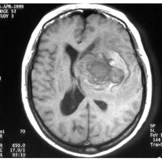
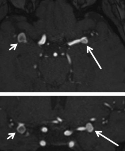
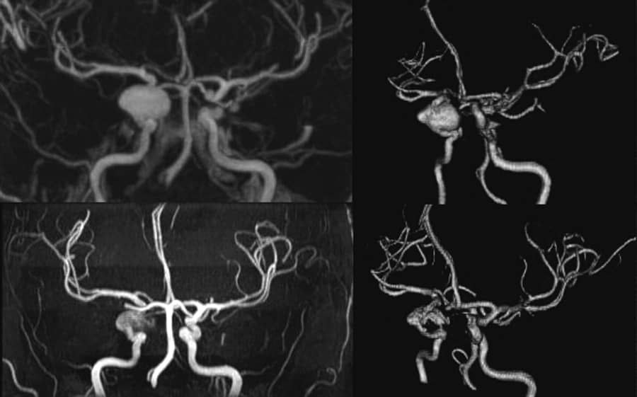
MRI revealed bulbous dilatation at the junction of the right internal carotid artery and posterior cerebral artery measuring approximately 9 mm in diameter. The lesion obscured the right posterior communicating artery likely originating from it. The lesion retained contrast on T1-Weighted imaging with gadolinium suggestive of an aneurysm. We observed compression of the oculomotor nerve just before the cavernous sinus portion. The ventricles appeared normal in size and showed no signs of raised intracranial pressure.Treatment
The patient was referred to neurosurgery for aneurysm coiling, but financial constraints prevented her from pursuing the recommended treatment. Instead, she received prescription medication and was advised to make lifestyle adjustments to manage her condition.Teaching points
1. Time-of-flight (TOF) is useful in the detection of berry aneurysms in patients with renal impairment. However turbulent flow within the aneurysm can make diagnosis difficult.2. Contrast-enhanced MR angiography has shorter acquisition times compared to TOF and does not suffer from signal loss due to turbulent or slow flow or spin saturation in larger scan volumes.4
3. Berry aneurysms yield hyperintense lesions on T1-weighted imaging with gadolinium-based contrast agents.
Conclusion
Magnetic resonance imaging plays a pivotal role in the detection of berry or saccular aneurysms. This case shows a multi-faceted approach to successfully enhancing patient care and highlights the need for public awareness surrounding intracranial berry aneurysms.Acknowledgements
Firstly, I give thanks to the God for granting me thestrength and wisdom. I would also pour out my heartfelt gratitude to Dr. petronella Samuels, DR. Abdul Nashirudeen
Mumuni, Dr. Mariam Salifu , Arhin Gabriel, Dr. Klenam Dzefi -Tettey , Abigail Frimpong, Dr. Akwesi Sarpong and Prof Derek Jones for their guidance during my write ups, my last thanks goes to Nsiah Donkor Anita and Ahmed Kanyiri Yakubu my true friend and co-authors who assisted in the write up.
References
1. Brisman, J.L., Song, J.K. and Newell, D.W. (2006). Cerebral aneurysms. New England Journal of Medicine, 355(9), 928-939.2. Hacien-Bey, L. and Provenzale, J.M. (2011). Current Imaging Assessment and Treatment of Intracranial Aneurysms. American Journal of Roentgenology. 196(1)
3. Jiang, Z., Chen, Y., Zeng, C., Feng, J., Wan, Y., & Zhang, X. (2020). Neurosurgical clipping versus endovascular coiling for patients with intracranial aneurysms: a systematic review and meta-analysis. World Neurosurgery, 138, e191-e222.
4. Gamal, G.H. (2015). Diagnostic accuracy of contrast enhancement MRI versus CTA in the diagnosis of intracranial aneurysm in patients with non-traumatic subarachnoid hemorrhage. The Egyptian Journal of Radiology and Nuclear Medicine, 46(1), 125-130.
Figures