5191
Effect of noise exposure during 7 Tesla MRI.1Department of Clinical Sciences, Faculty of Medicine, Lund University, Lund, Sweden, 2Department of Clinical Sciences Lund/Logopedics, Phoniatrics and Audiology, Faculty of Medicine, Lund University, Lund, Sweden
Synopsis
Motivation: Ensuring the proper use of hearing protection during extensive scanning at a 7T system with high acoustic noise levels is crucial.
Goal(s): We investigate if MRI examinations have acute and/or persisting effects on the hearing even if hearing protection is used.
Approach: Otoacoustic emissions (OAE) were used to evaluate the effects of noise exposure after two MRI scanning sessions in a single day. OAE measurements were conducted prior to and after each scanning and with a one-week follow-up.
Results: We found no significant differences in outer hair cell function between the baseline measurement and the first MRI and the follow-up OAE measurement.
Impact: Research protocols at a 7T system often involve long scanning sessions using sequences with high acoustic noise levels. When hearing protection is adequately used, MRI scanning can be performed without effects on the participant`s hearing.
Background
Acoustic noise emission during an MRI scanning session can exceed levels of 130 dBA(1). The noise contributes to patient discomfort(2) and can potentially cause cochlear damage on outer hair cells (3) if hearing protection is not adequately used. Furthermore, noise exposure can also result in tinnitus.The highest acoustic noise levels are found in sequences where multiple gradients are used simultaneously, for instance, three-dimensional acquisitions and gradients with rapid rise times or switching times(4) such as Echo planer Imaging used in for example functional MRI and diffusion tensor imaging (DTI).The limited inner space of head coils in 7Tesla (T) MRI systems reduces the possibility of using headphones(5), indicating the necessity to optimize methods for passive acoustic noise protection. This study assesses the potential impact of noise exposure during 7T brain MRI in healthy adults when using a two-barrier hearing protection.
Methods
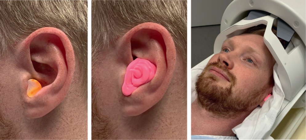
We used otoacoustic emission (OAE) to evaluate the effects of acoustic noise exposure in 39 healthy adults after MRI scanning sessions, using a 7T Achieva, Philips system. OAE generation depends on the physiological status of the cochlear outer hair cells and is a noninvasive measure(6, 7). Large OAE responses are recorded from normally functioning outer hair cells while substantially reduced or absent OAE responses indicate that the outer hair cell integrity is compromised. All participants were fitted with hearing protection consisting of earplugs placed in the ear canal. Furthermore, a sound-absorbing, impression paste (Otoform®Ak) was applied to the auricle for additional damping and fixation of the earplug (fig 1). The participants were enrolled in a research project involving two MRI scanning sessions on the same day. The MRI protocol had an effective duration of 50 minutes per scanning session and included three-dimensional T1- and T2-weighted image sequences, as well as resting-state functional MRI, flow mapping, DTI, and B0 and B1 mapping. OAE assessments were performed before and after each MRI scanning session, with a follow-up completed one week later. Statistical significance in the difference between baseline OAE and the follow-up OAE´s were assessed by paired sample T-tests. The significance was set to 5%.Results
Our analysis revealed no significant differences in outer hair cell function between the baseline measurements and the first MRI scanning session. A significant difference was observed at 1.5 kHz (p=.047) and 2 kHz (p=.016) in the left ear, as well as at 4 kHz in the right ear (p=.021) after the second MRI scan. However, the follow-up OAE measurement showed no significant difference compared to the baseline at any of the frequencies in either ear.Conclusion
Our study's findings suggest no lasting effects on outer hair cell function in adults who undergo two one-hour MRI scanning sessions in a single day while using two-barrier hearing protection. However, it is important to routinely consider adequate hearing protection and fit as it is important for patient comfort and to avoid permanent cochlear damage.Acknowledgements
This research did not receive any specific grant from funding agencies in the public, commercial, or not-for-profit sectors.
References
1. Foster JR, Hall DA, Summerfield AQ, Palmer AR, Bowtell RW. Sound-level measurements and calculations of safe noise dosage during EPI at 3 T. J Magn Reson Imaging. 2000;12(1):157-63.
2. Theysohn JM, Maderwald S, Kraff O, Moenninghoff C, Ladd ME, Ladd SC. Subjective acceptance of 7 Tesla MRI for human imaging. Magma. 2008;21(1-2):63-72.
3. Brummett RE, Talbot JM, Charuhas P. Potential hearing loss resulting from MR imaging. Radiology. 1988;169(2):539-40.
4. McJury MJ. Acoustic Noise and Magnetic Resonance Imaging: A Narrative/Descriptive Review. J Magn Reson Imaging. 2022;55(2):337-46.
5. Fagan AJ, Bitz AK, Björkman-Burtscher IM, Collins CM, Kimbrell V, Raaijmakers AJE. 7T MR Safety. J Magn Reson Imaging. 2021;53(2):333-46.
6. Kemp DT. Stimulated acoustic emissions from within the human auditory system. J Acoust Soc Am. 1978;64(5):1386-91.
7. Richardson M. Otoacoustic emissions. Arch Dis Child. 1995;73(4):284-6.
8. Hansson B, Björkman-Burtscher, I. M. Biological effects, patient experience, and occupational safety. In: Choi I-Y, Jezzard P, editors. Advances in Magnetic Resonance Technology and Applications. 4: Academic Press; 2021. p. 59-72.
Figures