5186
Abbreviated liver MRI (AMRI) as a surveillance method for hepatocellular carcinoma in high risk patients1MRI, Cambridge University Hospitals, Fulbourn, Cambridge, United Kingdom
Synopsis
Motivation: The low sensitivity of liver ultrasound precludes the diagnosis of hepatocellular carcinoma, which led to an increased interest in abbreviated liver mri as an alternative diagnostic tool
Goal(s): The goal of this research study was to assess the diagnostic performance of abbreviated liver MRI for the diagnosis of hepatocellular carcinoma
Approach: This study is a literature review, in which the latest available literature relevant to the topic was assessed with the intent of answering the research question
Results: Liver AMRI shows higher sensitivity than ultrasound for diagnosis of HCC and its diagnostic performance is comparable to that seen in a conventional protocol
Impact: In summary, liver AMRI should be considered as an altertative strategy to ultrasound for HCC surveillance. Future research including prospective studies, cost-effectiveness assessments and a consensus on an ideal AMRI protocol should be carried out.
Background
Hepatocellular carcinoma is the most prevalent primary liver cancer and its incidence rate has increased in recent decades. For this reason, early detection of HCC is essential to allow patients to be considered for curative therapies, improving survival rates (Park, Seo & Kim, 2022). Patients with chronic liver disease such as cirrhosis, are routinely monitored for liver malignancies through imaging surveillance. The current standard for liver surveillance is ultrasound which has notoriously low sensitivity, especially for patients with a large body habitus. Previous studies have shown that abbreviated liver MRI has higher sensitivity than ultrasound for the evaluation and follow-up of diffuse liver disease and the detection and follow-up of metastatic liver disease (Lee et al., 2023, Ringe, Yoon, 2023). To overcome the limitations of ultrasound, the interest in abbreviated liver MRI has risen in the latest years. Abbreviated MRI is a technique based in protocols with fewer sequences than the conventional ones to minimize scanning time and improve sensitivity and cost-effectiveness in the surveillance programs (Lee et al., 2023). The limited number of sequences that make an abbreviated protocol are tailored to a specific disease, making it possible to reduce acquisition times to 10 minutes or even less (Ringe, Yoon, 2023). The literature proposes three options of abbreviated liver MRI protocols: · Gadoxetic acid-enhanced MRI with hepatobiliary phase HBP imaging (referred to as HBP-AMRI): HBP, DWI and T2W · Dynamic contrast-enhanced MRI (referred to as DCE-AMRI): Pre and post contrast T1W (arterial, portal venous and delayed phases) · Non-enhanced MRI (referred to as NE-AMRI or NC-AMRI): DWI, T2W, optional T1 in and out-of-phase (Park, Seo & Kim, 2022). All three protocols present advantages and disadvantages to them and the examples of sequences mentioned above are commonly presented for such protocols but different clinical sites will have different protocols. HBP-AMRI is likely to have the highest sensitivity amongst the AMRI approaches due to the combination of the diffusion weighted and the hepatobiliary phase images, but it demonstrates a relatively high percentage of false-positive results. This approach allows for the patient to be injected with the contrast media outside the scanner room, which significantly improves scanning times. DCE-AMRI presents the possibility of not recalling patients for a full protocol liver MRI after observation of positive results and allows for evaluation of vascular thrombus, however, it does not acquire ancillary imaging features (Park, Seo & Kim, 2022). Both HBP and DCE-AMRI have the disadvantage of potential risks related to the contrast agent, such as gadolinium deposition in the tissues and nephrogenic systemic fibrosis. The advantages of the NC-AMRI are the avoidance of the contrast media injection and the best time resolution and cost-effectiveness amongst all three approaches. Here, like with the HBP-AMRI, patients must be recalled for a full protocol liver MRI if positive results are reported (Lee et al., 2023).Teaching point and clinical practice
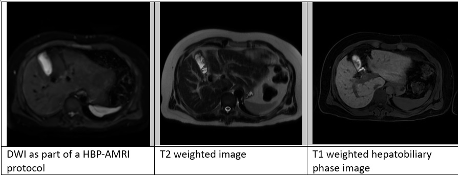
In the MRI unit at Cambridge University Hospital abbreviated MRI protocols are being used in patients with compromised ultrasound screening because of obesity, steatosis, parenchymal heterogeneity and other factors. In this site, the determination of a HBP- AMRI candidacy is made by a gastrointestinal radiology consultant. Once a patient is made a candidate for the AMRI surveillance program, they are scanned every six months. If surveillance imaging presents with new findings, patients will be referred for a complete liver MRI scan. Should those lesions turn out to be benign, patients return to the standard surveillance program, otherwise, follow-up is carried out with the conventional liver MRI. HBP-AMRI are allowed 15minutes slots which accounts for patient setup times and scanning times (around 10minutes). In regard to the protocol applied in this site, hepatobiliary contrast is administrated intravenously, outside the scanner room, and the actual exam is only initiated 15 minutes later. The abbreviated protocol includes four sets of images acquired in the axial plane which are a diffusion, balanced gradient echo, T2 and hepatobiliary phase T1 weighted sequences. These images are acquired with use of deep learning and parallel imaging techniques, which greatly improve image and time resolution.Conclusion
Overall, literature has reported values of sensitivity significantly higher for AMRI than ultrasound (Gupta et al., 2021) and a diagnostic performance comparable to that seen in full contrast-enhanced MRI studies (Barakat, Awadallah & Madkour, 2022). Based on these results, AMRI should serve as an alternative strategy for HCC surveillance in patients at risk due to chronic liver disease (Yokoo et al., 2023).Acknowledgements
I would like to thank Dr. Iain MacDonald, Senior Lecturer in Medical Imaging Science and MRI Postgraduate Programme Lead, for his extraordinary support and encouragement in this work. I would like to thank Dr. David Bowden, Gastrointestinal Radiology Consultant and MR Clinical Lead in Addenbrookes Hospital for his expert advice and collaboration throughout this study.References
· Barakat, M., Awadallah, S. & Madkour, S. 2022, "The role of three different contrast-enhanced, abbreviated MRI protocols as a screening tool of hepatocellular carcinoma in patients with chronic hepatitis C virus infection", Polish journal of radiology, vol. 87, no. 1, pp. 574-583. · Gupta, P., Soundararajan, R., Patel, A., Kumar-M, P., Sharma, V. & Kalra, N. 2021, "Abbreviated MRI for hepatocellular carcinoma screening: A systematic review and meta-analysis", Journal of hepatology, vol. 75, no. 1, pp. 108-119. · Kim, D.H., Choi, S.H., Lee, J.S. & Choi, J. 2022, "Inter-reader agreement of abbreviated magnetic resonance imaging for hepatocellular carcinoma detection: a systematic review and meta-analysis", Abdominal imaging, vol. 47, no. 1, pp. 123-132. · Lee, J.H., Kim, Y.K., Min, J.H., Cha, D., Hwang, J.A. & Ahn, S. 2023, "Comparison of noncontrast, dynamic, and hepatobiliary phase abbreviated MRI protocols for detection of hepatic malignancies", Clinical imaging, vol. 101, pp. 206-214. · Park, H.J., Seo, N. & Kim, S.Y. 2022, "Current Landscape and Future Perspectives of Abbreviated MRI for Hepatocellular Carcinoma Surveillance", Korean journal of radiology, vol. 23, no. 6, pp. 598-614. · Ringe, K.I. & Yoon, J.H. 2023, "Strategies and Techniques for Liver Magnetic Resonance Imaging: New and Pending Applications for Routine Clinical Practice", Korean journal of radiology, vol. 24, no. 3, pp. 180-189. · Yokoo, T., Masaki, N., Parikh, N.D., Lane, B.F., Feng, Z., Mendiratta-Lala, M., Lee, C.H., Khatri, G., Marsh, T.L., Shetty, K., Dunn, C.T., Al-Jarrah, T., Aslam, A., Davenport, M.S., Gopal, P., Rich, N.E., Lok, A.S. & Singal, A.G. 2023, "Multicenter Validation of Abbreviated MRI for Detecting Early-Stage Hepatocellular Carcinoma", Radiology, vol. 307, no. 2, pp. e220917.Figures