5185
3D Radial mDIXON Acquisition for Improved Breast Imaging1Philips, Cleveland, OH, United States
Synopsis
Motivation: Breast magnetic resonance imaging (MRI) is a sensitive technique for staging and screening for breast cancer, however, it is susceptible to motion artifacts and inhomogeneous fat saturation.
Goal(s): Demonstrate that 3D radial mDIXON acquisition provides robust motion suppression and homogenous fat saturation.
Approach: Compared image quality of 3D radial mDIXON to 3D Cartesian mDIXON and 3D Cartesian with spectral fat saturation. Advanced postprocessing was also performed on the 3D radial mDIXON to see if additional diagnostic information could be obtained.
Results: 3D radial mDIXON of the breast showed constantly high image quality with less motion artifacts and homogenous fat suppression.
Impact: 3D radial mDIXON provides many advantages over Cartesian spectrally fat saturated acquisitions. The radial acquisition is less sensitive to motion artifacts. mDIXON provides consistent fat suppression compared to spectral fat suppression. Advanced postprocessing of mDIXON images provides further diagnostic utility.
Background:
Patient and technical factors may lead to unwanted artifacts when performing breast MRI degrading image quality1 and impair diagnostic evaluation2. Common imaging artifacts seen during breast MRI include motion artifacts, inhomogeneous fat saturation, misregistration, and areas of high signal intensity close to the coil3. Motion induced artifacts are the most reported reason that affect image interpretation1-4. Movement artifacts can affect the entire study at the sequence and slice level and can come from bulk patient motion or physiological movements from breathing, cardiac cycle, and blood flow3. Inhomogeneous or failed fat saturation is secondary to motion artifacts when it comes to impacting the diagnostic evaluation of breast MRIs3. Fat suppression is crucial in breast MRI as the high signal from fat can obscure enhancing lesions5. Poor fat saturation can be caused by many factors including patient positioning, large FOV, and changes in B0 from respiration or movement. Motion artifacts and improper fat suppression lead to signal intensity variations, conspicuity of moving structures, as well as a general blurring. All of these can obscure real lesions or lead to false findings1. To address these two major sources affecting breast MRI diagnostic interpretation we evaluate the use of a 3D radial mDIXON technique. The 3D radial acquisition is less sensitive to motion artifacts compared to its 3D Cartesian counterpart4. Moreover, the use of mDIXON over spectral fat saturation techniques provides more consistent and homogenous fat saturation6. Utilizing an mDIXON approach also affords more advanced postprocessing options that could provide a higher diagnostic utility.Methods:
The 3D radial mDIXON acquisition was qualitatively and quantitively compared to the 3D Cartesian using a spectral fat suppression acquisition to assess image quality. Images were acquired on 1.5T scanner (Philips Ingenia, Best, Netherlands) using a dedicated 7 channel phased array breast coil. Qualitative image quality assessment was performed by 3 radiologists for motion artifacts and performance of fat saturation. Quantitative image quality metrics for signal to noise ratio (SNR) and contrast to noise ratio (CNR) along with indices for the presence of motion artifacts and quality of fat saturation (shading) were computed using MRQY7. Additional postprocessing was performed using the 3D radial mDIXON data to calculate fat fraction. This advanced postprocessing was performed offline using in-house written software (Philips Research Imaging Development Environment).Results:
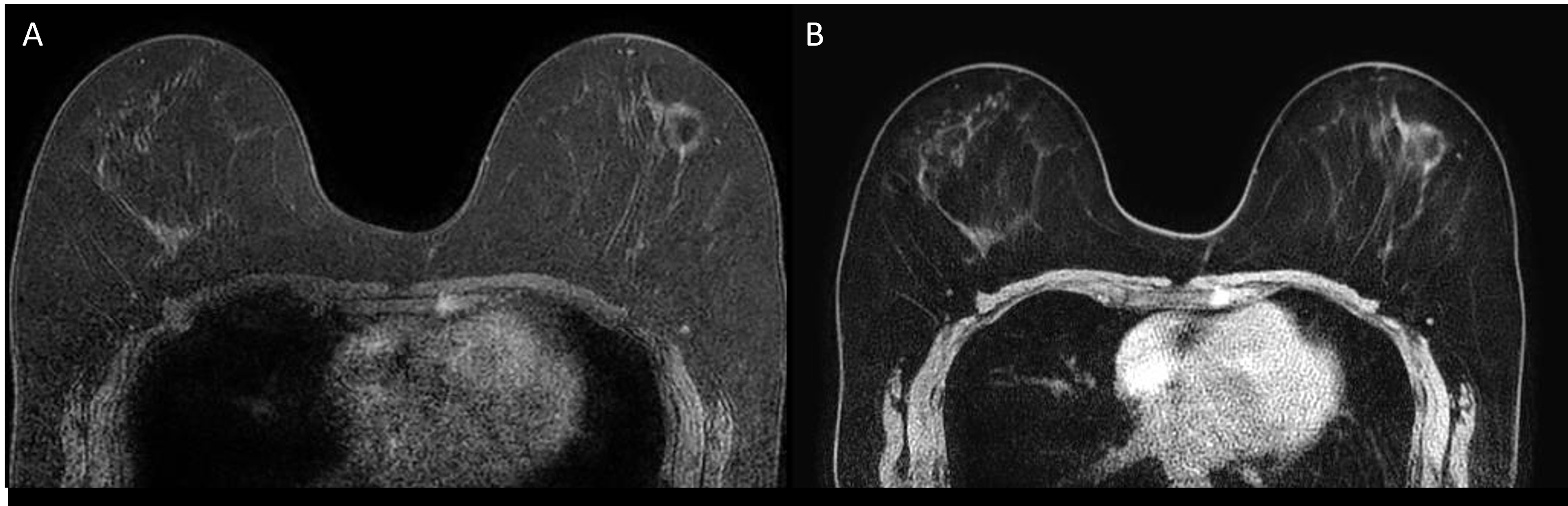
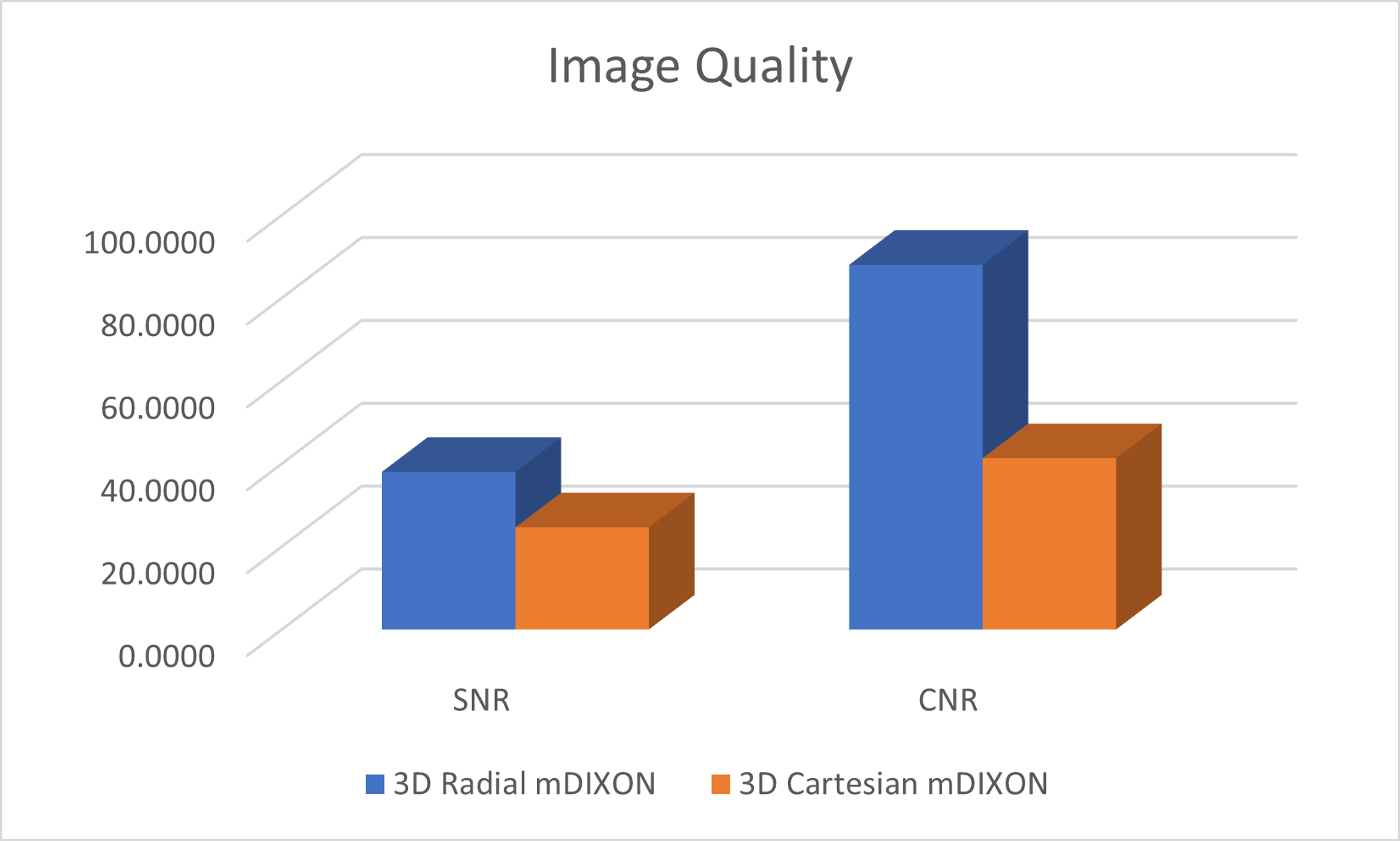
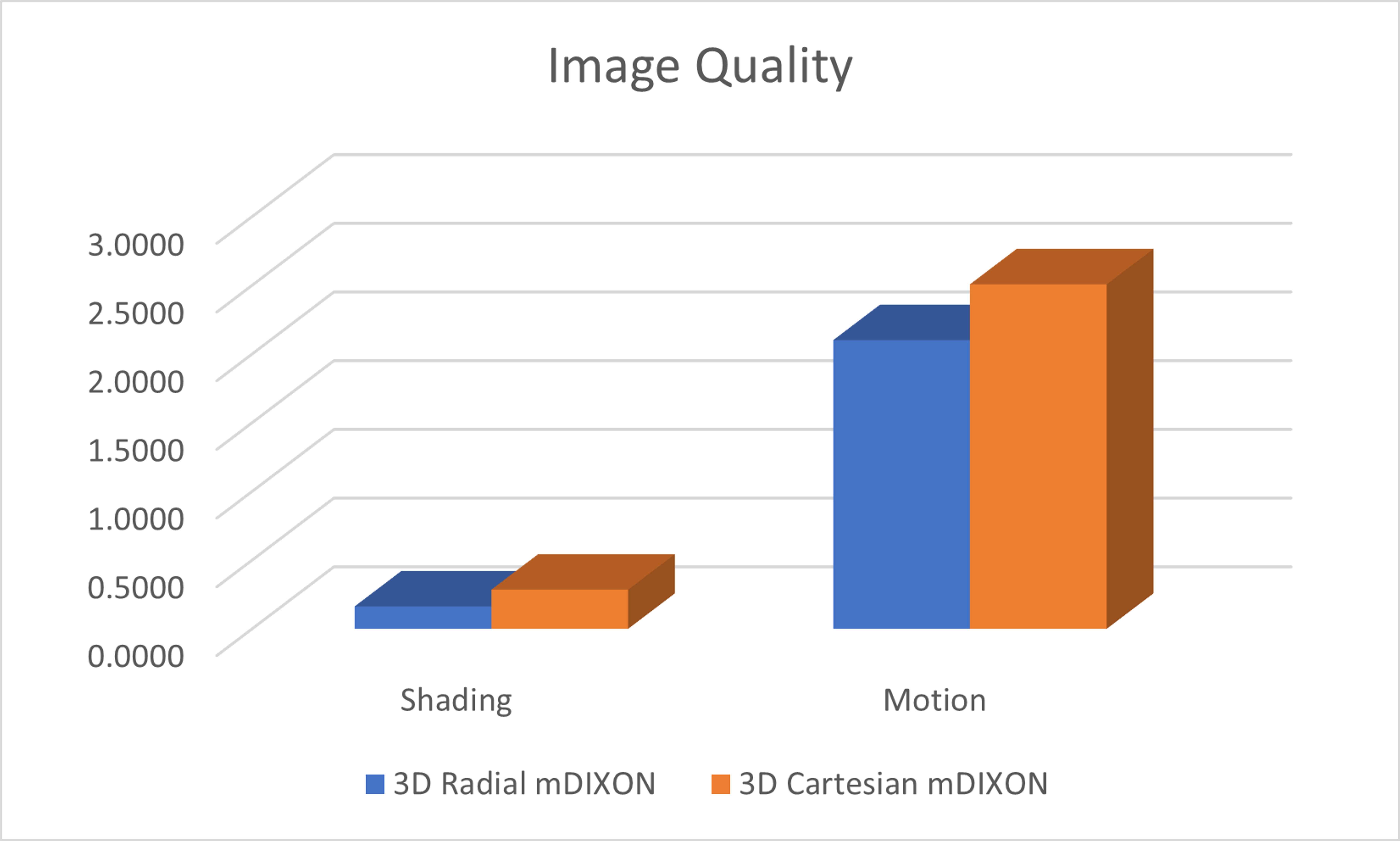
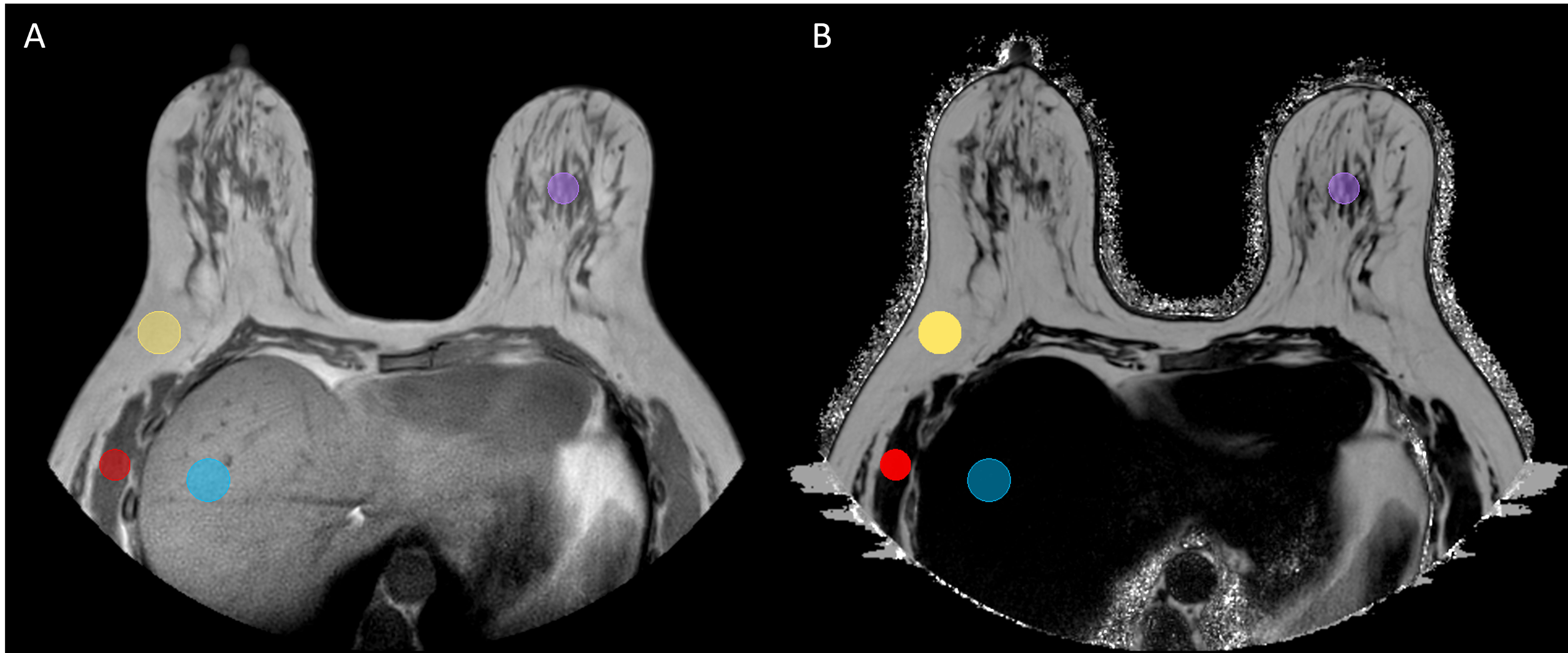
Qualitative assessment results showed higher and more consistent image quality for both metrics in the 3D radial mDIXON approach over the 3D Cartesian with spectral fat suppression (Figure 1). Quantitative results agreed with qualitative reports with the 3D mDIXON showing better image quality compared to the 3D Cartesian with spectral fat suppression. Specifically, the 3D radial mDIXON had higher SNR and CNR (Figure 2) while having lower motion and shading artifacts (Figure 3). Evaluation of fat fraction postprocessing showed consistent values for areas of fat, muscle, liver, and glandular tissue (Figure 4).Conclusion:
3D radial mDIXON showed higher and more consistent image quality compared to the 3D Cartesian with spectral fat suppression. This is consistent with other reports showing that radial acquisition of short-tau inversion recovery (STIR) had improved image quality compared to its Cartesian counterpart4. The ability of the 3D radial mDIXON to reduce motion artifacts and provide homogeneous fat saturation makes this an ideal technique to help mitigate the two major sources that affect diagnostic interpretation for breast MRI. Furthermore, the ability to calculate fat fraction with the mDIXON data provides additional diagnostically relevant information. Lymph node fat fraction from high-resolution 3D Dixon images has shown to be a promising quantitative indicator of metastases in axillary lymph nodes6. Overall 3D radial mDIXON provides a robust and more comprehensive breast imaging strategy.Acknowledgements
No acknowledgement found.References
1. Carbonaro LA, Schiaffino S, Clauser P, et al. Side of contrast injection and breast size correlate with motion artifacts grade and image quality on breast MRI. Acta Radiologica. 2021;62(1):19-26. 2. Clauser P, Dietzel M, Weber M, Kaiser CG, Baltzer PA. Motion artifacts, lesion type, and parenchymal enhancement in breast MRI: what does really influence diagnostic accuracy? Acta Radiologica. 2019;60(1):19-27.
3. Fiaschetti V, Pistolese C, Funel V, et al. Breast MRI artefacts: evaluation and solutions in 630 consecutive patients. Clinical Radiology. 2013;68(11):e601-e608.
4. Santucci D, Lee SS, Hartman H, et al. Comparison of Cartesian and radial acquisition on short-tau inversion recovery (STIR) sequences in breast MRI. Radiologia Brasileira. 2017;50:216-223.
5. Harvey JA, Hendrick RE, Coll JM, Nicholson BT, Burkholder BT, Cohen MA. Breast MR imaging artifacts: how to recognize and fix them. Radiographics. 2007;27(suppl_1):S131-S145.
6. Buus TW, Sivesgaard K, Fris TL, Christiansen PM, Jensen AB, Pedersen EM. Fat fractions from high-resolution 3D radial Dixon MRI for predicting metastatic axillary lymph nodes in breast cancer patients. European Journal of Radiology Open. 2020;7:100284.
7. Sadri AR, Janowczyk A, Zhou R, et al. MRQy—An open‐source tool for quality control of MR imaging data. Medical physics. 2020;47(12):6029-6038.
Figures