5182
Comparison of Magnetic Resonance Imaging–Based and Conventional Measurements for Proton Beam Therapy of Uveal Melanoma1Radiology, LUMC Leiden, Leiden, Netherlands, 2Radiology, LUMC, Leiden, Netherlands, 3Radiology and ophtalmology, LUMC, Leiden, Netherlands
Synopsis
Keywords:
Motivation: Proton therapy is a relatively new option for treatment of uveal melanomas. For adequate treatment, accurate measurement of tumour dimensions and tumour-marker distance is essential. The dedicated MRI protocol for uveal melanoma patients developed in our institution could be helpful.
Goal(s): In this study, we compared MRI-based measurements with conventional ophthalmic measurements.
Approach: Retrospectively, MRI based measurements of tumour dimensions and marker-tumour distances in 23 patients were compared with conventional ophthalmic measurements and evaluated.
Results: MRI allowed for the three-dimensional assessment of the tumour. In specific patients, it provided a more reliable measurement of tumour dimensions and marker-tumour distances.
Impact: Dedicated ocular MRI proved to be a reliable tool for measurements required in the planning of proton therapy in patients with uveal melanoma.
Background:
In recent years, proton therapy has become an important tool in the treatment of uveal melanomas. Also, a dedicated high resolution ocular MRI protocol has been developed in our institution. For adequate treatment, accurate measurement of tumour dimensions is essential, as well as measurement of the distance between the tumour and the markers used in proton therapy.Objective:
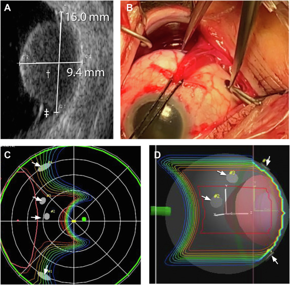
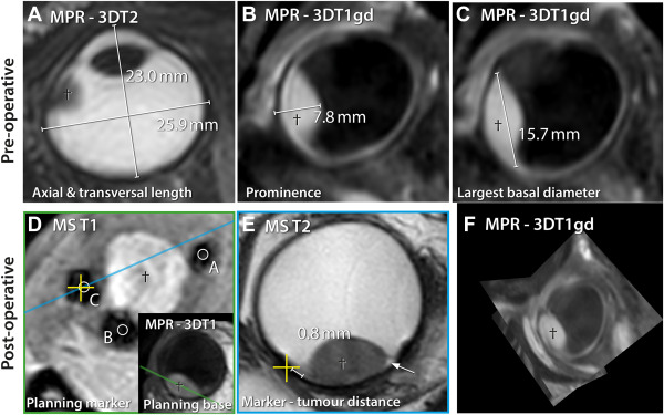
Conventionally, ocular proton therapy is planned using measurements obtained by an ophthalmologist using ultrasound, fundoscopy, biometry, and intraoperative assessments. Owing to the recent advances in magnetic resonance imaging (MRI) ¹ of uveal melanoma, it is possible to acquire high resolution 3-dimensional images of the eye, providing the opportunity to incorporate MRI in ocular proton therapy planning. In this study, we described how these measurements can be obtained using MRI, compared the MRI-based measurements with conventional ophthalmic measurements, and identified potential pitfalls for both modalities.Methods:
The study was designed as a cross-sectional study. Data from 23 consecutive patients with uveal melanoma treated with proton therapy were retrospectively evaluated.Magnetic resonance imaging based measurements of axial length, tumour height and basal diameter, and marker-tumour distances were compared with the conventional ophthalmic measurements, and discrepancies were evaluated in a multidisciplinary setting. Main Outcome Measures: Tumour prominence and basal diameters on MRI and ultrasound, axial length on MRI and biometry, tumour-marker distances on MRI and measured intraoperatively.
Results:
The mean absolute differences of the tumour height and basal diameter measurements between ultrasound and MRI were 0.57 mm and 1.44 mm, respectively. Larger absolute differences in height and basal diameter were observed when the full tumour extent was not visible on ultrasound (0.92 mm and 1.67 mm, respectively) compared with when the full tumour extent was visible (0.44 mm and 1.15 mm, respectively). When the full tumour was not visible on ultrasound, MRI was considered more reliable. Tumour marker distances measured using MRI and intraoperative techniques differed < 1 mm in 55% of the markers. For anteriorly located and mushroom-shaped tumours (25% of the markers), MRI provided more accurate measurements. In flat uveal melanoma (15% of the markers), however, it was difficult to delineate the tumour on MRI. The mean absolute difference in axial length between optical biometry and MRI was 0.50 mm. The presence of the tumour was found to influence optical biometry in 15 of 22 patients; the remaining patients showed a better agreement (0.30 mm). Magnetic resonance imaging based biometry was considered more reliable in patients with uveal melanoma.Conclusions:
Magnetic resonance imaging allowed for the 3 dimensional assessment of the tumour and surrounding tissue. In specific patients, it provided a more reliable measurement of axial length, tumour dimensions, and marker-tumour distances and could contribute to a more accurate treatment planning. Nevertheless, a combined evaluation remains advised, especially for flat uveal melanomas.Acknowledgements
This piece of research is one of the results of a multidisciplinary project, in which I was asked to take part as a senior MRI technician. I am grateful for this opportunity, so thank you:
Myriam G. Jaarsma-Coes MSc , Teresa A. Ferreira MD , Berit M. Verbist MD, (Department of Radiology, LUMC Leiden.
Marina Marinkovic MD , T.H. Khanh Vu MD, PhD , Luc van Vught MSc ,Gregorius P.M. Luyten MD, (Department of Ophtalmology, LUMC Leiden)
Myra F. Rodrigues MD, Yvonne L.B. Klaver MD, PhD, PhD , Coen R.N. Rasch MD, PhD, (Department of Radiation Oncology)
And of course: Jan-Willem M. Beenakker PhD (Departments of Radiology and Ophtalmology, LUMC Leiden)
References
1.Teresa A. Ferreira, Lorna Grech Fonk, Myriam G Jaarsma-Coes, Guido R van Haren, Marina Marinkovic, Jan Willem M Beenakker: MRI of Uveal Melanoma, Cancers 2019;11(3):377Figures