5181
Improving Image Quality Using the Pause Function Combination to PROPELLER Sequence in Brain MRI: a brain phantom study1Department of Radiological Sciences, School of Health Sciences, Fukushima Medical University, Fukushima, Japan, 2Chiba University Hospital, Chiba, Japan, 3Kameda General Hospital, Kamogawa-shi, Japan, 4Meiwa Hospital, Nishinomiya-shi, Japan, 5Hitachinaka General Hospital, Hitachinaka-shi, Japan, 6QST Hospital, Anagawa-shi, Japan
Synopsis
Motivation: Finding ways to use the PROPELLER sequence as an even more the robustness method to motion.
Goal(s): This study investigates whether repositioning the head after pausing during PROPELLER imaging, enhances image quality.
Approach: This study investigated whether image quality improved with repositioning the head after a pause during PROPELLER method with a brain phantom and driver system.
Results: We found that image quality improved at all rotational angles and that pausing multiple times was effective depending on the frequency of motion.
Impact: Incorporating a pause function into the PROPELLER method is expected to be clinically applicable as a practical means to further improve the robustness of the PROPELLER method to motion.
Purpose
The PROPELLER method is widely used because it oversamples the low-frequency domain and can reduce artifacts due to in-plane rotation and translational motion. However, this correction may be inadequate when large rotational or translational motions occur without a definitive solution. Some MRI systems have a "pause" function, and the PROPELLER method can be used with the pause function; however, studies on image quality improvement combining both techniques are lacking. This study aimed to verify whether the image quality can be improved by repositioning the head using the PROPELLER method in combination with a pause function for head motion.Method
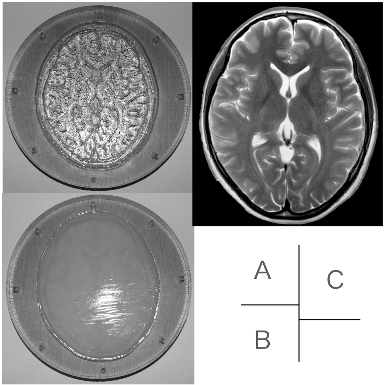
We used a brain phantom showing image contrast and construction similar to those of in vivo MRI (Fig. 1) and a driver system to rotate the phantom precisely in the head coil. All brain phantom images in this study were performed using a 3.0T MRI combined with an 8-channel head coil and acquired using single-slice, coronal, fast spin-echo T2WI-based PROPELLER with motion correction. For each slice, 18 blades were collected (i.e., TR = 18). Two studies were performed by combining the angle of rotational movement of the head phantom and the number of repositions after a pause. The first study included three trials, and the second included four trials. In all the subsequent trials, the phantom was set at the zero-degree position at the beginning of PROPELLER imaging.Study 1:Image quality when paused and repositioned at and near the original angle (Trial 1–3) (Fig. 2a)
Study 2: Image quality according to the number of repositions after pausing (Trial 1–4) (Fig. 2b)
Two radiologists and two radiological technologists rated the "degree of artifact contamination in the image" on a 5-point scale: excellent: 4 points; good: 3 points; fair: 2 points; poor: 1 point; very poor: 0 point. Furthermore, we quantitatively evaluated the original motionless image and the correlation coefficient between each image.
Results
Figure 3a shows the results of the reconstructed T2WIs with motion correction, and figure 3b and 3c show quantitative and visual evaluation in trials 1–3 of study 1. When no repositioning was performed (Trial 1), few artifacts were observed at a rotation angle of 20°, with the highest score of 4 points. As the rotation angle increased beyond 30°, the artifacts gradually increased and the score gradually decreased (30°: 3 points, 40°: 2 points, and 50°: 1 point). However, when the head phantom was repositioned to its original (zero-degree) angle (Trial 2), few artifacts were observed at all rotation angles, and all scores were 4, which was the highest possible. Furthermore, when the head phantom was repositioned near its original angle (10°), the score was 4, which was the highest score for all rotation angles, although there was a slight increase in artifacts compared to the results of Trial 2. Figure 4a shows the results of the reconstructed T2WIs with motion correction, and figure 4b and 4 show quantitative and visual evaluation in trials 1–4 of study 2. When no repositioning was performed (Trial 1), artifacts were noticeable even at a rotation angle of 20°; as the rotation angle increased, artifacts increased further, and the scoring gradually decreased (20°: 3 points, 30°: 2 points, 40°: 1 point, and 50°: 0 points). When the head phantom was repositioned only once out of the two movements (Trials 2 and 3), artifacts were reduced and scores increased at all angles compared with Trial 1 (20°: 4 points, 30°: 3 points, 40°: 3 points, and 50°: 2 points). When the head phantom was moved twice and repositioned (Trial 4), a few artifacts were observed at all rotation angles, and all scores were 4, which was the highest possible.Conclusion
The utility of incorporating a pause function into the PROPELLER method has not yet been investigated. This study investigated whether image quality improved with repositioning the head after a pause during the PROPELLER method with a brain phantom and driver system. We found that image quality improved at all rotational angles and that pausing multiple times was effective depending on the frequency of motion. This technique is expected to be clinically applicable as a practical means to further improve the robustness of the PROPELLER method to motion.Acknowledgements
The authors thank Ms. Kanae Moriyama and Mr. Kenichi Kazahari (GE Healthcare) for providing information on the PROPELLER technique. We also thank Editage (www.editage.com) for English language editing.References
Saotome K, Matsushita A, Matsumoto K, et al. A brain phantom for motion-corrected PROPELLER showing image contrast and construction similar to those of in vivo MRI. Magn Reson Imaging 2017; 36:32–39.Figures
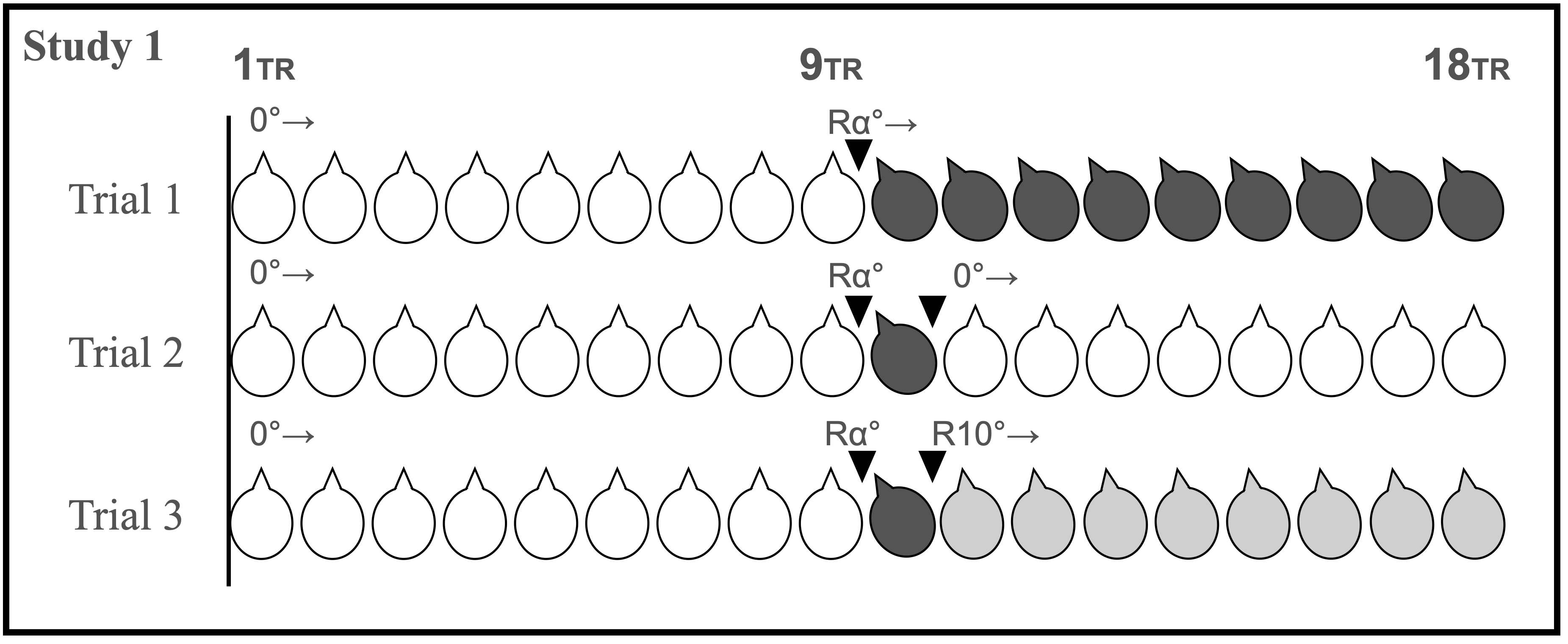
Phantom angles and pause timing for Study 1 (including 3 trials) are shown. Black arrowheads indicate when the imaging was paused.
R: right, L: left, TR: repetition Time, α = 20, 30, 40, and 50 degrees
Trial 1: Case in which patient (brain phantom) moved once but did not repositioning.
Trial 2: Case in which imaging was stopped and repositioning was performed immediately after the patient (brain phantom) moved once.
Trial 3: Case in which imaging was stopped immediately after the patient (brain phantom) moved once and repositioned to an angle slightly away from the original position.
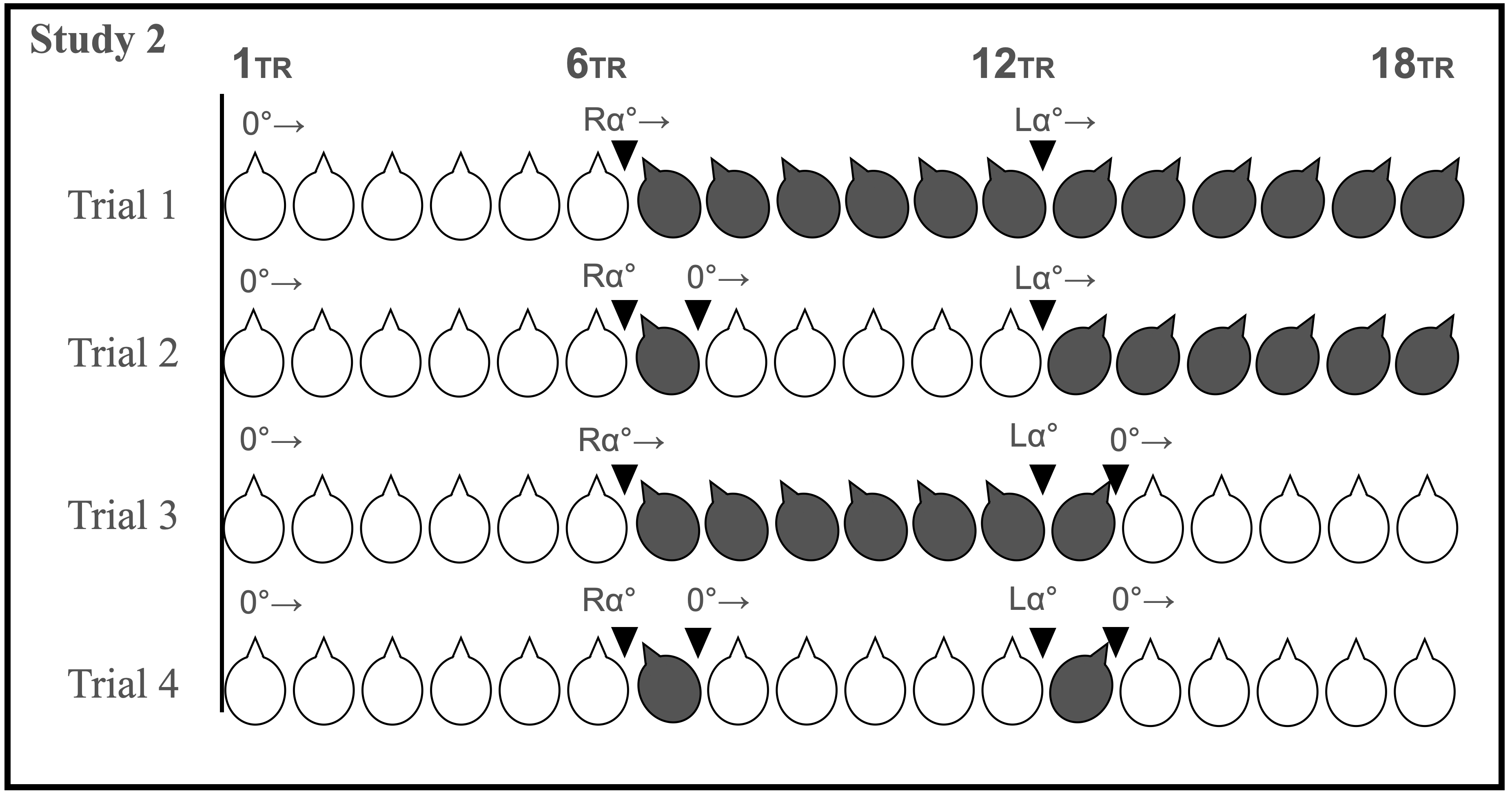
Trial 1: Case in which the patient (brain phantom) moved twice but did not reposition.
Trial 2: Case in which the patient (brain phantom) moved twice, but only the first time repositioning occurred immediately after the movement.
Trial 3: Case in which the patient (brain phantom) moved twice, but only the second time repositioning occurred immediately after the patient moved.
Trial 4: Case in which the patient (brain phantom) moved twice, but both times repositioning occurred immediately after the movement.
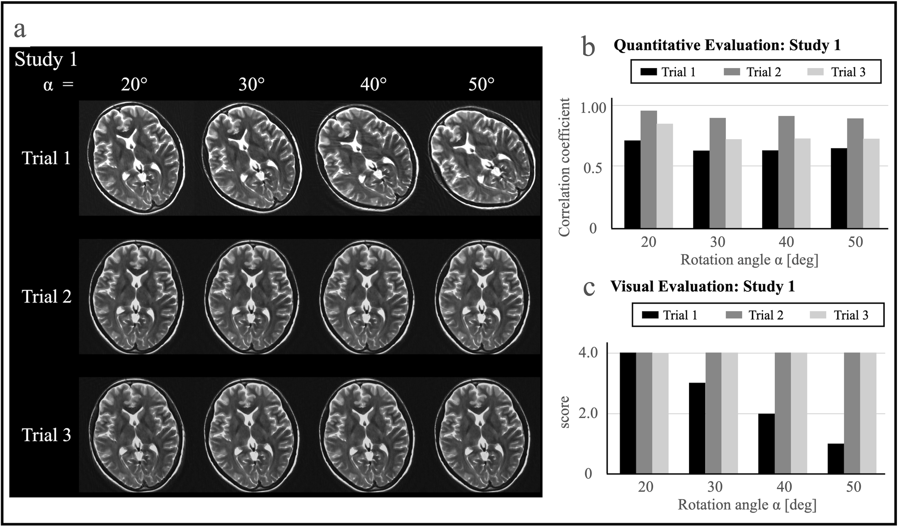
a) Reconstructed T2-weighted images of Study 1
b) Quantitative Evaluation
c) Visual Evaluation
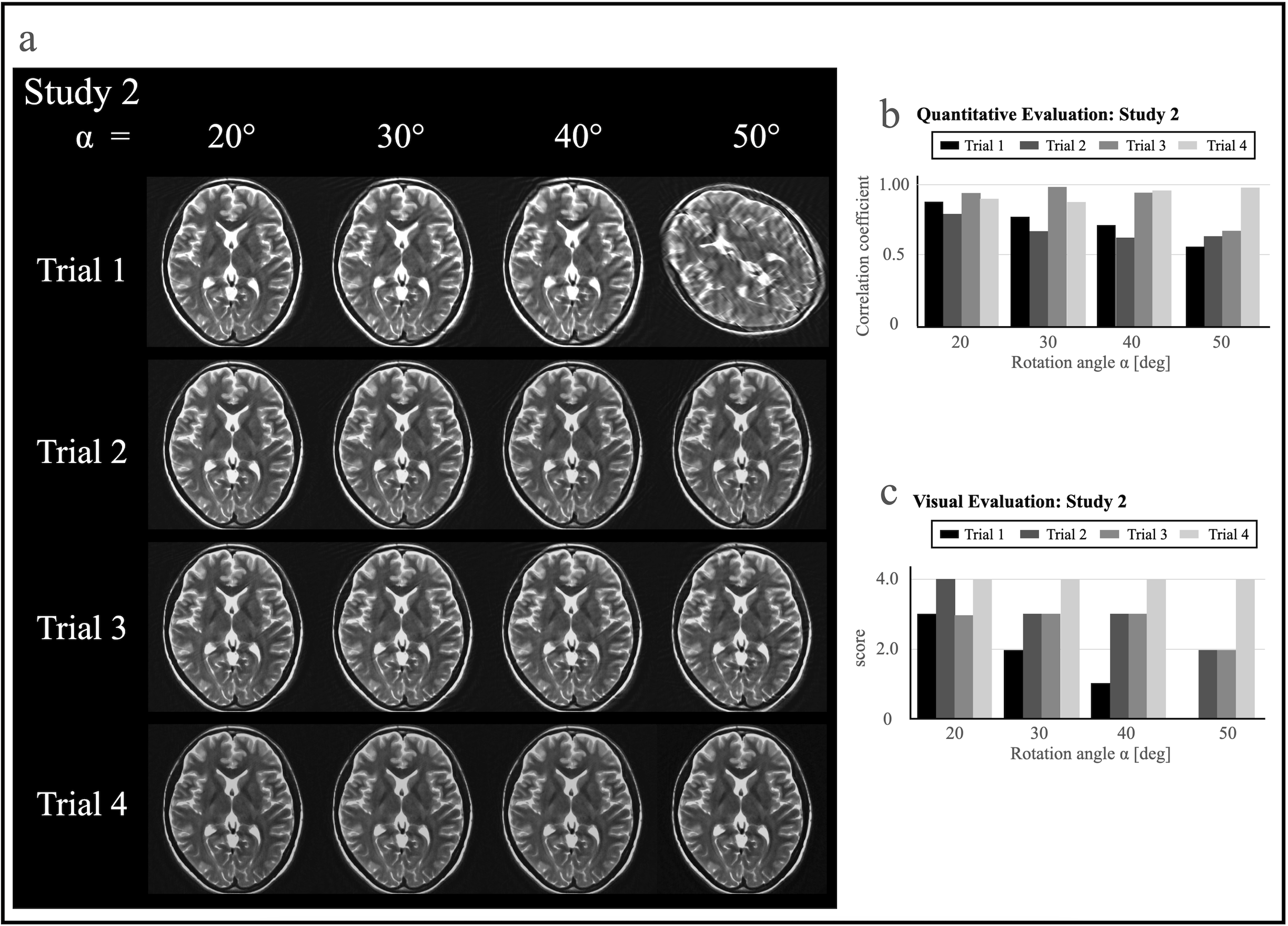
a) Reconstructed T2-weighted images of Study 2
b) Quantitative Evaluation
c) Visual Evaluation