5180
A Novel Inherited Modeling Structure of Automatic Brain Tumor Segmentation from MRI1Najran University, Najran, Saudi Arabia
Synopsis
Motivation: Auto segmentation of the affected part is needed to facilitate radiologists
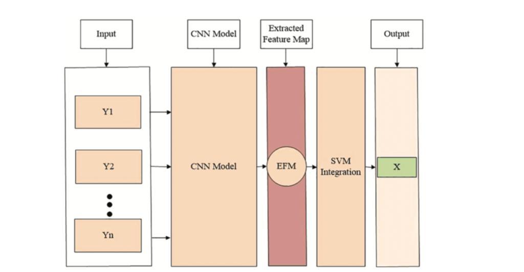
Goal(s): we have considered a hybrid model that inherits the convolutional neural network (CNN) properties to the support vector machine (SVM) for the auto-segmented brain tumor region
Approach: we have considered a hybrid model that inherits the CNN properties to the SVM for the auto-segmented brain tumor region
Results: An accuracy value of 0.98, which is most prominent than existing techniques
Impact: The proposed approach is more suitable for medical experts to diagnose the early stages of the brain tumor.
Abstract
Brain tumor is one of the most dreadful worldwide types of cancer and affects people leading to death. Magnetic resonance imaging methods capture skull images that contain healthy and affected tissue. Radiologists checked the affected tissue in the slice-by-slice manner, which was timeconsuming and hectic task. Therefore, auto segmentation of the affected part is needed to facilitate radiologists. Therefore, we have considered a hybrid model that inherits the convolutional neural network (CNN) properties to the support vector machine (SVM) for the auto-segmented brain tumor region. The CNN model is initially used to detect brain tumors, while SVM is integrated to segment the tumor region correctly. The proposed method was evaluated on a publicly available BraTS2020 dataset. The statistical parameters used in this work for the mathematical measures are precision, accuracy, specificity, sensitivity, and dice coefficient. Overall, our method achieved an accuracy value of 0.98, which is most prominent than existing techniques. Moreover, the proposed approach is more suitable for medical experts to diagnose the early stages of the brain tumor.Acknowledgements
Authors would like to acknowledge the support of the Deputy for Research and Innovation-Ministry of Education, Kingdom of Saudi Arabia for funding this research through a Project (NU/IFC/ENT/01/014) under the institutional funding committee at Najran University, Kingdom of Saudi Arabia.References
1- M. A. Khan, I. Ashraf, M. Alhaisoni, R. Damasevi ˇ cius, R. Scherer ˇ et al., “Multimodal brain tumor classification using deep learning and robust feature selection: A machine learning application for radiologists,” Diagnostics, vol. 10, no. 8, pp. 565–584, 2020
2- S. Iqbal, M. U. Ghani Khan, T. Saba, Z. Mehmood, N. Javaid et al., “Deep learning model integrating features and novel classifiers fusion for brain tumor segmentation,” Microscopy Research and Technique, vol. 82, no. 8, pp. 1302–1315, 2019.
3- M. Sajjad, S. Khan, K. Muhammad, W. Wu, A. Ullah et al., “Multi-grade brain tumor classification using deep CNN with extensive data augmentation,” Journal of Computational Science, vol. 30, no. 1, pp. 174– 182, 2019.
4- V. Wasule and P. Sonar, “Classification of brain MRI using SVM and KNN classifier,” in Proc. 2017 Third Int. Conf. on Sensing, Signal Processing and Security (ICSSS), Chennai, India, pp. 218–223, 2017.
5- K. B. Vaishnavee and K. Amshakala, “An automated MRI brain image segmentation and tumor detection using SOM-clustering and proximal support vector machine classifier,” in Proc. 2015 IEEE Int. Conf. on Engineering and Technology (ICETECH), Coimbatore, India, pp. 1–6, 2015
Figures