5175
EPI Distortion Correction using FSL TOPUP for Diffusion Tensor Imaging of the Human Heart1Fuwai Hospital, Beijing, China, 2Philips Healthcare, Beijing, China
Synopsis
Motivation: Cardiac diffusion tensor imaging (cDTI) enables the characterization of the myocardial microstructural environment. However, B0 inhomogeneity around the heart can lead to EPI distortion and signal loss in cDTI.
Goal(s): To investigate the feasibility of using FSL TOPUP for EPI Distortion Correction in cDTI.
Approach: This proposed approach could be seamlessly integrated into cDTI postprocessing, significantly improving the accuracy and reproducibility of cDTI, particularly when researchers are analyzing metrics alongside other cardiac image parameters.
Results: The FSL TOPUP method could correct EPI distortion in both diffusion-weighted images (b0 and b400), following which kinds of cDTI metrics are reconstructed successfully after the correction.
Impact: The FSL TOPUP algorithm has been successfully integrated into the cDTI postprocessing in order to correct EPI distortion effectively.
Purpose
This study aims to investigate the feasibility of using FSL TOPUP for EPI Distortion Correction in cDTI.Introduction
Cardiac diffusion tensor imaging (cDTI) could characterize the myocardial microstructural environment by measuring the diffusion of water within an imaging voxel. It has attracted more and more attention in cardiac research, such as hypertrophic cardiomyopathy1,2, myocardial Infarction3, and dilated cardiomyopathy4. Cardiac and respiratory motion are the most challenging factors in cDTI. Second-order motion compensation (SOMC) and stimulated echo acquisition mode (STEAM) have been proposed to enhance the accuracy and reproducibility of cDTI combined with ECG and respiratory navigator5. Recently, a free-breathing SOMC cDTI method has been established using multitasking respiratory motion correction6. Although many attempts have been made to reduce the motion effect, the B0 inhomogeneity around the heart has been neglected, which leads to EPI distortion and signal loss in cDTI. The FSL TOPUP algorithm is one promising method to reduce the distortion with high time efficiency, developed in brain imaging7. In this study, we aimed to investigate the feasibility of using FSL TOPUP for EPI Distortion Correction in cDTI.Methods
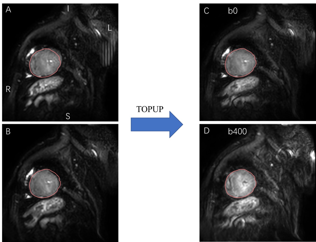
CMR was undertaken on a 3T scanner (Ingenia, Philips Healthcare, Best, The Netherlands), equipped with 32 channel anterior and posterior array coils). All scans were performed at end-expiratory and radio-frequency (RF) shimming was used adaptively to reduce the B1 inhomogeneity. In every scanning session first and second-order shimming and frequency adjustments were performed over a local volume (box) encompassing the whole heart. cDTI data were acquired using an electrocardiography-gated second-order motion-compensated single-shot spin-echo echoplanar imaging sequence with asymmetric bipolar diffusion waveforms and a respiratory navigator. The main DT-CMR protocol parameters[PS1] were as follows: b values = 0 s/mm2 and 400 s/mm2 in 16 diffusion encoding directions; repetition time TR = 2 RR intervals = 1,710 ms (assuming a heart rate of 70 beats/min); echo time of 79 msec; flip angle, 90°; field of view, 310 x 310 mm; acquisition matrix, 104 × 101; acquired in-plane resolution, 2.98 × 3.06; slice thickness, 8 mm; reconstructed voxel size, 1.7 × 1.7 × 8 mm; sensitivity encoding acceleration, 2. The proposed scheme was shown in Fig 1. In this scheme, the blip-up images were acquired for only the b value equals 0. The blip-down images were acquired for all b values used for DTI reconstruction. The module of FSL TOPUP was used to realize the EPI correction using the images of blip-up and blip-down b0 image, which is then applied to all diffusion-weighted images.Results
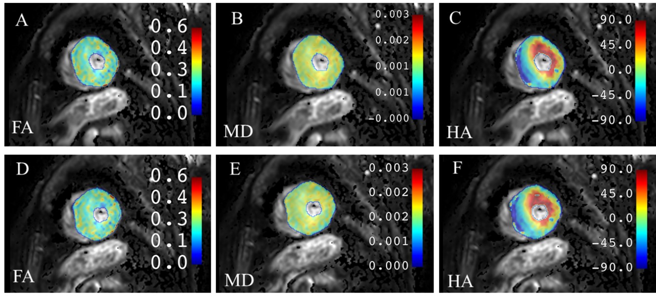
As shown in Fig. 1, before TOPUP correction, the b0 image of the short-axis of left ventricle acquired with a negative phase-encoding gradient in the RL direction was contracted due to B0 inhomogeneity (Fig. 1A), while after inversing the polarity of phase-encoding gradient, the direction EPI distortion correction was flipped (Fig. 1B). The FSL TOPUP method could correct EPI distortion in both diffusion-weighted images (b0 and b400), following which kinds of cDTI metrics are reconstructed successfully after the correction.Discussion and conclusions
cDTI has been attracting great research attention in kinds of cardiac diseases because of its capabilities in measuring myocardial microstructural environment. However, it is easily affected by cardiac and respiratory motion, which degrade image quality and thus accuracy and reproducibility of cDTI. Since the main magnetic field around the heart is inhomogeneity, EPI distortion might be severe enough to hinder quantitative measurement. Our pilot results indicate the FSL TOPUP algorithm can reverse EPI distortion effectively and kinds of cDTI metrics are reconstructed successfully after the correction. The proposed scheme could be integrated into the cDTI postprocessing, which could enhance the accuracy and reproducibility of cDTI, especially when researchers need to analyze metrics from cDTI and other different cardiac image parameters.Summary of Main Findings
In this study, the FSL TOPUP algorithm has been successfully integrated into the cDTI postprocessing in order to correct EPI distortion effectively. Kinds of cDTI metrics are reconstructed successfully after the correction, which might enhance the accuracy and reproducibility of cDTI.Acknowledgements
No acknowledgement.References
1 Joy, G. et al. Microstructural and Microvascular Phenotype of Sarcomere Mutation Carriers and Overt Hypertrophic Cardiomyopathy. Circulation 148, 808-818 (2023). https://doi.org:10.1161/circulationaha.123.063835
2 Ariga, R. et al. Identification of Myocardial Disarray in Patients With Hypertrophic Cardiomyopathy and Ventricular Arrhythmias. Journal of the American College of Cardiology 73, 2493-2502 (2019). https://doi.org:10.1016/j.jacc.2019.02.065
3 Moulin, K. et al. MRI of Reperfused Acute Myocardial Infarction Edema: ADC Quantification versus T1 and T2 Mapping. Radiology 295, 542-549 (2020). https://doi.org:10.1148/radiol.2020192186 4 von Deuster, C. et al. Studying Dynamic Myofiber Aggregate Reorientation in Dilated Cardiomyopathy Using In Vivo Magnetic Resonance Diffusion Tensor Imaging. Circulation. Cardiovascular imaging 9, e005018 (2016). https://doi.org:10.1161/CIRCIMAGING.116.005018
5 Nielles-Vallespin, S. et al. Cardiac Diffusion: Technique and Practical Applications. Journal of magnetic resonance imaging : JMRI 52, 348-368 (2020). https://doi.org:10.1002/jmri.26912
6 Nguyen, C. T. et al. Free-breathing diffusion tensor MRI of the whole left ventricle using second-order motion compensation and multitasking respiratory motion correction. Magnetic resonance in medicine 85, 2634-2648 (2021). https://doi.org:10.1002/mrm.28611
7 Holland, D., Kuperman, J. M. & Dale, A. M. Efficient correction of inhomogeneous static magnetic field-induced distortion in Echo Planar Imaging. NeuroImage 50, 175-183 (2010). https://doi.org:10.1016/j.neuroimage.2009.11.044
Figures