5174
Development in Intrinsic Neural Timescale of Hippocampal Subfields during Childhood and Adolescence1Beijing Advanced Innovation Center for Biomedical Engineering, School of Biological Science & Medical Engineering, Beihang University, Beijing, China, 2State Key Laboratory of Cognitive Neuroscience and Learning, Beijing Normal University, Beijing, China
Synopsis
Keywords:
Motivation: The hippocampus plays a pivotal role in cognition, hosting an intrinsic neural timescale hierarchy.
Goal(s): How does this intrinsic neural timescale hierarchy within the hippocampal subfield evolve throughout childhood and adolescence?
Approach: Our study encompasses 300 healthy children (6-14 years). We characterized the intrinsic neural timescale by assessing the decay of the temporal autocorrelation function (ACF) and utilized mixed-effect model to chart the developmental trajectories.
Results: Group-level intrinsic timescales within hippocampal subfields show clear temporal hierarchies along the long-axis and medial-lateral axis, and show significant correlations with hippocampal functional connectome gradients. Developmental trajectories reveal significant maturation and reorganization during childhood and adolescence.
Impact: Our research findings offer valuable insights into the unique patterns of INT within various hippocampal subfields. Furthermore, they shed light on the significant maturation and reorganization of the temporal integration and segregation structure that occurs during the onset of puberty.
Introduction
The hippocampus plays a crucial role in various cognitive functions and behaviors, encompassing declarative memory (Penfield W and Milner B, 1958), socioaffective processing(Zheng J et al., 2017), and imagination(Schacter DL et al., 2017). Recent studies have demonstrated that the brain displays a hierarchy of intrinsic neural timescale (INT), which may play a vital role in the temporal integration and segregation of input streams, and mediation of behavior, cognition, and mental features (Wolff A et al., 2022). Furthermore, a recent investigation has confirmed the presence of this temporal hierarchy within the hippocampus, showing distinctions along its long-axis (Raut RV et al., 2020). Here, we leverage a substantial dataset containing longitudinal multimodal MRI scans from a cohort of healthy children to explore common and unique INT patterns across different hippocampal subfields. Our research also delves into charting the developmental trajectory of this critical temporal hierarchy.Methods
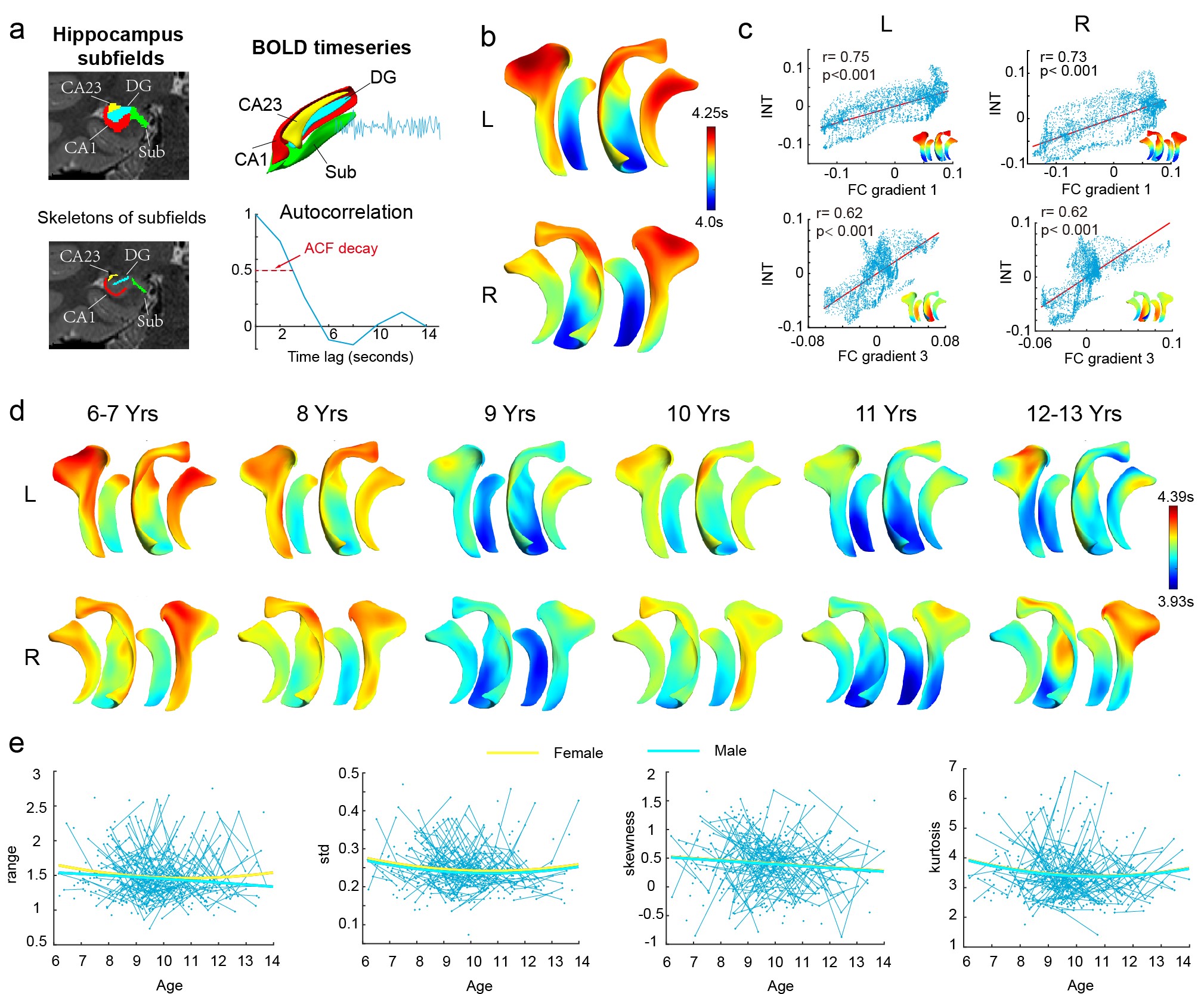
Our study encompassed 300 healthy children throughout childhood and early adolescence, comprising 140 females with ages ranging from 6 to 14 years. In total, we collected 478 scans, with 165 children undergoing a single scan, 92 children undergoing two scans, and 43 children undergoing three scans. The intervals between scans were approximately one year. For data acquisition, we utilized T1 and T2-weighted imaging in addition to resting-state functional MRI scans. These scans were performed on 3T SIEMENS Prisma scanners. Subsequently, all MR images were subjected to HCP minimum preprocessing procedures (Glasser MF et al., 2013), with several necessary modifications to tailor the process to the pediatric population.The segmentation of hippocampal subfields, including Sub, CA1, CA23, and DG, was accomplished using a registration-based multi-atlas segmentation algorithm known as ASHS (Schlichting ML et al., 2019) Subsequently, the boundary surface of each subfield was fitted using a spherical harmonic description and transformed into a triangulated mesh (Styner M et al., 2006). To further our analysis, a skeleton surface was created, traversing the centers of each hippocampal subfield. Spherical parameters on the subfield boundaries were then transferred onto the skeleton surface through the application of a Laplace method (Jones SE et al., 2000). Finally, we sampled blood oxygen level-dependent (BOLD) time series data at each vertex along the skeleton surface (see Fig. 1a).
In accordance with a previously established methodology(Raut RV, Snyder AZ and Raichle ME, 2020), we characterized the intrinsic timescale at each vertex. This characterization was achieved by assessing the decay of the temporal autocorrelation function (ACF). The quantification involved determining the time required for the ACF to decay to a value of 0.5, which is equivalent to half of the full width at half maximum (see Fig. 1a). Additionally, we computed the hippocampal-neocortical functional connectome gradient (FC gradient) to investigate its relationship with intrinsic timescales (INT). To quantitatively assess the developmental changes in hippocampal INT, we employed a mixed-effect model. This approach allowed us to identify the developmental trajectories of several essential characteristics that provide insight into the distribution of INTs within the hippocampus.
Results
At the group level, the INT within the hippocampal subfields exhibited a discernible temporal hierarchy, both along the long-axis and the medial-lateral axis (Fig. 1b). Furthermore, the INT displayed significant correlations with the hippocampal-neocortical functional connectome gradients (FC gradient1 for long-axis differentiation and FC gradient3 for medial-lateral differentiation), as illustrated in Fig. 1c.Regarding the development of INT, our findings revealed a noteworthy U-shaped trajectory in the range of INT in females (p=0.036, t_905=-2.102 for age^2×sex effect, p=0.055, t_905=1.923 for age×sex effect, p=0.051, t_905=1.955 for age^2 effect, and p=0.038, t_905=-2.075 for age effect). Interestingly, the skewness of INT demonstrated a significant decreasing trend associated with age (p=0.028, t_928=-2.192 for age effect), while the kurtosis of INT displayed a significant U-shaped trajectory (p=0.020, t_919=2.330 for age^2 effect, and p=0.012, t_919=-2.515 for age effect). These results indicate that the temporal integration and segregation structure within hippocampal subfields evolves from a relatively homogeneous state in childhood to a more differentiated and refined state during adolescence, reflecting the maturation and reorganization of this critical aspect of neural functioning.
Conclusions
Our research findings offer valuable insights into the unique patterns of INT within various hippocampal subfields. Furthermore, they shed light on the significant maturation and reorganization of the temporal integration and segregation structure that occurs during the onset of puberty.Acknowledgements
Shuyu Li is supported by NSFC (32271146) and the Startup Funds for Top-notch Talents at Beijing Normal University. Qiongling Li is supported by NSFC (82202245).
References
Glasser MF, Sotiropoulos SN, Wilson JA, Coalson TS, Fischl B, Andersson JL, Xu J, Jbabdi S, et al. (2013), The minimal preprocessing pipelines for the Human Connectome Project. Neuroimage 80:105-124.
Jones SE, Buchbinder BR, Aharon I (2000), Three‐dimensional mapping of cortical thickness using Laplace's equation. Human brain mapping 11:12-32.
Penfield W, Milner B (1958), Memory deficit produced by bilateral lesions in the hippocampal zone. AMA archives of Neurology & Psychiatry 79:475-497.
Raut RV, Snyder AZ, Raichle ME (2020), Hierarchical dynamics as a macroscopic organizing principle of the human brain. Proceedings of the National Academy of Sciences 117:20890-20897.
Schacter DL, Benoit RG, Szpunar KK (2017), Episodic future thinking: mechanisms and functions. Current Opinion in Behavioral Sciences 17:41-50.
Schlichting ML, Mack ML, Guarino KF, Preston AR (2019), Performance of semi-automated hippocampal subfield segmentation methods across ages in a pediatric sample. NeuroImage 191:49-67.
Styner M, Oguz I, Xu S, Brechbühler C, Pantazis D, Levitt JJ, Shenton ME, Gerig G (2006), Framework for the Statistical Shape Analysis of Brain Structures using SPHARM-PDM. The insight journal:242-250.
Wolff A, Berberian N, Golesorkhi M, Gomez-Pilar J, Zilio F, Northoff G (2022), Intrinsic neural timescales: temporal integration and segregation. Trends in cognitive sciences.
Zheng J, Anderson KL, Leal SL, Shestyuk A, Gulsen G, Mnatsakanyan L, Vadera S, Hsu FP, et al. (2017), Amygdala-hippocampal dynamics during salient information processing. Nature communications 8:1-11.
Figures