5173
From Neural Networks to Neuroimaging: Evaluation of Deep Learning-Based MRI Reconstruction to Accelerate, Enhance and Improve Patient Outcomes1MRI, Hospital of the University of Pennsylvania, Philadelphia, PA, United States, 2Perelman School of Medicine at the University of Pennsylvania, Philadelphia, PA, United States, 3MRI, Children's Hospital of Pennsylvania, Philadelphia, PA, United States
Synopsis
Keywords:
Motivation: The duration of MRI examinations can prove taxing for patients, leading to incomplete studies and compromised image quality due to motion. Critically ill patients requiring monitoring, those with MRI scanning time restricted implanted devices, and patients with altered mental status may be affected by lengthy scans.
Goal(s): We aim to reduce scan times in neuroradiologic studies while maintaining or improving image quality.
Approach: We applied accelerated acquisition and deep learning-based reconstruction to our current protocols. The images were assessed for signal-to-noise ratio and quality by two neuroradiologists.
Results: Scan times were drastically reduced, some more than twofold, with simultaneous improvement of image quality.
Impact: Deep learning-based reconstruction not only reduces MRI scanning time in common neuroradiologic examinations, but it also improves overall image quality. This empowers clinical sites to manage a higher workload while also diminishing potential for patient safety incidents.
Background
Successfully completing MRI examinations can be crucial for prompt diagnosis, treatment, and prognostication. However, lengthy MRI studies can be daunting, anxiety-inducing ordeals for many patients. Unlike the brief stillness required for x-ray, holding still throughout the duration of an MRI can be extremely challenging. Patient motion is estimated to happen on 10% to 42% of adult MRI sequences and happens most often in inpatient/emergency room patients1. Many patients receiving MRIs are acutely ill which exacerbates the difficulty of prolonged immobility. Lengthy MRI examinations can also increase the risk of patient safety incidents. This can be due to limitations in MR compatible clinical monitors, or from MRI related complications such as thermal injury. Finally, some FDA-approved MRI conditional implanted medical devices impose strict time limitations for MRI scans. For instance, Medtronic’s Activa deep brain stimulation system allows only 30 minutes of active scanning within a 90-minute window2. These time restrictions can result in truncated protocols or interrupted repeated examinations.The application of AI and machine learning techniques to MRI scanning offers great promise for obtaining faster scans without compromising imaging quality. Historically, acquiring high quality MRI imaging came at the expense of prolonged scan duration. However, deep learning derived image reconstruction has the potential to reduce scanning time while maintaining or improving image quality3.
Methods
We augmented our current neuroradiologic protocols using a set of fine-tuned accelerated image acquisition parameters and deep learning image reconstruction with the proprietary software Deep Resolve Boost (DRB; Siemens Healthineers, USA). DRB is described as “raw-data-to-image deep learning reconstruction enabling strong denoising for strong acquisition acceleration”4. We are conducting an ongoing prospective study involving patients undergoing both deep learning derived and conventional MRI on a 3T field strength scanner (MAGNETOM Vida Siemens Healthineers, Germany). All images are rated by two blinded neuroradiologists and assessed for multiple parameters including perceived signal-to-noise ratio and image resolution.Results
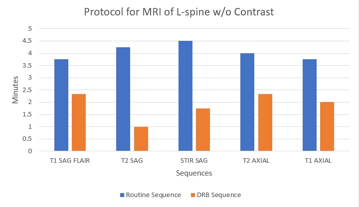
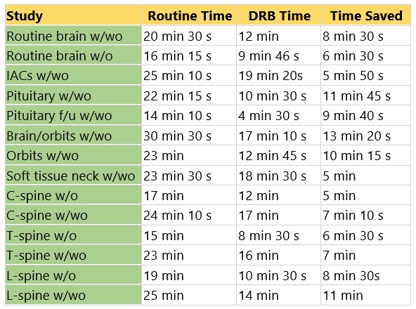
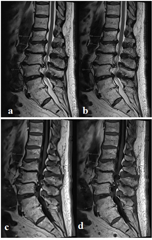
Use of DRB markedly enhanced scan efficiency. By pairing deep learning ability to process low signal raw data with alterations in scanning parameters such as a lower number of averages, and increases in the acceleration factor, we managed to achieve an impressive scanning acceleration. Reducing the number of averages allows the opportunity to accelerate by 30-60%, and modifying acceleration factor provides an additional 30-50% acceleration5. Image optimization required careful adjustment through trial and error rather than using a one-size-fits-all approach. 2D TSE T1 and T2 imaging responded best to greater parameter alterations and scan time could be significantly reduced. For example, scan time for a sagittal T2 of the lumbar spine decreased from 4 minutes and 14 seconds to just over a minute (Figure 1). Inversion recovery sequences like STIR and FLAIR needed more delicate alterations in scan parameters, and required changes in denoising strength to preserve image quality and avoid an overly synthetic appearance. Overall, these modifications yielded significant scan time reduction throughout the range of neuroradiologic protocols assessed (Figure 2).Use of DRB also resulted in improved image resolution as well as perceived signal to noise ratio and image quality. Maintaining matrix sizes and optimizing our protocols with DRB resulted in a doubling of the inherent resolution through interpolation. Use of DRB not only achieved our objective of reducing scan time, but also significantly improved MRI image quality (Figure 3).
Summary
Deep learning-based MRI scanning provides the ability to achieve higher quality imaging at a fraction of the time compared to conventional scanning. Faster scanning improves patient tolerance and decreases the incidence of motion degradation. It reduces the probability of a patient safety event and decreases the amount of medication needed to tolerate exams. Expedited scanning permits patients with MR-conditional devices to receive full exams or multiple MRIs in adherence with manufacturer defined time constraints. This technology has significant promise in increasing diagnostic efficiency, patient safety, and improved outcomes contributing to a superior client experience.Acknowledgements
No acknowledgement found.References
1. Andre, J. B., Bresnahan, B. W., Mossa-Basha, M., Hoff, M. N., Smith, C. P., Anzai, Y., & Cohen, W. A. (2015). Toward quantifying the prevalence, severity, and cost associated with patient motion during clinical MR examinations. Journal of the American College of Radiology, 12(7), 689–695. https://doi.org/10.1016/j.jacr.2015.03.007
2. Medtronic. (2021). MRI guidelines for Medtronic deep brain stimulation systems. Minneapolis, MN: Medtronic, Inc. https://www.medtronic.com/content/dam/emanuals/neuro/M929535A_b_074_view.pdf
3. Bash, S., Wang, L., Airriess, C., Zaharchuk, G., Gong, E., Shankaranarayanan, A., & Tanenbaum, L. N. (2021). Deep Learning Enables 60% Accelerated Volumetric Brain MRI While Preserving Quantitative Performance: A Prospective, Multicenter, Multireader Trial. AJNR. American journal of neuroradiology, 42(12), 2130–2137. https://doi.org/10.3174/ajnr.A7358
4. Siemens Healthineers USA. (n.d.). Deep resolve boost. https://www.siemens-healthineers.com/en-us/magnetic-resonance-imaging/options-and-upgrades/clinical-applications/deep-resolve-boost
5. Bash, S., & Tanenbaum, L. (2021, January 19). Deep learning: Promising to revolutionize image reconstruction. Applied Radiology. https://appliedradiology.com/articles/deep-learning-promising-to-revolutionize-image-reconstruction
Figures