5172
Altered Functional Connectivity of the Olfactory Cortex in Post-COVID-19 Brain Network1First clinical Medical College, Guangzhou University of Chinese Medicine, Guangzhou, China, 2Department of Diagnostic Radiology & Nuclear Medicine, University of Maryland,Baltimore, Baltiomore, MD, United States, 3The Department of Radiology, The first Affiliated Hospital of Guangzhou University of Chinese Medicine, Guangzhou, China
Synopsis
Keywords:
Motivation: To investigate the persistent effects on the brain following recovery from COVID-19.
Goal(s): To examine differences in brain’s functional connectivity between post-COVID-19 subjects and healthy controls.
Approach: To use resting-state fMRI data to identify brain functional networks.
Results: Post-COVID subjects exhibited higher functional connectivity in a brain network including the left olfactory cortex as the main hub connected to other brain regions.
Impact: We observed that post-COVID-19 subjects continue to exhibit significant anomalies in the olfactory cortex of the brain. These results support previous findings and highlight the relevance of research from a fMRI perspective for investigating the effects on brain of COVID-19.
Introduction
Brain impairment and neuropsychiatric symptoms are frequently documented post-COVID-19 infection1. Many patients suffered from symptoms such as anosmia, cognitive impairment, anxiety, depression, and psychosis2, 3, but the mechanisms behind these symptoms are unclear. Data from non-human primates showed that SARS-CoV-2 primarily invades the central nervous system (CNS) via the olfactory bulb and subsequently spreads to other functional areas4. Other research showed that SARS-CoV-2's neurotropism can access the nervous system by crossing the neural-mucosal interface within the olfactory mucosa3. A PET study has found reduced metabolisms in the olfactory gyrus and interconnected limbic/paralimbic regions, with the effect extending to the brainstem and the cerebellum, which were correlated with memory/cognitive impairment5. Previous neuroimaging studies of individuals who experienced olfactory disturbances following COVID-19 infection have revealed significant brain changes, typically involving the olfactory cortex. Among these, SARS-CoV-2 infected subjects repeatedly exhibited a significant increase in both structural and functional brain connectivity6. SARS-CoV-2 infected subjects also exhibited modified olfactory network connectivity linked to the severity of hyposmia and neuropsychological performance7. To gain more specific insight on the early neurosensory impact of COVID-19, here we performed a resting-state fMRI (rs-fMRI)-based study examining brain functional connectivity. While many related studies focused on participants reporting olfactory dysfunction, our study observed increased brain connectivity in the olfactory cortex among post-COVID-19 participants, including those without significant olfactory dysfunction.Method
108 subjects were enrolled (19–33 years old, M = 24.81, SD = 3.04, 31 males and 77 females). 50 participants reported a COVID-19 infection history within the previous 3 months (post-Covid-19 group), while 58 individuals were recruited as healthy controls. Inclusion criteria for the post-Covid group were: a positive result on the nasopharyngeal swab for SARS-CoV-2 RNA, a diagnosis of pneumonia of SARS-CoV-2 and/or self-reported positive Polymerase Chain Reaction (PCR) result from a SARS-CoV-2 test kit in the previous 3 months. Resting-state fMRI (rs-fMRI) was acquired from each participant and pre-processed using the Data Processing Assistant and Resting-State FMRI (DPARSF) toolbox running on MATLAB. Functional connectivity (FC) analyses were performed using Nilearn (https://nilearn.github.io/) and the Automated Anatomical Labelling (AAL) atlas8. Post-COVID-19 vs control difference of FC matrix was assessed using the Network Based Statistics (NBS) toolbox9. For each element of the FC matrix, a general linear model (GLM) including the group factor (post-COVID group = 1; Control group = -1), age, and sex was estimated. Then, the effect of the group factor was tested (post-COVID vs Control; Control vs post-COVID). Statistical threshold was t>=4 (p-FWE <= 0.05).Result
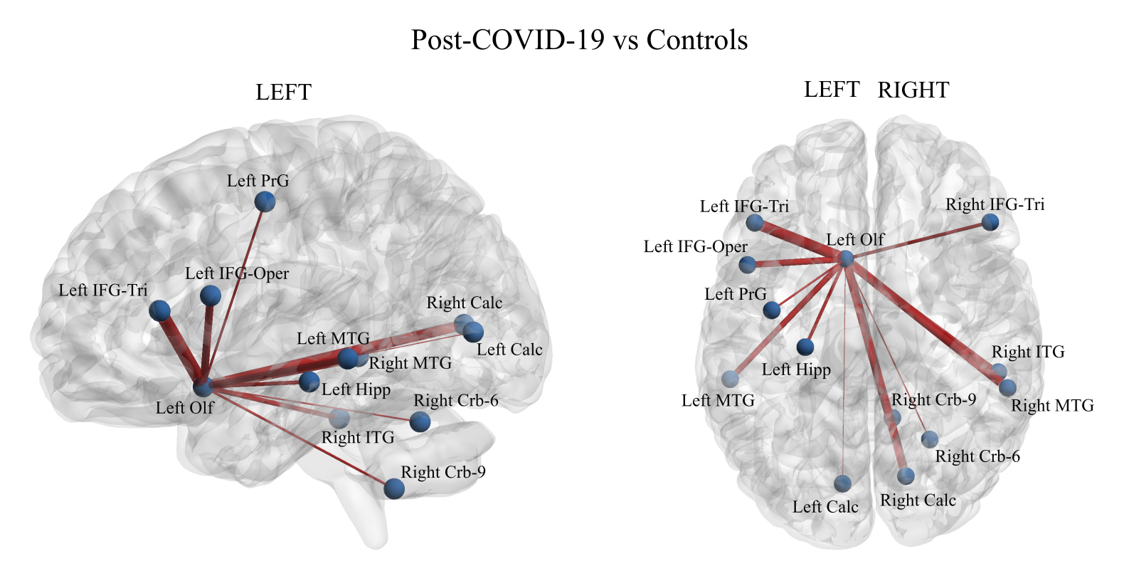
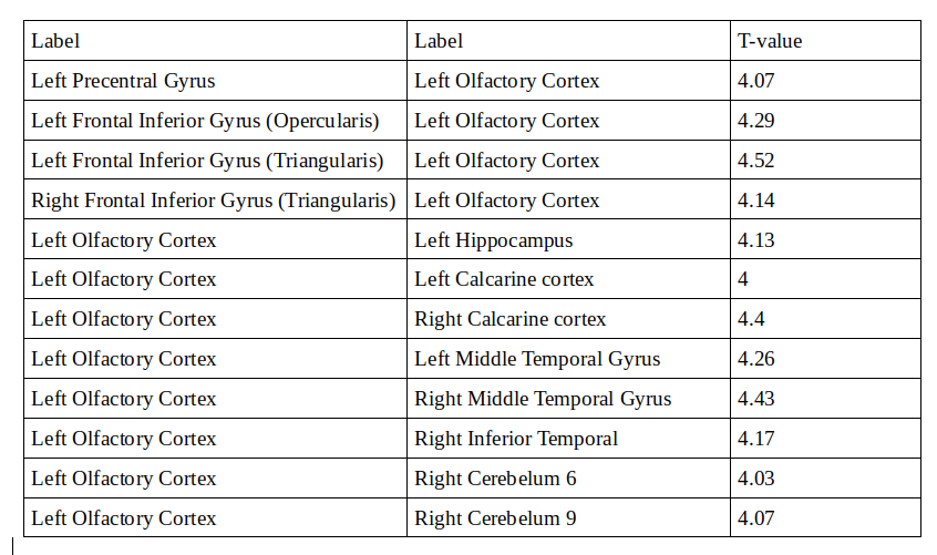
Compared to controls, post-COVID subjects exhibited higher FC in a brain network including the left olfactory cortex as main hub connected with frontal, temporal, occipital and cerebellar regions (Figure 1). A list of all significant connections included in the network is reported in Table 1. The opposite contrast (Controls vs post-COVID) did not yield significant results.Discussion
In our post-COVID group, 24% of the 50 participants reported experiencing olfactory dysfunction. However, the whole group, compared to controls, exhibited increased FC between the left olfactory cortex and numerous other brain regions. This may suggest that even in the absence of evident clinical symptoms, functional pairings between the olfactory cortex and its associated brain regions have still been subject to some form of alteration. In our study, we observed that some brain regions connected with the olfactory cortex overlap with the functional pathway of the olfactory system as defined by human fMRI studies10, 11. This may indicate that in pathological conditions, abnormal transmission may utilize similar information pathways as in a physiological state. Furthermore, while certain regions such as the cerebellum lack direct anatomical connections to the olfactory mucosa, our findings suggest increased connectivity between the olfactory cortex and cerebellum in the post-COVID compared to the control group. This observation may be relevant, especially considering the presence of SARS-CoV-2 RNA has been detected in the cerebellum of certain individuals3.Conclusion
We have discovered that the olfactory cortex and its associated brain regions are significantly affected following COVID-19 infection, that this impact persists for a period of time after recovery, and it is not limited to subjects who exhibited prominent olfactory dysfunction symtoms.Acknowledgements
We thank our research participants for their time and efforts.References
1. Boldrini M, Canoll PD, Klein RS. How COVID-19 Affects the Brain. JAMA Psychiatry. 2021;78(6):682–683. 2. Woo, Marcel S et al. Frequent neurocognitive deficits after recovery from mild COVID-19. Brain communications vol. 2,2 fcaa205. 23 Nov. 2020. 3. Meinhardt, Jenny et al. Olfactory transmucosal SARS-CoV-2 invasion as a port of central nervous system entry in individuals with COVID-19. Nature neuroscience vol. 24,2 (2021): 168-175. 4. Jiao, Li et al. The olfactory route is a potential way for SARS-CoV-2 to invade the central nervous system of rhesus monkeys. Signal transduction and targeted therapy vol. 6,1 169. 24 Apr. 2021. 5. Guedj, E et al. 18F-FDG brain PET hypometabolism in patients with long COVID. European journal of nuclear medicine and molecular imaging vol. 48,9 (2021): 2823-2833. 6. Esposito, Fabrizio et al. Olfactory loss and brain connectivity after COVID-19. Human brain mapping vol. 43,5 (2022): 1548-1560. 7. Muccioli, Lorenzo et al. Cognitive and functional connectivity impairment in post-COVID-19 olfactory dysfunction. NeuroImage. Clinical vol. 38 (2023): 103410. 8. Tzourio-Mazoyer, N et al. “Automated anatomical labeling of activations in SPM using a macroscopic anatomical parcellation of the MNI MRI single-subject brain.” NeuroImage vol. 15,1 (2002): 273-89. 9. Zalesky, Andrew et al. “Network-based statistic: identifying differences in brain networks.” NeuroImage vol. 53,4 (2010): 1197-207. 10. Zhou, Guangyu et al. Characterizing functional pathways of the human olfactory system. eLife vol. 8 e47177. 24 Jul. 2019, doi:10.7554/eLife.47177 11. Katata, Keita et al. Functional MRI of regional brain responses to 'pleasant' and 'unpleasant' odors. Acta oto-laryngologica. Supplementum ,562 (2009): 85-90. doi:10.1080/00016480902915715Figures