5171
3D T2-Weighted Imaging of the Breasts in the Supine Position1Radiology, Stanford University, Stanford, CA, United States, 2Coils, GE HealthCare, Aurora, OH, United States, 3MRI Clinical Solutions and Research Collaboratins, GE HealthCare, Menlo Park, CA, United States, 4Global MR Applications and Workflow, GE HealthCare, Boston, MA, United States
Synopsis
Keywords:
Motivation: Despite its importance in breast MRI, T2-weighted supine imaging is understudied, risking image quality due to motion and prolonged respiratory-triggered scans.
Goal(s): To evaluate effectiveness of 3D supine T2-weighted breast imaging using a 60-channel flexible breast coil.
Approach: Three individuals underwent prone and supine breast imaging using two parallel imaging settings. An MRI Radiologist evaluated four images in a blinded study of techniques.
Results: Increased acceleration available with the 60-channel breast coil, achieved high 3D T2-weighted image quality in the supine position within a clinically feasible scan time.
Impact: Supine positioning in breast MRI, facilitated with flexible blanket coils and fast, high-performing protocols, has the potential to improve the comfort and efficiency of the exam, allowing breast MRI to be accessible to a much wider population of women.
Introduction
Breast MRI is a highly sensitive method for detecting breast cancer. However, there are several challenges that the exam. A number of these challenges are due to the breast coil. Conventional breast MR uses prone positioning which reduces respiratory and cardiac motion thereby reducing image artifacts. However, lying prone on the hard coil casing often becomes uncomfortable, making the patient susceptible to motion to relieve pain. Further, set up of the patient in the coil is time consuming and requires expertise. Supine breast MRI has been intermittently investigated but is limited by the lack of coils well-suited for the position [1]. The emergence of flexible blanket coils has greatly increased the potential for wider investigation and clinical adoption of supine breast MRI [2]. Along with the potential to improve the comfort and efficiency of the exam itself, supine positioning improve alignment with surgical procedures. Supine positioning, however, does present some challenges, most notably, increased motion. T2-weighed imaging is an essential component of breast MRI but acquiring T2-weighted imaging with high-resolution and respiratory triggering may lead to clinically unfeasible scan times. In this work we investigate 3D T2-weighted imaging of the breasts in the supine position in comparison to 3D T2-weighted imaging in the breasts in the prone position. Further, we investigate an increased degree of parallel imaging with a high channel count flexible breast coil to reduce scan times of respiratory-triggered 3D T2-weighed supine breast MRI.Methods
3 healthy female volunteers (ages 25-65) were recruited to be scanned with three 3D T2-weighted protocols. Recruitment followed IRB polices and all subjects provided written informed consent. All scans were performed on a 3T Signa Premier Scanner (GE HealthCare, Waukesha, WI). A 3D T2-weighted acquisition was performed in the prone position and two 3D T2-weighted acquisitions were performed in the supine position. Resolution ( 1 mm x 1 mm x 2 mm) was the same for all three protocols. The prone protocol and the first supine protocol used 3x2 parallel imaging. The second supine protocol used 4x4 parallel imaging. The prone protocol was acquired with a 16-channel Sentinelle Breast Coil (In Vivo Corp, Gainsville, FL) while the supine protocols were scanned with a 60-channel flexible AIR Breast Coil [3]. Supine protocols were acquired with respiratory-triggering while the prone protocol was performed under free breathing. Protocol positioning followed standard clinical procedures. For supine positioning, subjects were placed on the MRI table feet first and instructed to raise their arms above their head so as not to include their arms in the imaging. A respiratory gating bellows was placed around the abdomen. A Velcrowrap was loosely wrapped around the flexible supine coil and patient to ensure coils were conforming to torso of each patient.A radiologist with extensive breast MRI expertise performed a blinded observer study of the three protocols. Two slices from each volunteer/protocol (slice location +/- 5 slices from central slice) were included in the study. All images were randomized as a single group. The radiologist rated four features for each image (sharpness, noise, other artifact, overall image quality) on a scale of 1 to 5 with 1 representing lowest performance and 5 representing highest performance for each feature.Results
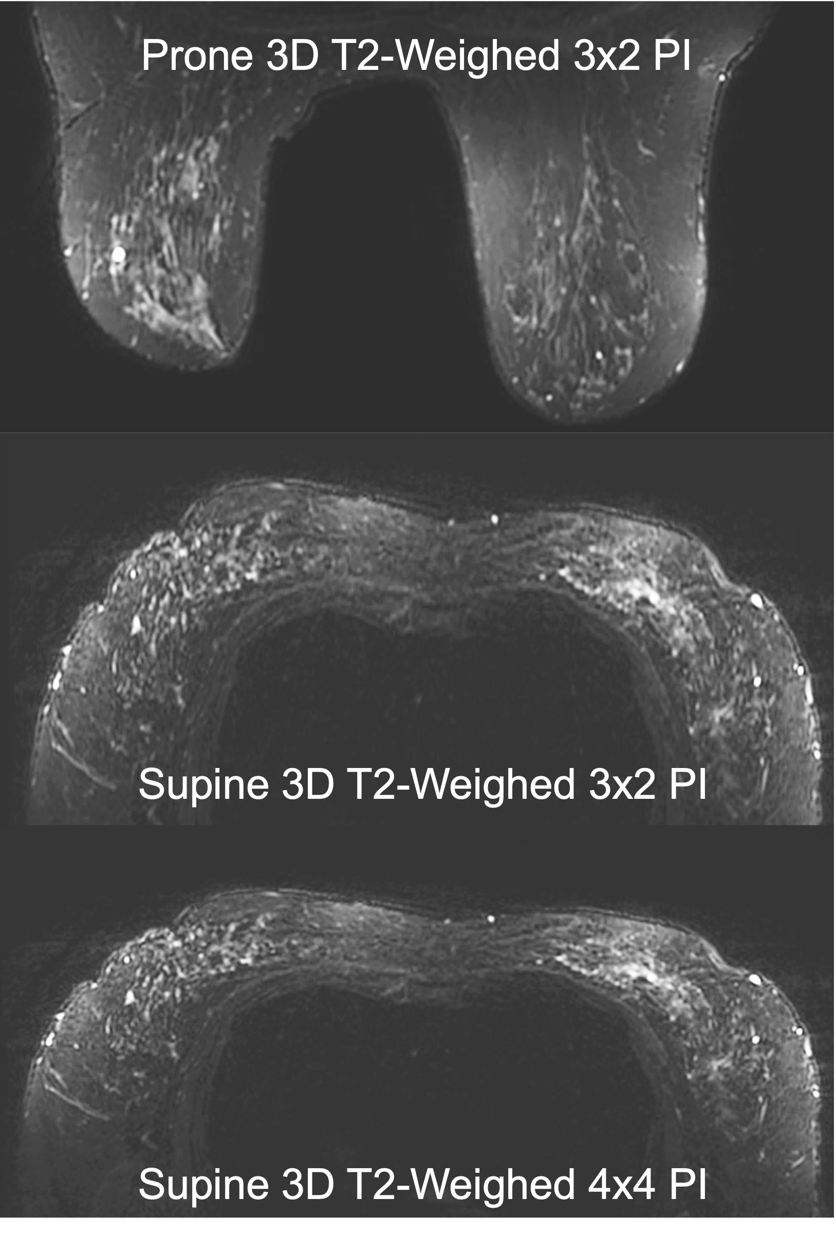
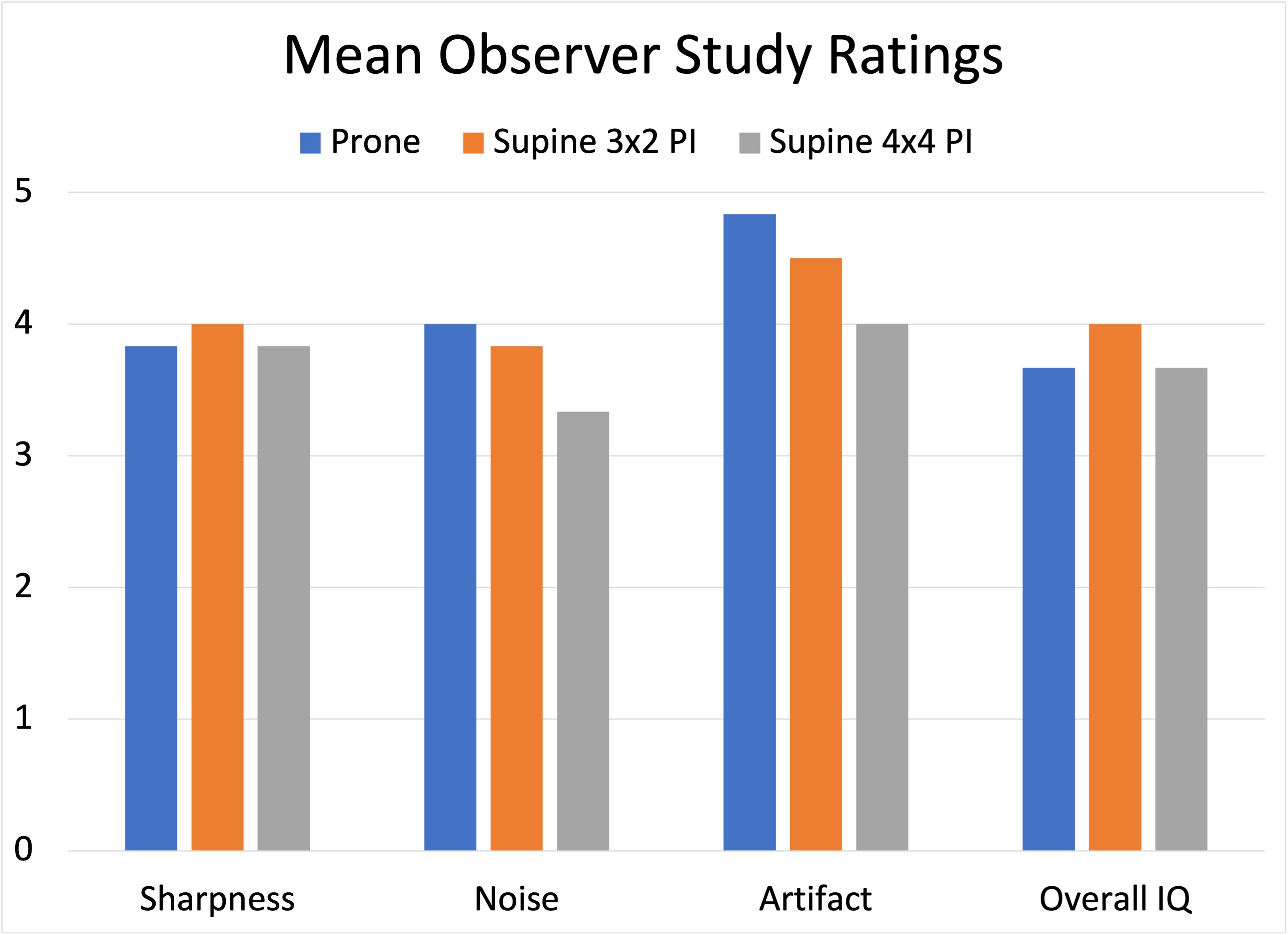
All ratings were 3 or higher for all four features for each of the three protocols indicating high performance for all protocols as ratings 3, 4 and 5 represented adequate, high and exceptional performance respectively (Figures 1 and 2). While the mean ratings for the supine 4x4 protocols were slightly lower than the prone 3x2 for all four images features, the mean ratings were still 3 or higher. For two of the features, sharpness and overall image quality, mean ratings with supine 3x2 PI were slightly higher than prone 3x2.Discussion and Conclusion
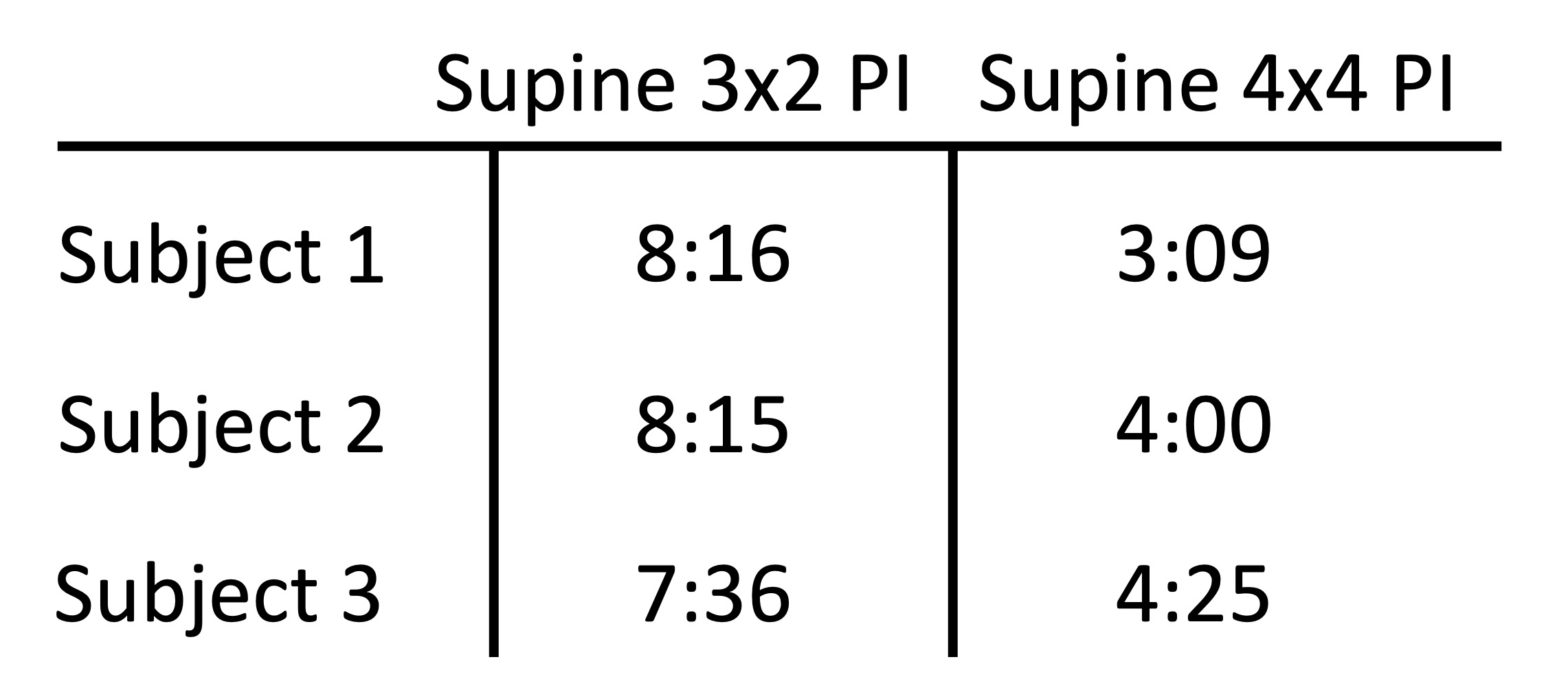
All three protocols achieved high ratings for all four image features. Though further investigation with a higher number of patients and observers is needed to confirm these findings, these initial results are encouraging for the continued pursuit of supine breast MRI as T2-weighted imaging is an essential component of breast MRI. With respiratory triggering in the supine position, the image quality of the supine and prone 3D acquisitions were comparable and most importantly the higher coil count of the flexible breast coil allowed for higher parallel imaging factors, greatly shortening the increased scan time due to respiratory triggering (Figure 3). Further studies will also include assessments of patient comfort and set-up times between prone and supine positioning.Acknowledgements
NIH/NIBIB R01 EB009055, NIH/NCI R01 CA249893 and GE HealthcareReferences
1. Fausto A, Fanizzi A, Volterrani L, et al. Feasibility, Image Quality and Clinical Evaluation of Contrast-Enhanced Breast MRI Performed in a Supine Position Compared to the Standard Prone Position. Cancers (Basel). 2020;12.
2. Moran CJ, Middione MJ, Mazzoli V, McKay-Nault JA, Guidon A, Waheed U, Rosen EL, Poplack SP, Rosenberg J, Ennis DB, Hargreaves BA, Daniel BL. Multishot Diffusion-Weighted MRI of the Breasts in the Supine vs. Prone Position. J Magn Reson Imaging, 2023; 58(3): 951-962.
3. Vincent J, Follante C, Bayram E, et al. Ultra-Flexible, High-Resolution, 60-Channel RF Coil for Supine Breast Imaging. In Proceddings of the ISMRM and SMRT Annual Meeting and Exhibition. 2021:abstract 1591.
Figures