5160
Impact of Deep Resolve Sharp & Gain AI software on patient throughput and image quality at 3.0 Tesla: A preliminary study in knee.1Radiology, UCHealth, Highlands Ranch, CO, United States, 2Radiology, University of Colorado School of Medicine, Aurora, CO, United States, 3Colorado School of Public Health, Aurora, CO, United States
Synopsis
Keywords:
Motivation: To quantify the impact of Deep Resolve Sharp and Gain (DRSG) Artificial Intelligence (AI) software on patient throughput and image quality of knee MRI exams at 3.0 Tesla.
Goal(s): To study the clinical the impact of DRSG on image quality and examination time.
Approach: Thirty patients underwent examinations without or with DRSG enabled. An attending musculoskeletal radiologist blindly reviewed exams and the quality of structures was assessed in routine diagnostic planes. Exam times were recorded.
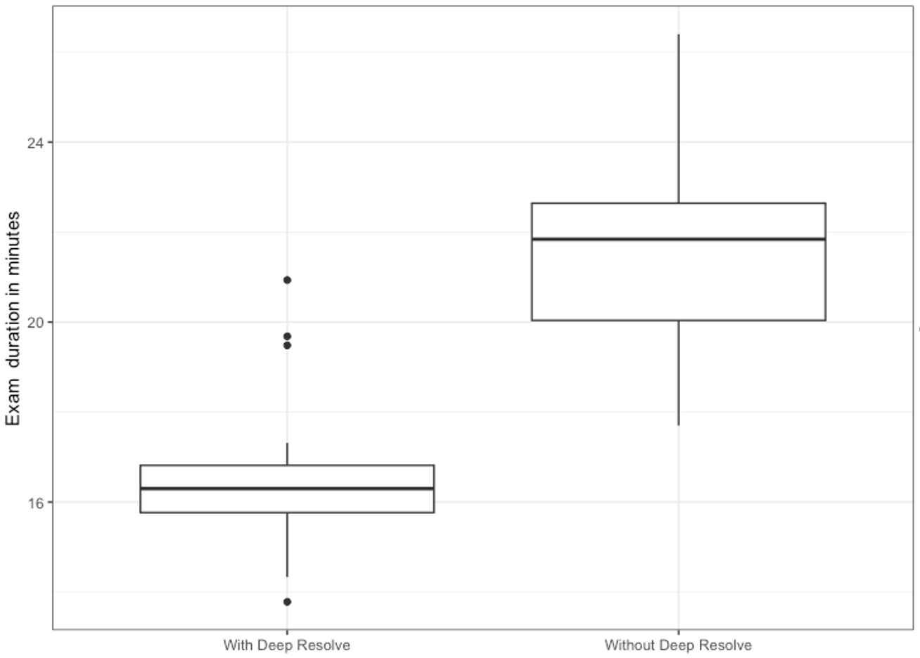
Results: Knee structures showed no statistical difference in image quality. Meanwhile, a statistically significant reduction in mean exam time was observed with DRSG-enabled protocols of 24% (p<0.001).
Impact: Siemens Healthineers Deep Resolve Sharp & Gain is a clinically useful tool to significantly reduce MR knee exams times without compromising image quality at 3.0 Tesla.
Introduction
Deep Resolve Sharp & Gain is an artificial intelligence-based software that can be deployed on Siemens Healthineers MRI scanners to accelerate image acquisition. This study aims to investigate the performance of Deep Resolve Sharp & Gain (DRSG) on magnetic resonance (MR) knee exams.Hypothesis
Use of DR results in a reduction of image acquisition time while maintaining diagnostic image quality compared to protocols without DR enabled.Methods
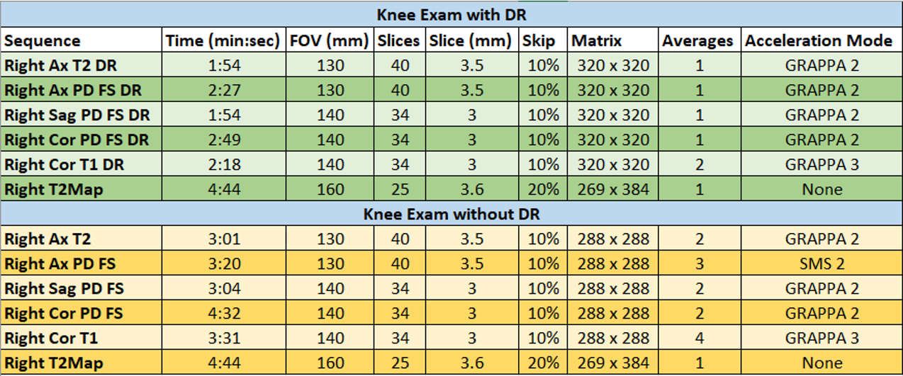
Thirty patients underwent MR knee examinations. Exams were acquired on 3.0T Magnetom Vida (Siemens Healthineers, software: XA31). In the pre-implementation period (January 24th - 31st, 2023), sixteen patients received standard-of-care knee examinations without DRSG enabled. Clinical deployment of DRSG-enabled examinations occurred after a trial period (February 1st -14th, 2023) to allow for testing, optimization, review, and approval as the standard of care. In the post-implementation period (February 14th -24th, 2023), fourteen patients received knee examinations with DRSG enabled. Sequences used in the examinations included: axial T2, axial Proton Density Fat Suppression (PD FS), sagittal PD FS, coronal T1, and coronal PD FS. Image quality analysis was blinded and performed by a fellowship-trained MSK attending radiologist practicing for six years exclusively in musculoskeletal imaging. The anatomical structures judged included fibrocartilage, hyaline cartilage, joint fluid, bone marrow, and other commonly assessed tissues, evaluated in planes typically used in clinical practice. Structures were graded on a 1-4 point scale (1-major artifacts, 2-moderate artifacts with low image quality, 3-minor artifacts with good image quality, and 4-no artifacts with excellent image quality). The anticipated clinical impact of the artifacts was also judged on 0-3 scale (0-no impact, 1-minor impact, 2-major effect, 3-uninterpretable). Imaging protocols, with and without DRSG enabled, are detailed in figure 1. A two sample t-test was performed to compare mean acquisition times in both study groups.Results
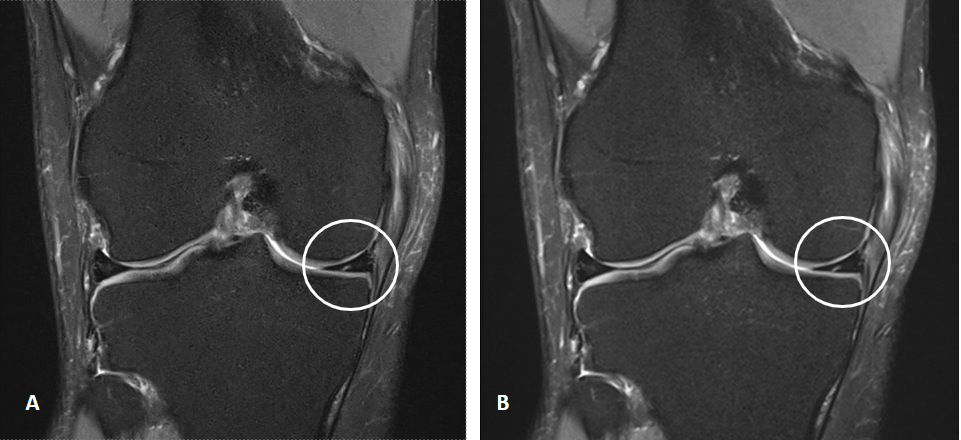
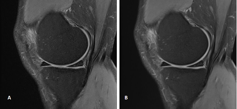
Image quality differences were minimal and did not appear systematic between images acquired with DRSG enabled compared to conventional techniques. No significant anticipated clinical impact was identified. Meanwhile, DRSG-enabled protocols had a mean exam time of 16.3 minutes, significantly shorter than the mean exam time of 21.4 minutes for the conventional protocol (p<0.001).Acknowledgements
Jack Pattee Ph.D., Christopher Allen R.T.(R)(MR)(CT), Leslie Sisney R.T.(R)(MR)(CT)
References
Hahn, S., MD, Yi, J., MD, & Lee, H. J., MD, et al (2021). Image Quality and Diagnostic Performance of Accelerated Shoulder MRI With Deep Learning–Based Reconstruction. American Journal of Roentgenology, 218(3). https://doi.org/10.2214/AJR.21.26577
Chazen, J. L., Tan, E. T., Fiore, J., Nguyen, J. T., Sun, S., & Sneag, D. B. (2023). Rapid lumbar MRI protocol using 3D imaging and deep learning reconstruction. Skeletal Radiology, 52, 1331–1338. https://doi.org/10.1007/s00256-022-04268-2
Kiryu, S., MD, PhD, Akai, H., MD, PhD, & Yasaka, K., MD, PhD, et al (2023). Clinical Impact of Deep Learning Reconstruction in MRI. RadioGraphics, 43(6). https://doi.org/10.1148/rg.220133
Herrmann, J., Koerzdoerfer, G., Nickel, D., et al (2021) Feasibility and Implementation of a Deep Learning MR Reconstruction for TSE Sequences in Musculoskeletal Imaging. Diagnostics,11, 1484. https://doi.org/10.3390/diagnostics11081484
Siemens Healthineers (n.d.). Deep Resolve MRI-Faster than ever before. Retrieved June 1, 2023, from https://www.siemens-healthineers.com/en-us/magnetic-resonance-imaging/technologies-and-innovations/deep-resolve
Figures