5157
Value Assessment of Orthogonal Phase Encoding Reduction Artifacts, O.P.E.R.A.1Radiology, Azienda USL Toscana Centro, Empoli, Florence, Italy, 2FTGM Fondazione Toscana Gabriele Monasterio, Pisa, Italy, 3Biomedical Engineering, University of Pisa, Pisa, Italy, 4Bluesotware, Poggibonsi, Siena, Italy, 5Radiology, Ausl Le Scotte, Siena, Siena, Italy, 6Radiology, Area Vasta Toscana Sud-EST, Siena, Siena, Italy, 7Neonatal Intense Care Unit, AUSL Le Scotte,Siena, Siena, Italy
Synopsis
Keywords:
Motivation: Artifacts are common in MR and reduce the image quality, and led to a degradation of the radiographer's work. We developed a retrospective method, Orthogonal Phase Encoding Reduction Artifacts (OPERA) which works on DICOM images.
Goal(s): To test the value assessment of OPERA on several anatomic regions in a Clinical Study on a Public General Hospital.
Approach: A total of 604 MR exams were acquired on 585 patients by using an 1.5T scanner.
The OPERA-images were compared with standard images per Artifacts, Signal-Noise-Ratio, and scan-time by two radiologists.
Results: OPERA algorithm reduced the repeated sequences of 5.9% and the additional sequences of 2.17%.
Impact: Opera
requires no hardware modifications. OPERA is a post-processed software and
works on DICOM, so it is applicable on any type of MRI scanner.
OPERA reduce Motion Artifact, and consequently could reduce the number of
repeated sequences on uncooperative patient.
Background
In Magnetic Resonance Imaging (MRI) artifacts are an important problem leading to reduced image quality1, loss of diagnostic information, and time consuming. These problems led to a degradation of the radiographer's work. Artifacts can be reduced by using several algorithms, most of them specific for a single anatomical region, working at K-Space reconstruction level, and often leading to prolongation of the scan time2,3.We previously introduced a method (OPERA: Orthogonal Phase Encoding Reduction of Artifact4). It is a post-process software which works on DICOM images for the automatic correction of artifacts generated by motion and wrap-around.
Purpose
This prospective study were tested the MR value of OPERA on the reduction of artifacts, the reduction of the number of re-scan sequences due to severe artifacts and the reduction of additional sequences required by radiologists.Method
In MRI the position of ghosts, motion and aliasing artifacts is predictable along columns or rows of pixels, by knowing the phase encoding direction. OPERA algorithm compares the intensity values of the pixels of two DICOM images acquired on the same anatomy by using the same parameters but with orthogonal phase-encoding directions. In order to avoid prolongation of the exam time, the Number of Signal Averages (NSA), (or similar) can be exploited by making an even NSA, with half of them being distributed along one phase encoding direction and the remaining half being distributed along the opposite one.This study was conducted in a medium-sized general public hospital by using an 1.5T MR scanner by comparing 3 full calendar weeks (MR exams n=302: Group A) with the following 3 weeks where OPERA procedure was applied to all MR sequences imaging of non-moving organs (MR exams n=302; Group B). A total of 604 MR exams were acquired on 585 patients (54.8% females; mean age 52.7±17.2 years)]. Imaged anatomical regions were: brain, n=182; spine, n=138; shoulder, n=64; elbow, n= 26; pelvis, n=96; knee, n=68; ankle, n=30.
During the first 3 weeks (Group A), the sequences were acquired with the Standard parameters required by good clinical practice. For the next 3 weeks (Group B),OPERA procedure was used. Each sequence was acquired with vertical phase encoding direction and with horizontal phase encoding direction and NSAOPERA=NSASTD/2. Both sequences were sent to the PACS and the pair was processed in a remote-server by OPERA algorithm to obtain a third sequence with artifact-reduced images that were returned to the PACS.
For both A and B Groups, the radiographer noted the sequences with several motion artifacts that were been repeated, and the additional sequences, over the standard protocol, required by radiologists for the diagnosis. Radiographers and radiologists have a long-standing MRI expertise (>15 years.)
The MedCalc 17.2 statistical software was used for statistical analysis.
Results
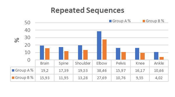
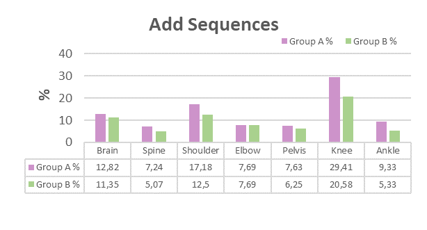
Significant motion artifacts were identified on sequences in 18.42 % (n= 279) of Group A, and in 13.34% (n= 202) Group B ( Fig 1). Additional sequences were required by radiologist in 12.48 % (n= 189) of Group A, and in 9.7% (n= 147) of Group B. (Fig 2).Overall, as compared to the standard procedure, OPERA algorithm consistently reduced the percentages of repeated sequences of 5.9% (two tailed p value= 0.0639) while reducing the percentage of additional sequences of 2.17% (Hodges-Lehmann median difference 5,9200; 95% Confidence interval -1,2900 to 13,3700; Mann-Whitney U, 10,00; two tailed p value= 0.3379).
Conclusion
The present study confirms the potential of OPERA algorithm in reducing both the need for MR sequence repetition due to severe artifacts and MR sequences addiction for better diagnosis. Even the software could be usable on uncooperative patient or patients with pathology which do not allow motionless posture during the MR examination. The method was demonstrated to be applicable to several sequences and anatomical regions. Elbow studies were characterized by severe motion artifacts due to the patient position and the extremely pain in post-traumatic cases.The remarkable number of sequences “not-repeat” could potentially have a positive side effect in saved time. The peculiarities of the present algorithm allows a wide applicability to different commercially available MRI machines.
OPERA would simplify the radiographer work by eliminating the parameter “ phase encoding direction”, oversampling, or saturation regions.
Acknowledgements
We sincerely thank:
all the radiographers, and medical radiologists of San Giuseppe Hospital in Empoli for the opportunity to acquire the images during the clinical practice sessions;
Francesco De Milato for valuable technical advice;
Radiographers Luca Bartalini (MR specialist) for help and technical support in the preliminary stages;
Professor Davide Caramella (Radiologist, Cisanello Hospital Pisa, Italy) for advice in the early development of the main idea about “orthogonal-correction method”.
References
1 . Andre JB, Bresnahan BW, Mossa-Basha M, Hoff MN, et al.; Toward Quantifying the Prevalence, Severity, and Cost Associated With Patient Motion During Clinical MR Examinations. J Am Coll Radiol. 2015 Jul;12(7):689-95. doi: 10.1016/j.jacr.2015.03.007.
2. Kruger DG, Glenn SS, et al. ; An orthogonal correlation algorithm for ghost reduction in MRI. MRM 1997; 38: 678-686;
3. Forbes KP, Pipe JG, Bird CR, Heiserman JE (2001) PROPELLER MRI: clinical testing of a novel technique for quantification and compensation of head motion. J Magn Reson Imaging 14:215–222
4. Dell’Orso, A., Positano, V., De Felice C. et al. OPERA: a novel method to reduce ghost and aliasing artifacts. Magn Reson Mater Phy 34, 451–467 (2021). https://doi.org/10.1007/s10334-020-00881-1
Figures