5182
Real-time hyperpolarized 13C-pyruvate CMRI imaging pipeline for monitoring of cardiotoxicity1Department of Internal Medicine, University of Texas Southwestern Medical Center, Dallas, TX, United States, 2Philips Healthcare, Gainesville, FL, United States, 3Advanced Imaging Research Center, University of Texas Southwestern Medical Center, Dallas, TX, United States, 4ETH Zurich, Institute for Biomedical Engineering, Zürich, Swaziland, 5Dallas VA Medical Center, Dallas, TX, United States, 6Harold C. Simmons Comprehensive Cancer Center, Dallas, TX, United States
Synopsis
Motivation: Real-time imaging protocol based on hyperpolarized 13C pyruvate to probe metabolic changes in patients undergoing standard-of-care chemotherapy for breast cancer, with cardiotoxic potential.
Goal(s): An imaging protocol that results in high-quality reproducible B0 heart shimming. To implement an echo-shifted mDIXON acquisition with spatial-spectral excitation to detect pyruvate and its major byproducts. A processing pipeline for reconstruction of metabolic images.
Approach: Single-shot EPI acquisitions with shifted echo times (n=6) were acquired following a SpSp excitation, as to generate signal for mDIXON / IDEAL reconstructed images of these metabolites.
Results: We have established a real-time 13C-hyperpolarization and imaging protocol and reconstruction pipeline
Impact: Noninvasive real-time metabolic imaging using hyperpolarized 13C may aid clinical evaluation of possible cardiac toxicity for breast cancer patients
Abstract
Hyperpolarized (HP) [1-13C] pyruvate magnetic resonance spectroscopic imaging (MRSI) is a powerful tool for investigating cardiac metabolism in real-time, by detecting the downstream metabolic products of pyruvate in the myocardium. The identification of [1-13C] pyruvate along with its derivatives, namely [1-13C] lactate and 13C-bicarbonate, provides a means to assess the myocardium's metabolic profile. In this study, we employed a spectral-spatial (SPSP) excitation pulse, followed by an echo planar imaging (EPI) readout to distinguish and capture each metabolite distinctly. The goal of this research is to establish a MATLAB-based analysis pipeline for 13C-MRI image processing and visualize myocardial bicarbonate, lactate, and pyruvate metabolism.Methods:
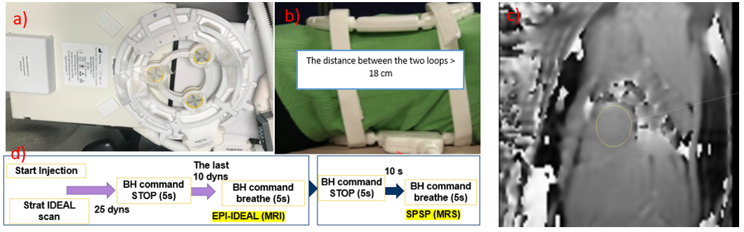
A total of 49 patients with breast cancer were enrolled in the study. All patients tolerated the hyperpolarized 13C MR procedure well. Hyperpolarized pyruvate aliquots were prepared and analyzed using SPINlab™ (GE Healthcare). A 13C transmit/receive Helmholtz loop-pair coil from PulseTeq (UK) is used for hyperpolarized pyruvate imaging, using a 3T scanner (Philips Healthcare) (see Fig 1.a). Positioning the heart between the two loops maximizes the reception of the net magnetic field, allowing for optimal excitation and reception of the hyperpolarized pyruvate signal, resulting in high-quality images (see Fig 1.b). To achieve a uniform magnetic field in the region of interest, first-order shimming followed by a 1H B0 map sequence is applied. A shimming plan for the entire heart is crucial because a non-uniform magnetic field can cause image distortion, artifacts, and frequency shifts, which can affect the MRI results. The 1H B0 map is obtained prior to 13C imaging (see Fig 1.c). A pulse sequence of a SPSP with a single-resonance excitation is developed to excite a specific frequency of the pyruvate metabolites, followed by an EPI readout to generate separate images from each downstream metabolic products of [1-13C] pyruvate (see Fig 1.d). The 13C data is acquired from a 10-cm long-axis slice, ECG-triggered in end-systole. The field of view of 220x220 mm2 with the slice thickness 30 mm is selected to record 5x5x30 mm3 in-plane resolution. The offset frequency 4320Hz is selected to center pyruvate signal for detecting the 13C bicarbonate signal. Additionally, a Half-Scan of k-space is applied to reduce the acquisition matrix size to 44x43 mm2 and decrease the readout duration to 30 ms.Image processing steps:
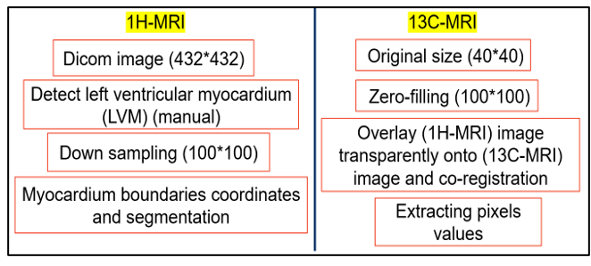
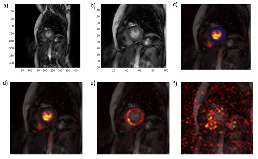
The 1H-MRI and 13C-MRI images were visualized and analyzed using MATLAB R2021a as we represented in Fig.2. The left ventricle (LV) myocardium was detected manually in the DICOM images of 432×432 voxels. The detected contours and the DICOM images were downsampled to pair with the size of the 13C-MRI images in 100×100 voxels. The LV myocardium contours are overlaid onto the 13C-MRI images, followed by registration. Consequently, the pixel values and their boundaries coordinates on the line of the area were recorded. Finally, the mean signal of each frame is calculated and plotted. In Figure 3, we displayed bicarbonate, lactate, and pyruvate metabolism images (see Fig 2). Moreover, the 13C-MRI images are recorded every ~3 seconds for 44-time points images (see Fig 3).Results and Conclusions:
Figure 3 exemplifies the metabolic images obtained in this study. As seen, a high SNR pyruvate signal, contained to the ventricle was detected. Subsequently, a bicarbonate image localized to the myocardium was also reconstructed, albeit with decreased SNR. Still, based in d) and e) we are able to calculate bicarbonate-to-pyruvate ratios in these patients. The uncertainty in detecting the lactate is seen in figure f), suggesting that additional technical improvements will be needed if we are to report on the lactate-to-bicarbonate ratios. We are still in the phase of recruiting additional subjects, to allow us to calculate group-based values for these metabolites.Conclusions:
The established acquisition and postprocessing allow for cardiac metabolic imaging in women undergoing chemotherapy for breast tumors. The impact of this research would be to allow for early detection of cardiotoxicity in this patient cohort.
Acknowledgements
VGZ received support from the Cancer Prevention Research Institute of Texas, award RP180404. Research reported in this publication was supported by the National Center for Advancing Translational Sciences of the National Institutes of Health under award Number UL1 TR003163. The content is solely the responsibility of the authors and does not necessarily represent the official views of the NIH (National Institutes of Health).References
1- Park JM, Reed GD, Liticker J, Putnam WC, Chandra A, Yaros K, Afzal A, MacNamara J, Raza J, Hall RG, Baxter J, Derner K, Pena S, Kallem RR, Subramaniyan I, Edpuganti V, Harrison CE, Muthukumar A, Lewis C, Reddy S, Unni N, Klemow D, Syed S, Li H, Cole S, Froehlich T, Ayers C, de Lemos J, Malloy CR, Haley B, Zaha VG. Effect of Doxorubicin on Myocardial Bicarbonate Production From Pyruvate Dehydrogenase in Women With Breast Cancer. Circ Res. 2020 Dec 4;127(12):1568-1570. doi: 10.1161/CIRCRESAHA.120.317970. Epub 2020 Oct 14. PMID: 33054563; PMCID: PMC7874930.Figures