5150
Impact of radiographer-led change on cMRI protocol in porcine stem cell study1Westmead Hospital, Sydney, Australia, 2Sectra, Sydney, Australia, 3Sydney University, Sydney, Australia
Synopsis
Keywords:
Motivation: Knowledge that a suboptimal acquisition protocol was being implemented in a large and expensive animal research study.
Goal(s): Demonstration of improved image quality in a porcine study investigating remuscularisation effects of stem cell treatment in diseased and injured hearts.
Approach: Implementation of a FB protocol in tandem with a BH protocol in a subset of porcine subjects (n=38) who had undergone percutaneous induction of myocardial infarction.
Results: Superior image quality and greater reproducibility of BH techniques with shorter scan times results in the need for remarkably smaller sample sizes to show clinically significant treatment effects.
Impact: MRI radiographers are an untapped resource in many sites performing research. This study demonstrated the impact of radiographer expertise in protocol development and subsequent improvement in research data quality and outcomes in a large and expensive animal study.
Background/Purpose
Our MRI research department undertook a porcine cardiac study to assess remuscularisation effects of stem cell treatment in diseased and injured hearts. A known complication of this treatment approach is the inducement of ventricular arrhythmias and cMRI is the modality of choice for further investigation. A free-breathing (FB) protocol inherited from a previous porcine study at another site was initially implemented. Following radiographer-led data quality discussions with the Principal Investigator about this study’s endpoint, a sub-study of 38 animals was undertaken comparing two methods of data acquisition, FB versus breath-held (BH) protocol. Advantages of FB are the ability to scan without use of general anaesthesia and mechanical ventilation and its potential effects on cardiovascular physiology. However, FB data is compromised by breathing motion artifacts, reducing data quality and reproducibility. Conversely, although image quality is much improved with the BH technique, an anaesthetist and specialist equipment are required. We sought to assess the reproducibility of cMR metrics between FB and BH cMR in a porcine model of ischaemic cardiomyopathy.Method
A total of 38 adult female porcine subjects underwent FB or BH cMR, weighing 32.6±8.3kg at 16.2±12.2days post percutaneous induction of myocardial infarction (Table 1). The first 19 subjects underwent a FB cMR protocol under light sedation, where mechanical ventilation was not employed and animals were allowed to breathe freely throughout the duration of the scan. The second group of 19 underwent a BH cMR protocol under general anaesthesia where breath was held in end-expiration for all image acquisitions. All cMR examinations were performed on a Siemens 3T Prisma system, utilising an 18-channel body array with spine array coils and a 4-lead electrocardiogram gating (Image 1). Two experienced, blinded observers independently assessed and calculated Left Ventricular (LV) and Right Ventricular (RV) End Diastolic Volume (EDV), End Systolic Volume (ESV) and Ejection Fraction (EF) according to standard reporting guidelines (Table 1). All study protocols were approved by the Animal Ethics Committee of Western Sydney Local Health District.Results
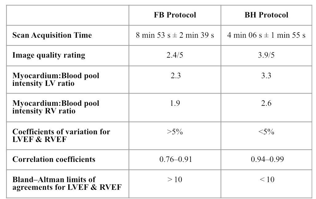
Subjective image quality was significantly improved in the BH cohort in comparison with the FB cohort as demonstrated by the image quality scores of 3.9 vs. 2.4 (p<0.0001) respectively. The majority of BH scans were rated as having above average image quality, as assessed by clarity of endocardial borders and ease of volumetric contouring. Free-breathing scans, on the other hand, were largely rated as having poor image quality and two scans were rated as uninterpretable by both observers (Figure 1A). Objective image quality was assessed by calculating the ratio of image intensity between myocardium and blood pool in both the left and right ventricular chambers. This assessment showed a greater image intensity ratio in BH scans as compared to FB scans (LV ratio: 3.3 vs. 2.3; p<0.001, RV ratio: 2.6 vs. 1.9; p<0.001), confirming crisper delineation at the tissue: blood interface in BH scans (Figure 1B & 1C). The time taken to acquire the cine short axis stack was significantly shorter in the BH as compared to the FB cohort (4 min 06 s±1 min 55 s vs. 8 min 53 s±2 min 39 s; p<0.0001), resulting in reduced time under anaesthesia; an important animal welfare consideration. Intra- and inter-observer reproducibility of volumetric analysis was substantially improved in the BH cohort. Correlation scatter and Bland–Altman plots show clearly improved linear associations, tighter grouping of data points and reduced bias in the BH cohort (Table 2). An unexpected but very meaningful finding was the significant effect on sample sizes. We showed that they could be reduced by greater than 85% due to the superior accuracy of the BH protocol. This is highly significant from both a cost perspective as well as an animal welfare perspective.Conclusion
As predicted, BH protocol produces superior image quality, shorter scan acquisition and greater reproducibility to show clinically significant treatment effects as compared to FB protocol. A further very significant benefit is cost and sample size reductions, warranting the utilisation of of anaesthetic staff and specialist equipment. Experienced MRI radiographers are technical experts in MRI data acquisition and protocol optimisation and are positioned to make significant positive contributions to imaging research.Acknowledgements
No acknowledgement found.References
Selvakumar, D., Deshmukh, T., Foster, S., Sanaei, N., Min, A., Grieve, S., Pathan, F., Chong, J. (2022). Comparative assessment of motion averaged free-breathing or breath-held cardiac magnetic resonance imaging protocols in a porcine myocardial infarction model. Scientific reports, 12(1), 3727. https://doi.org/10.1038/s41598-022-07566-wFigures
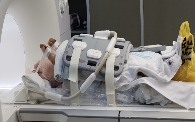
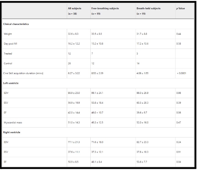
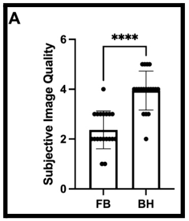
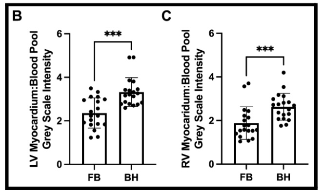
Figure 1B: Objective assessment of image quality - Left ventricular myocardium to blood pool image intensity ratio.
Figure 1C: Objective assessment of image quality - Right ventricular myocardium to blood pool image intensity ratio. ***p<0.001