5149
Left Ventricular Dyssynchrony in Anterior ST-Elevation Myocardial Infarction1Radiology and Nuclear Medicine, Capital Medical University XuanWu Hospital, Beiing, China
Synopsis
Motivation: Since the septal function is critical to the global LV function and prognosis, we suggest further refining global and segmental septal viability analysis.
Goal(s): This study aimed to characterize myocardial strain alterations by CMR-feature tracking for patients with anterior STEMI.
Approach: The size of the infarction and the extent of injury, to investigate if any changes in septal function would affect the balance of strain.
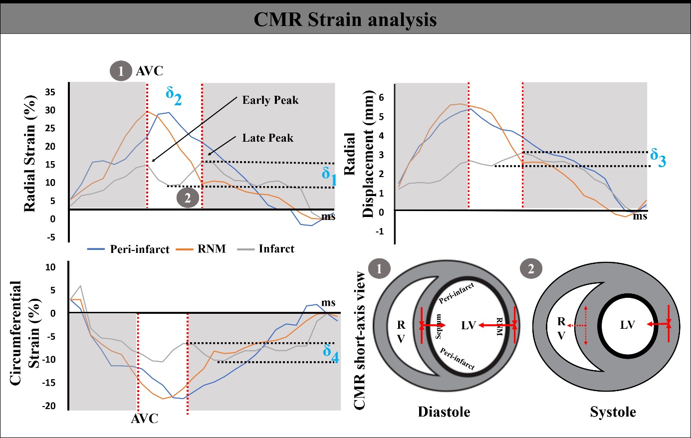
Results: The infarcted septum swings in a bimodal mode, and myocardial injury reduces the radial strain contractility. A more than 75% transmural degree was the septal strain-contraction reserve cut-off point.
Impact: The combination of CMR-LGE and strain puts forward an efficient risk stratification. This technique can help clinicians make accurate decisions, especially for patients with LBBB, avoiding the LV lead placed in the infarcted zone.
Objectives
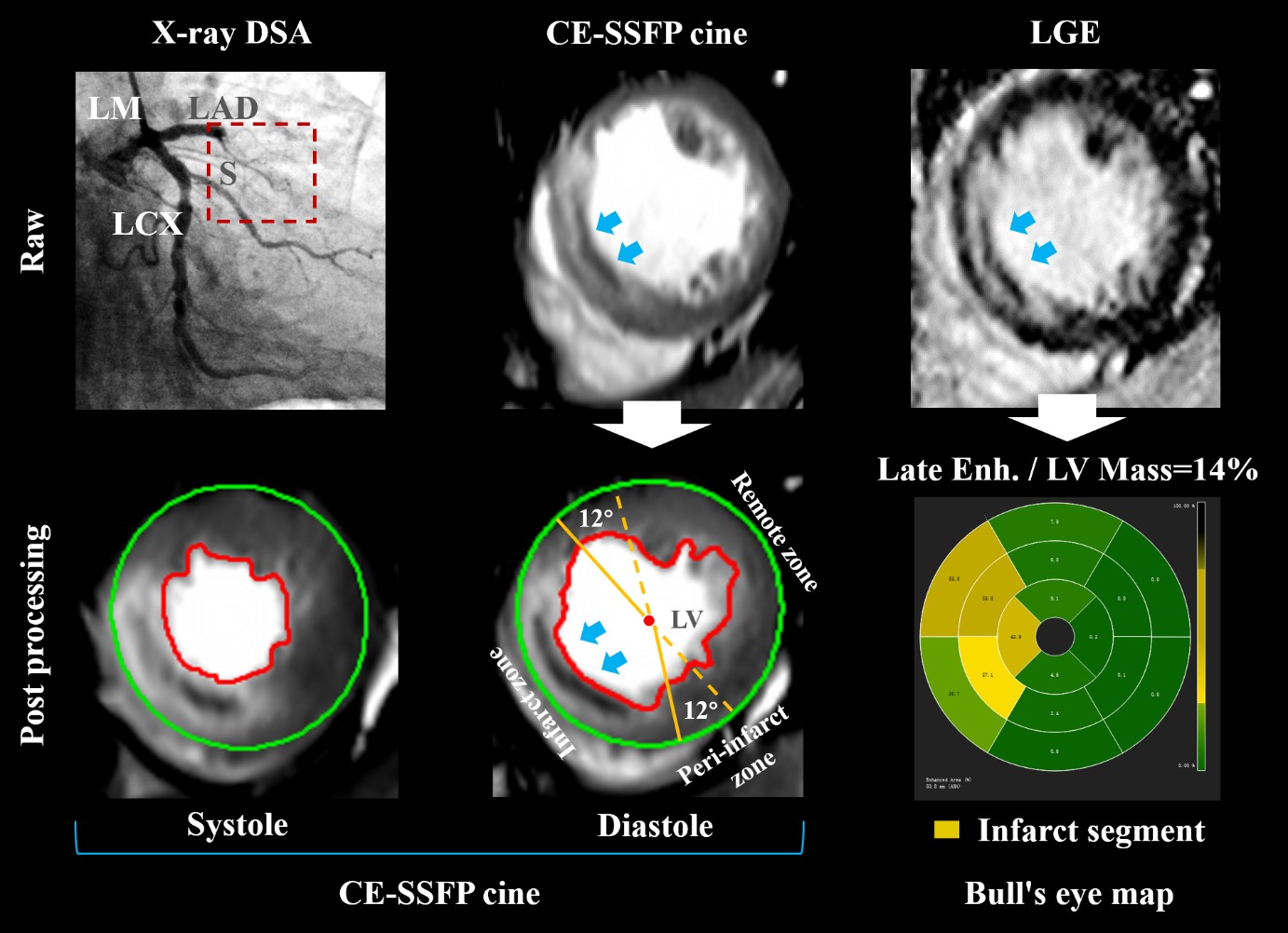
Cardiovascular magnetic resonance-feature tracking (CMR-FT) enables quantification of myocardial deformation and may be used as an objective measure of myocardial involvement in ST-elevation myocardial infarction (STEMI). We sought to investigate the associations between myocardial dyssynchrony parameters and myocardium damage for STEMI.Methods
We analyzed 65 patients (45-80 years old) with anterior STEMI after primary percutaneous coronary intervention during 3-7 days (observational [STEMI] group) and 60 healthy volunteers (normal control [NC] group). Myocardial dyssynchrony parameters were derived, including global and regional strain, radial rebound stretch and displacement, systolic septal time delay, and circumferential stretch.Results
CMR characteristics, including morphologic parameters such as left ventricular ejection fraction (LVEF) (45.3% ±8.2%) and myocardium damage in late gadolinium enhancement (LGE) (19.4% ±4.7% LV), were assessed in the observation group. The global radial strain (GRS) and global longitudinal strain (GLS) substantially decreased in anterior STEMI compared with the NC group (GRS: 19.4% ±5.1% vs. 24.8% ±4.0%, P < 0.05; GLS: -10.1% ±1.7% vs. -13.7% ±1.0%, P < 0.05). Among 362 infarcted segments, radial and circumferential peak strains of the infarcted zone were the lowest (14.4% ±3.2% and -10.7% ±1.6%, respectively). The radial peak displacement of the infarct zone significantly decreased (2.6 ±0.4 mm) (P < 0.001) and manifested in the circumferential displacement (3.5° ±0.7°) in the STEMI group (P < 0.01). As microvascular occlusion (MVO) was additionally present, some strain parameters were significantly impaired in LGE+/MVO+ segments (radial strain [RS]: 12.2% ±2.1%, circumferential strain [CS]: -9.6% ±0.7%, longitudinal strain [LS]: -6.8% ±1.0%) compared to LGE+/MVO- (RS: 14.6% ±3.2%, CS: -10.8% ±1.8%, LS: -9.2% ±1.3%) (P < 0.05). When the extent of transmural myocardial infarction is greater than 75%, the parameter of the systolic septal delay (mean, 148ms) was significantly reduced compared to fewer degrees of infarction (P < 0.01).Conclusion
In anterior STEMI, the infarcted septum swings in a bimodal mode, and myocardial injury reduces the radial strain contractility. A more than 75% transmural degree was the septal strain-contraction reserve cut-off point.Acknowledgements
Not applicable.References
1. Calle S, Kamoen V, De Buyzere M, et al. A strain-based staging classification of left bundle branch block-induced cardiac remodeling. JACC Cardiovasc Imaging (2021) 14:1691-1702.
2. Wouters PC, Leenders GE, Cramer MJ, et al. Acute recoordination rather than functional hemodynamic improvement determines reverse remodeling by cardiac resynchronisation therapy. Int J Cardiovasc Imaging (2021) 37:1903-1911.
3. Zweerink A, Nijveldt R, Braams NJ, et al. Segment length in cine (SLICE) strain analysis: A practical approach to estimate potential benefit from cardiac resynchronization therapy. J Cardiovasc Magn Reson (2021) 23:4.
4. Prinzen FW, Augustijn CH, Allessie MA, et al. The time sequence of electrical and mechanical activation during spontaneous beating and ectopic stimulation. Eur Heart J (1992) 13:535-543. 5. Risum N, Tayal B, Hansen TF, et al. Identification of typical left bundle branch block contraction by strain echocardiography is additive to electrocardiography in prediction of long-term outcome after cardiac resynchronization therapy. J Am Coll Cardiol (2015) 66:631-641.
6. Stankovic I, Prinz C, Ciarka A, et al. Relationship of visually assessed apical rocking and septal flash to response and long-term survival following cardiac resynchronization therapy (PREDICT-CRT). Eur Heart J - Cardiovasc Imaging (2016) 17:262-269.
7. Menet A, Bernard A, Tribouilloy C, et al. Clinical significance of septal deformation patterns in heart failure patients receiving cardiac resynchronization therapy. Eur Heart J - Cardiovasc Imaging (2017) 18:1388-1397.
8. Parsai C, Bijnens B, Sutherland GR, et al. Toward understanding response to cardiac resynchronization therapy: Left ventricular dyssynchrony is only one of multiple mechanisms. Eur Heart J (2009) 30:940-949.
9. Gjesdal O, Remme EW, Opdahl A, et al. Mechanisms of abnormal systolic motion of the interventricular septum during left bundle-branch block. Circ Cardiovasc Imaging (2011) 4:264-273.
10. Aalen JM, Remme EW, Larsen CK, et al. Mechanism of abnormal septal motion in left bundle branch block: Role of left ventricular wall interactions and myocardial scar. JACC Cardiovasc Imaging (2019) 12:2402-2413.
11. Leenders GE, Lumens J, Cramer MJ, et al. Septal deformation patterns delineate mechanical dyssynchrony and regional differences in contractility: Analysis of patient data using a computer model. Circ Heart Fail (2012) 5:87-96.
12. Ghadban R, Alpert MA, Dohrmann ML, et al. A QS pattern in leads V1 and V2 is associated with septal scarring independent of scar etiology - A cardiac magnetic resonance imaging study. J Electrocardiol (2018) 51:577-582.
13. Konstam MA, Abboud FM. Ejection fraction: Misunderstood and overrated (changing the paradigm in categorizing heart failure). Circulation (2017) 135:717-719.
14. Li H, Qu Y, Metze P, et al. Quantification of Biventricular Myocardial Strain Using CMR Feature Tracking: Reproducibility in Small Animals. Biomed Res Int (2021) 2021:8492705.
15. Amzulescu MS, De Craene M, Langet H, et al. Myocardial strain imaging: Review of general principles, validation, and sources of discrepancies. Eur Heart J - Cardiovasc Imaging (2019) 20:605-619.
16. Palmisano A, Benedetti G, Faletti R, et al. Early T1 Myocardial MRI Mapping: Value in Detecting Myocardial Hyperemia in Acute Myocarditis. Radiology (2020) 295(2):316-325.
17. Collet J, Thiele H, Barbato E, et al. 2020 ESC Guidelines for the management of acute coronary syndromes in patients presenting without persistent ST-segment elevation. Eur Heart J (2021) 42:1289-1367.
18. Sun Z, Zhang Q, Zhao H, et al. Retrospective assessment of at-risk myocardium in reperfused acute myocardial infarction patients using contrast‐enhanced balanced steady‐state free‐precession cardiovascular magnetic resonance at 3T with SPECT validation. J Cardiovasc Magn Reson (2021) 23:25.
19. Goransson C, Ahtarovski KA, Kyhl K, et al. Assessment of the myocardial area at risk: Comparing T2-weighted cardiovascular magnetic resonance imaging with contrast-enhanced cine (CE-SSFP) imaging-a DANAMI3 substudy. Eur Heart J - Cardiovasc Imaging (2019) 20:361-366.
20. Kidambi A, Mather AN, Swoboda P, et al. Relationship between myocardial edema and regional myocardial function after reperfused acute myocardial infarction: An MR imaging study. Radiology (2013) 267:701-708.
21. Ibrahim T, Hackl T, Nekolla SG, et al. Acute myocardial infarction: serial cardiac MR imaging shows a decrease in delayed enhancement of the myocardium during the 1st week after reperfusion. Radiology (2010) 254:88-97.
22. Cha MJ, Lee JH, Jung HN, et al. Cardiac magnetic resonance-tissue tracking for the early prediction of adverse left ventricular remodeling after ST-segment elevation myocardial infarction. Int J Cardiovasc Imaging (2019) 35:2095-2102.
23. Tan YT, Wenzelburger F, Lee E, et al. The pathophysiology of heart failure with normal ejection fraction: Exercise echocardiography reveals complex abnormalities of both systolic and diastolic ventricular function involving torsion, untwist, and longitudinal motion. J Am Coll Cardiol (2009) 54:36-46.
24. Sengupta PP, Tajik AJ, Chandrasekaran K, et al. Twist mechanics of the left ventricle: Principles and application. JACC Cardiovasc Imaging (2008) 1:366-376.
25. Vo HQ, Marwick TH, Negishi K. MRI-derived myocardial strain measures in normal subjects. JACC Cardiovasc Imaging (2018) 11:196-205.
26. Nagel E, Chandrashekhar Y. A Strain by any other tracking would perform as sweet? JACC Cardiovasc Imaging (2018) 11:288-289.
27. Stoumpos S, Rankin A, Hall Barrientos P, et al. Interrogating the haemodynamic effects of haemodialysis arteriovenous fistula on cardiac structure and function. Sci Rep (2021) 11:18102.
28. Kelle S, Giusca S, Buss SJ, et al. BMI does not influence the prediction of cardiac events using stress CMR. Int J Cardiol (2015) 179:31-33.
29. Gavara J, Rodriguez-Palomares JF, Rios-Navarro C, et al. Longitudinal strain in remote non-infarcted myocardium by tissue tracking CMR: Characterization, dynamics, structural and prognostic implications. Int J Cardiovasc Imaging (2021) 37:241-253.
30. Pahlm US, Ubachs JF, Heiberg E, et al. Regional wall function before and after acute myocardial infarction; an experimental study in pigs. BMC Cardiovasc Disord (2014) 14:118. 31. Fukamachi D, Yamada A, Ohgaku A, et al. Protective effect of the Impella on the left ventricular function after acute broad anterior wall ST elevation myocardial infarctions with cardiogenic shock: Cardiovascular magnetic resonance imaging strain analysis. BMC Cardiovasc Disord (2022) 22:201.
32. Boe E, Russell K, Eek C, et al. Non-invasive myocardial work index identifies acute coronary occlusion in patients with non-ST-segment elevation-acute coronary syndrome. Eur Heart J - Cardiovasc Imaging (2015) 16:1247-1255.
33. Aalen JM, Donal E, Larsen CK, et al. Imaging predictors of response to cardiac resynchronization therapy: Left ventricular work asymmetry by echocardiography and septal viability by cardiac magnetic resonance. Eur Heart J (2020) 41:3813-3823.
34. Delicce, AV, Makaryus, AN. Physiology, Frank Starling Law. In StatPearls. StatPearls (2023). Publishing.
35. Prinzen FW, Lumens J. Investigating myocardial work as a CRT response predictor is not a waste of work. Eur Heart J (2020) 41:3824-3826.
36. Zweerink A, van Everdingen WM, Nijveldt R, et al. Strain imaging to predict response to cardiac resynchronization therapy: A systematic comparison of strain parameters using multiple imaging techniques. ESC Heart Fail (2018) 5:1130-1140.
37. Podlesnikar T, Pizarro G, Fernández-Jiménez R, et al. Left ventricular functional recovery of infarcted and remote myocardium after ST-segment elevation myocardial infarction (METOCARD-CNIC randomized clinical trial substudy). J Cardiovasc Magn Reson (2020) 22:44-56.
38. Feisst A, Kuetting DLR, Dabir D, et al. Influence of observer experience on cardiac magnetic resonance strain measurements using feature tracking and conventional tagging. Int J Cardiol Heart Vasc (2018) 18:46-51.
39. Kelle S, Bourantas CV, Korosoglou G. Editorial: Insights in cardiovascular imaging: 2022. Front Cardiovasc Med (2023) 10:1231842.
Figures