5147
Challenges in setting up an ex-vivo brain MRI protocol1Brain Imaging Centre, Douglas Research Centre, Douglas Research Centre, Montreal, QC, Canada, 2Department of Psychiatry, McGill University, Montreal, QC, Canada
Synopsis
Keywords:
Motivation: Help MRI operators and other professionals who plan to acquire ex-vivo brain MRI
Goal(s): What are the challenges in setting up an ex-vivo brain MRI protocol?
Approach: This abstract shows the challenges faced during the establishment of an ex-vivo brain MRI protocol in a 3-tesla scanner.
Results: We organized 2 categories of challenges our team faced: Dealing with motion and with contrast. We have detailed and illustrated each challenge and provided the learning points we thought were important.
Impact: Scanning of ex-vivo specimens poses challenges. The literature doesn't present the problems faced in preparation phase. We provide realistic view of these challenges from the perspective of MRI scientists, to help other teams approach this problem with the proper orientation.
Introduction
MRI is a powerful tool for neuroscience. Despite the improvements in sequences, hardware, and software, MRI protocols involving long experiments sometimes can not be tolerated by participants. Scanning ex-vivo specimens can be an alternative to explore the full potential of MRI. Sequences can be built to last for hours or days, extrapolating spatial resolution and contrast mechanisms. At first sight, scanning ex-vivo specimens seems to be an easy task for the MRI operator. However, many variables can impact the image quality. Familiarizing the operators with how ex-vivo scans look can prevent mistakes and differentiate real image characteristics from artifacts. We sought to provide MRI operators and other professionals information to help them go through the process of protocol establishment in a more guided way, by detailing the challenges and how to deal with them.Methods
We used brains from the Douglas-Bell Canada Brain Bank. One hemisphere is kept in plastic containers, immersed in a 4% formaldehyde solution at room temperature. All hemispheres were scanned in the 3T Prisma system and 64-channel coil, located at the Cerebral Imaging Center of Douglas Hospital (Montreal, Canada) as part of a developing protocol led by Dadar and Zeighami et al.Results
We organized two categories of challenges that can occur during ex-vivo MRI acquisition: motion and contrast. We provide representative examples, explain the underlying causes, and offer potential solutions.Discussion
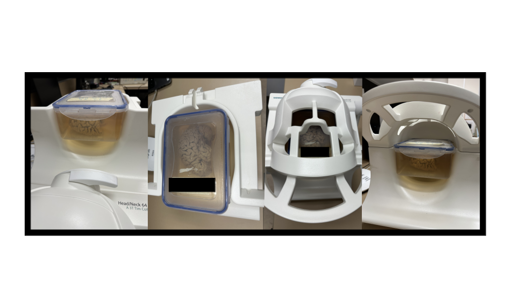
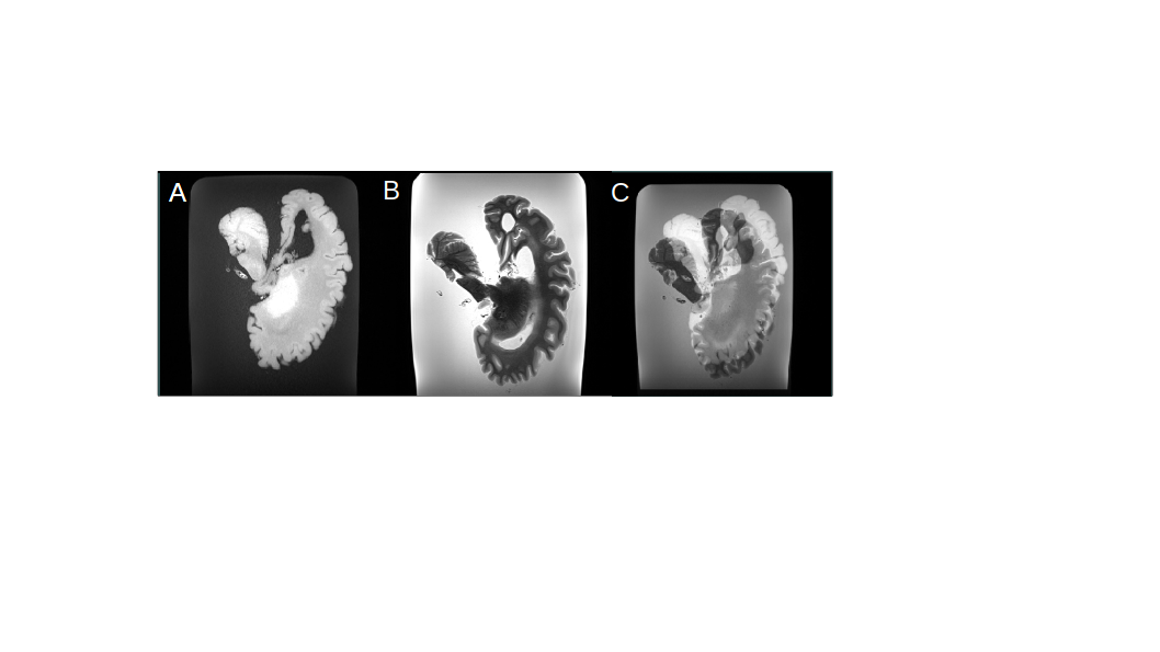
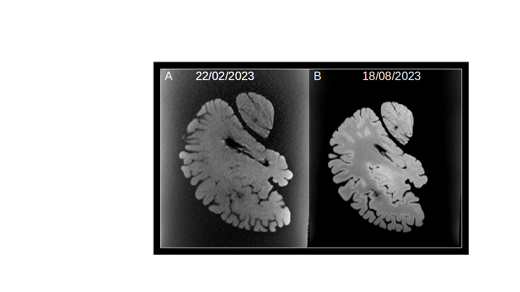
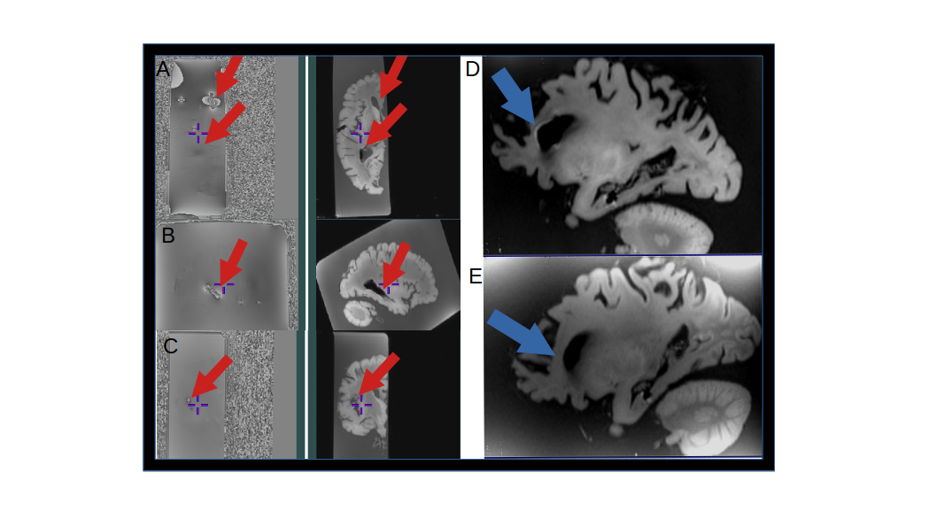
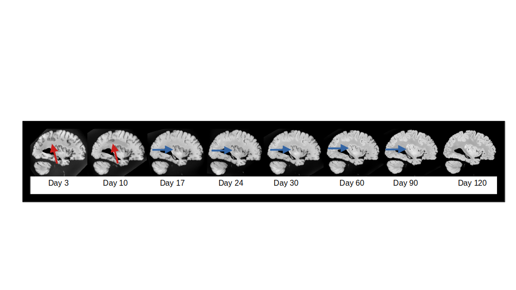
Motion: Yes, ex-vivo brains move. Motion severity depends on the type of container, the specimen is placed in and even the specimen’s specific pathology. Special containers can be built to accommodate the specimens based on anatomy. It needs to be coil-shaped, most are 3D-printed. In our case, we kept the hemisphere in the original container to prevent air contact or contamination. Luckily, it fits perfectly on the 64-channel head-neck coil, avoiding container motion. However, some brains can float between the sequences. It can happen if the specimen is not yet fixed or has a high atrophy grade. Learning points: Take the time to position the container. Carefully avoid tilting, or the specimen will drift. Start already with the table up and advance to the isocenter inside slowly, using a manual way. Wait at least 1-2 minutes to start the sequences. Plan localizers between sequences using high-power of the gradient. It helps to verify if the specimen didn’t move and lost the plan. Due to the fast change of gradients, leave diffusion for last. Contrasts:Temperature: It has a high impact on T1 images and less on T2. The T1 images look degraded. Also, quantitative measures must be corrected if the aim is to compare with in vivo data. Learning point: Leave the specimen at room temperature for at least 4 hours before the scan. In case the specimen needs to be kept at a low temperature, give preference to T2 sequences and keep monitoring the temperature for possible corrections. Fixatives: The substance used to fix the brain changes the tissue properties. The most common agent is formaldehyde. In high concentrations (10%), it fixes tissues fast but has high toxicity. Alternatively, specimens fixed with a solution of salt, 0.8% formaldehyde, and 3 alcohols were tested. The protocol set for the 4% formaldehyde did not work, presenting a SAR above the first level. We hypothesized the solution has a different conductivity, changing the radiofrequency patterns. Another challenge is that, as a water dilution, the fixative presents high signals in MRI images. Learning point: Knowing the fixative solution of the specimen can help to optimize the contrast. Also, when possible, scan the specimen immersed in a proton-free substance, which has no MR signal and susceptibility matched with brain tissue. Bonus: In the course of the fixation process, a line of different contrast appears 3 days after fixation, for approximately 16 days. Also, the WM/GM contrast is flipped at 90 days. Air bubbles: It depends on the way the specimen is stored and wrapped. In the case of container + fixative solution, air bubbles can appear inside ventricles or between enlarger gyri. Learning point: Specific capsules or vacuum pumps can be used to prevent air bubbles. In case you cannot change the container, slowly turning the container a few times and exposing the ventricles can help dislodge the trapped air.Conclusion
We summarize the challenges our team faced in setting an ex-vivo brain protocol. The current literature does not provide adequate information on the challenges and solutions for ex vivo preparation protocols We hope to provide important information for MRI personnel in this field.Acknowledgements
Healthy Brains For Healthy Lives (HBHL)
Fonds de recherche du Québec
Natural Sciences and Engineering Research Council of Canada
Dr Josefina Maranzano
Ève-Marie Frigon
Dominique Mirault
References
Miller KL, Stagg CJ, Douaud G, Jbabdi S, Smith SM, Behrens TEJ, Jenkinson M, Chance SA, Esiri MM, Voets NL, Jenkinson N, Aziz TZ, Turner MR, Johansen-Berg H, McNab JA. Diffusion imaging of whole, post-mortem human brains on a clinical MRI scanner. Neuroimage. 2011 Jul 1;57(1):167-181. doi: 10.1016/j.neuroimage.2011.03.070. Epub 2011 Apr 5. PMID: 21473920; PMCID: PMC3115068.
De Barros A, Arribarat G, Combis J, Chaynes P and Péran P. Matching ex vivo MRI With Iron Histology: Pearls and Pitfalls. Front. Neuroanat. 2019;13:68. doi:10.3389/fnana.2019.00068
Shatil AS, Uddin MN, Matsuda KM, Figley CR. Quantitative Ex Vivo MRI Changes due to Progressive Formalin Fixation in Whole Human Brain Specimens: Longitudinal Characterization of Diffusion, Relaxometry, and Myelin Water Fraction Measurements at 3T. Front Med (Lausanne). 2018 Feb 20;5:31. doi: 10.3389/fmed.2018.00031. PMID: 29515998; PMCID: PMC5826187.
Post-mortem brain temperature and its influence on quantitative MRI of the brainBerger C, Bauer M Wittig H, Scheurer E, Lenz C. Magnetic Resonance Materials in Physics, Biology and Medicine 2022; 35:375–387 https://doi.org/10.1007/s10334-021-00971-8
Figures