5145
The Significance of fMRI in Surgical Planning for Tumour Resection and Epilepsy Surgery: Optimal Language Paradigms for Language Area Mapping1Hunter Medical Research Institute, New Lambton Heights, Australia
Synopsis
Motivation: The Significance of fMRI in Surgical Planning for Tumour Resection and Epilepsy Surgery: Optimal Language Paradigms for Language Area Mapping
Goal(s): This paper explores the importance of fMRI in surgical planning and the most effective language paradigms for language area mapping.
Approach: fMRI allows neurosurgeons to pinpoint critical brain regions responsible for language, motor function, and sensory perception. This information is pivotal in minimizing postoperative neurological deficits
Results: For epilepsy surgery, it aids in delineating the epileptogenic zone as data clearly shows increase in seizure freedom with detailed imaging identifying the true extent of the lesion.
Impact: Resource for radiographers on how to perform a fMRI examination, sequence parameters and corresponding language paradigms.
Introduction
Functional Magnetic Resonance Imaging (fMRI) has enhanced neurosurgical planning, particularly in cases of tumour resection and epilepsy surgery. By providing invaluable insights into the brain's functional organization, fMRI has become an important tool in optimizing surgical outcomes. However, this technique is not readily available in the clinical setting due to the expertise and specialised setup required, disadvantaging patients and their physicians. This paper explores the importance of fMRI in surgical planning and the most effective language paradigms for language area mapping. It also provides a resource for radiographers on how to perform a fMRI examination, sequence parameters and corresponding language paradigms.The significance of surgical planning
Preoperative localization and lateralisation: fMRI allows neurosurgeons to pinpoint critical brain regions responsible for language, motor function, and sensory perception. This information is pivotal in minimizing postoperative neurological deficits. In the context of tumour resection and epilepsy surgery, identifying and preserving brain areas are of paramount importance.Personalized surgical strategies: each patient's brain organization is unique. fMRI empowers surgeons to tailor their approach to individual patients, ensuring maximal tumour removal while minimizing damage to essential brain regions. For epilepsy surgery, it aids in delineating the epileptogenic zone as data clearly shows increase in seizure freedom with detailed imaging identifying the true extent of the lesion.
Intraoperative Navigation: Real-time fMRI integration with neuro-navigation systems enables precise intraoperative guidance. Surgeons can monitor brain function during the procedure, verifying the effectiveness of their approach and adjusting as necessary.
Language Paradigms for Language Area Mapping
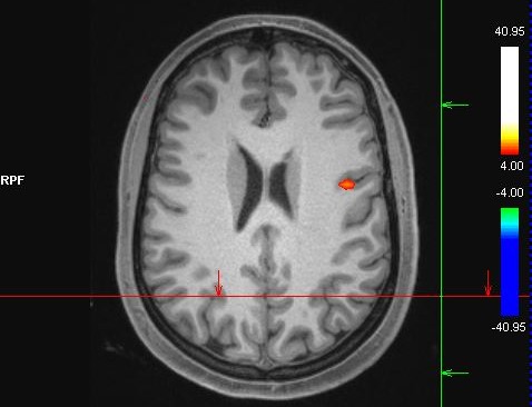
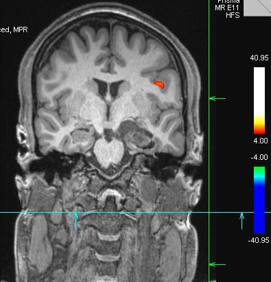
Task-Based Paradigms:a. Verb Generation Task: This task requires patients to generate verbs associated with visually presented nouns. It effectively activates the left inferior frontal gyrus (Broca's area) and is essential for identifying language areas.
b. Auditory Comprehension Task: Listening to sentences and judging their correctness engages the superior temporal gyrus and posterior superior temporal sulcus, revealing areas responsible for auditory comprehension.
c. Reading and Sentence Completion Tasks: Engaging patients in reading and completing sentences activates the angular gyrus and supramarginal gyrus, associated with semantic processing and sentence comprehension.
Resting-State fMRI:
Resting-state fMRI analyses intrinsic brain connectivity in the absence of a specific task. It has proved valuable for language area mapping by identifying the default mode network, which includes language regions such as the angular gyrus and posterior cingulate cortex.
Multimodal Approaches: Combining task-based and resting-state fMRI offers a comprehensive view of the language network. Multimodal paradigms allow surgeons to create more precise maps of language areas.
Electrocortical Stimulation (ECS) Validation: ECS remains the gold standard for mapping cortex during surgery. fMRI data can be validated through ECS, ensuring the highest level of accuracy in language area identification.
A comprehensive resource for performing fMRI/language mapping including sequence parameters and language paradigms can be found on https://hmri.org.au/hmri-imaging-centre.
Conclusion
In summary, fMRI's importance in surgical planning for tumour resection and epilepsy surgery cannot be overstated. It empowers surgeons to make informed decisions, optimizing patient outcomes. The most effective language paradigms for language area mapping provide a comprehensive understanding of the brain's language network, enhancing the precision of surgical procedures. This synergy between fMRI and advanced language paradigms has the potential to enhance the field of neurosurgery and improve the quality of life for patients with brain pathologies for personalised medical care.Acknowledgements
No acknowledgement found.References
Figures