5142
Operating MRI 7 Tesla Ultra High Field (UHF) in Clinical and Research Practice1Translational Imaging Center, Houston Methodist Hospital Research Institute, Houston, TX, United States, 2Radiology, Houston Methodist Hospital, Houston, TX, United States
Synopsis
Motivation: Clinical and research patients can be imaged safely at 7T while providing improved quality and resolution enabling better detection of lesions while maintaining a safe working environment for our patients and technologists.
Goal(s): The goal is to guide new operator of MRI 7T on how to prepare themselves and patient for advance imaging exam.
Approach: Informative approaches for equipment, safety, side effects of 7T MRI for patients and technologist.
Results: Operating a 7T MRI is similar to other field strengths, but technologist should be well trained and understand the environment in UHF scanners.
Impact: 7T MRI is an advance imaging technology that is very beneficial for patient care. This guidelines will provides MRI technologist some insight on the equipment, safety, and how to prepare the patient for 7T MRI studies.
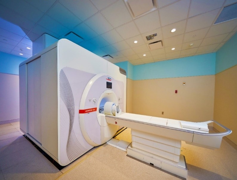
Figure 1: MRI 7T Siemens Magnetom Terra
MRI 7T Magnetic Field Effects Preparation: With the stronger magnetic field at 7T, the static magnetic effect is a safety consideration for patient and operator. The patient needs to be aware of the static magnetic field effects that cause a temporary imbalance of the vestibular system in which they may feel vertigo, dizziness, nausea, and nystagmus when the table is moving the head inside scanner bore. The side effect symptoms can vary from light to severe depending on patient sensitivity. Technologists operating the 7T may feel vertigo, walking imbalance, and metallic taste as they approach the scanner bore. He/she needs to walk slowly inside the 7T scanner room while taking care of patients. All patient preparation instructions for 7T exams should be done outside the scanner room to limit the time spent near the magnetic field.
Patient Safety Preparation: The higher magnetic field causes higher SAR values than for lower field strengths, particularly for spin-echo sequences. In addition, 7T has a strong angle of defection and pulling force on objects, and higher RF deposition in the body. Additional safety screening is necessary for the patient with any passive and active implants prior to undergoing a 7T exam. With a conservative approach, patients with active implants not tested for 7T should not be scanned at this field strength. Also, most of the passive implants that are MR safe and conditional at 3T are also considered conditional at 7T, such as spinal fixation devices, orthopedic, and dental implants that are made of titanium, aluminum, platinum, gold, and silver materials. Vascular implants such as stents, coils, filters or aneurysm clips that are conditional at 3T will need additional approval by a radiologist prior to 7T imaging, generally excluded if the implant is within the transmit RF coil range, for example at the shoulders and above for neuroimaging with a head coil.
Summary: Operating at 7T is very similar to operating other standard clinical 1.5T and 3T MRI scanners. Nevertheless, the operator of 7T should be well trained and understand the MRI environment. Safety related to the patient, equipment, and implants is critical for the technologist to master, prior to taking care of patient in the 7T environment.
Acknowledgements
N/AReferences
Hoff, M. N., McKinney IV, A., Shellock, F. G., Rassner, U., Gilk, T., Watson Jr, R. E., ... & Kanal, E. (2019). Safety considerations of 7-T MRI in clinical practice. Radiology, 292(3), 509-518.
Okada, T., Akasaka, T., Thuy, D. H., & Isa, T. (2022). Safety for human MR scanners at 7T. Magnetic Resonance in Medical Sciences, 21(4), 531-537.
Figures