5140

Optimized MRI workflow in colon cancer scan
SHA LIU1, NAN SUN1, ZHEN GUAN1, XIAO TING LI1, YU XIN YANG2, and KE XUE2
1Peking University Cancer Hospital & Institute, BEI JING, China, 2MR Collaboration, United Imaging Research Institute of Intelligent Imaging, BEI JING, China

Synopsis

Keywords:

Motivation: Torso enhanced CT is the standard method for colon cancer staging. MRI is a complimentary method. Our purpose is to develop a MR Colon cancer scanning method with simple process and short scan time.

Goal(s): develop a new T2WI breath-hold scanning technique that eliminates the need for bowel preparation and reduces scan time


Approach: compared the image quality between trans-axial T2WI and axial T2WI in visualization of anatomical structure

Results: Patient examination time for our colon MRI is less than 10min, compared with conventional MRI, our approach offered a simplified and expedited means of visualizing lesion details with superior clarity.

Impact: Our research focused on MRI scan optimization. Without bowel preparation, using breath-hold, our creative trans-axial imaging plan becomes highly manageable and enables efficient repetition of the entire process.

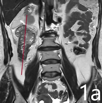
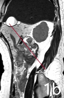
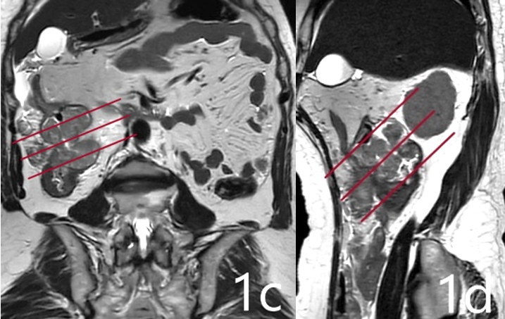
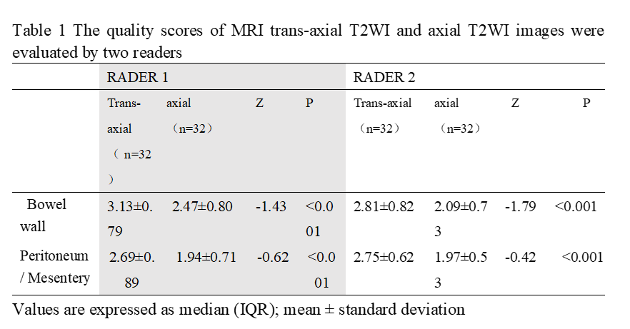
Introduction Contrast-enhanced CT (CE-CT) is the primary imaging modality for evaluating colon cancer (CC). However, its soft tissue contrast is often suboptimal and may inadequately fulfill diagnostic requirements. MRI can provide excellent soft tissue contrast, and several studies [1] have evaluated the performance of T2-weighted imaging (T2WI) in CC staging and validated its critical role, especially for locally advanced CC. However, challenges such as complex patient preparation, long scan time, and unstable image quality associated with colonic T2WI limit its wide application in clinical practice. Thus it is currently used to screen patients with liver metastasis or exhibiting allergies to iodine-based contrast agents. Thus our study aimed to optimize the T staging colon T2WI scan by obtain the tumor trans-axial image from dual oblique plane by optimizing the scan process by eliminating bowel preparation, using breath hold instead of triggered scan. We also proposed to compare the image quality of tumor trans-axial T2WI with axial T2WI to approve the effectiveness of the MRI workflow. Methods From September 2022 to July 2023, totally 32 patients (14 males, age: median 57 years old, 34-79 years old) were retrospectively enrolled. Inclusion criteria: patients with biopsy confirmed colon cancer; the ability to hold the breath in 20sec; complete image information, and the image information should be uniformly transmitted into our hospital's impact filing and communication system (PACS). Exclusion criteria: Patients who experience claustrophobia, had a colonoscopy within the past month, or have refused MRI. MRI was performed on a 3.0T scanner (uMR790, United Imaging Healthcare) with a 24-channel super flexible body coil. All T2WI was acquired with the FSE sequence combined with AI-assisted Compressed Sensing (ACS) technology. We regularly scanned full abdomen breath-hold (BH) coronal T2WI with a scan time of 15s, triggered axial DWI with a scan time of 2 minute 40 s, BH axial T1 with a scan time of 15s. For the trans-axial scan, the following steps were employed (Figure 1): (1) obtain the first oblique T2WI by aligning positioning line parallel to the long axis of tumor-located colon on coronal T2WI (fig 1a), scan time=8s; (2) obtain the second oblique T2WI by making positioning line parallel to the long axis of tumor-located colon on the first oblique T2WI (fig 1b), scan time=8s; (3) obtain trans-axial T2WI by drawing positioning line perpendicular to the long axis of tumor-located colon on both first and second oblique T2WI (fig 1c,1d). The scan parameters were as follows: TR/TE=2400ms/110ms, FOV=280*280mm2, pixel size=0.7*0.7mm2, slice thickness=3mm, 20-30 slices, scan time=30-45s. A full abdomen coverage BH axial T2WI were acquired with following parameters: TR/TE=4400ms/110ms, FOV=320*300mm2, pixel size=0.8*0.8mm2, slice thickness=5mm, 50 slices, scan time=45s, this sequence and parameters were commonly used for colon MRI but with respiratory trigger in the past literature. The colon MRI scan time was no more than 5min, resulting in a total patient examination time of approximately 10min. Patient preparation entailed a minimum 4-hour fasting period with no bowel preparation and the administration of intramuscular injection antispasmodic agents. Two experienced radiologists independently evaluated image quality parameters for trans-axial T2WI and axial T2WI, including the bowel wall visualization and peritoneum or mesentery clarity. Clarity was scored on a scale from 1 to 4, where 1 represented the structure was indistinguishable, 2 represented the structure was visible with blurring outline 3 represented the structure was clearly displayed but the outline was smooth, and 4 represented the structure was clearly displayed and the outline was sharp. A third experienced radiologist was introduced for Arbitrition. SPSS 22.0 IBM statistical software was used. A Wilcoxon Signed Ranks Test was applied to compare the difference of clarity scores in trans-axial T2WI and conventional axial T2WI, with p<0.05 indicating statistical significance. Results MRI trans-axial T2WI images showed better visualization of the bowel wall (2.81-3.13v.s.2.09-2.47, P<0.001), and the peritoneum or mesentery display (2.69-2.75v.s.1.94-1.97, P<0.001) than axial T2WI. Discussion and Conclusion Axial T2WI was main scan plane in the literature, some author used single oblique plane. Neither was perfect for structure description because of partial volume effect [2]. Our optimized workflow for colon T2WI significantly improved the image quality, effectively mitigating the partial volume effect and offering radiologists high-quality images for more accurate diagnosis. Besides, compared with conventional axial T2WI, our workflow requires no specific patient preparation, reduces scan time in 10 minutes for expeditious imaging, and eliminates the need for contrast enhancement, thereby minimizing renal burden. This study excluded patients who were unable to hold their breath, as the optimization of scanning protocols for such cases is the subject of other ongoing studies.

Acknowledgements

No acknowledgement found.

References

[1]Soo Yeun Park,Seung Hyun Cho,Min A.Lee Ghilsuk YoonY, et al. Diagnostic performance of MRI- versus MDCT-categorized T3cd/T4 for identifying high-risk stage II or stage III colon cancers: a pilot study. Abdominal Radiology(2019) 44:1675-1685doi: https://doi.org/10.1007/s00261-018-1822-7.

[2]Seren R Rafaelsen , Claus Dam , Chris Vagn-Hansen, et al. CT and 3 Tesla MRI in the TN Staging of Colon Cancer: A Prospective, Blind Study. European Journal of Radiology 121 (2019) 108741:2022, 29(2), 1069-1079;doi: https://doi.org/10.3390/curroncol29020091.
Figures

Table 1 The quality scores of MRI trans-axial T2WI and axial T2WI images were evaluated by two readers

(1) obtain the first oblique T2WI by aligning positioning line parallel to the long axis of tumor-located colon on coronal T2WI (fig 1a)

((2) obtain the second oblique T2WI by making positioning line parallel to the long axis of tumor-located colon on the first oblique T2WI (fig 1b),

(3) obtain trans-axial T2WI by drawing positioning line perpendicular to the long axis of tumor-located colon on both first and second oblique T2WI (fig 1c,1d)

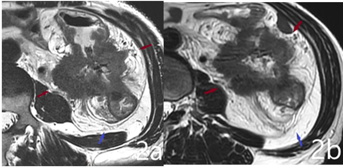
Fig 2a Dual-oblique T2WI images clearly show that the tumor has broken through the intestinal wall to the retroperitoneal adipose tissue (red arrow), and the adjacent peritoneum is thickened and clearly displayed (blue arrow head). Fig 2b Axis T2WI images showed that the tumor broke through the intestinal wall into the retroperitoneal adipose tissue, and the adjacent peritoneum was thickened and poorly displayed.

Proc. Intl. Soc. Mag. Reson. Med. 32 (2024)
5140
DOI: https://doi.org/10.58530/2024/5140