5137
Measurement and analysis of objective indicators of ossification development in MRI of knee joint1the First Affiliated Hospital of Xi'an Jiaotong University, Xi'an,, China, 2the First Affiliated Hospital of Xi'an Jiaotong University, Xi'an, Shaanxi province, China, 3the First Affiliated Hospital of Xi'an Jiaotong University, xi'an, China
Synopsis
Keywords:
Motivation:
Adolescents have high sensitivity to X-ray, and MRI has the advantage of non-radiation, which has not been perfected in the assessment of adolescent bone age.
Goal(s): To analyze the predictive value of MRI ossification development of knee joint with gender and age.
Approach: The indexes representing epiphyseal development of distal femur and proximal tibia were selected for measurement, and the differences of epiphyseal development of distal femur and proximal tibia between different gender and different age groups were explored.
Results: Males performed better on the distal femur and females on the proximal tibia for age prediction, which could be evaluated separately if necessary.
Impact: The objective indicators of ossification development in MRI of knee joint have certain predictive value with gender and age, which helps to evaluate bone age objectively and accurately, and provides a new idea for epiphysis evaluation
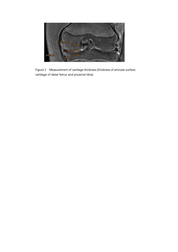
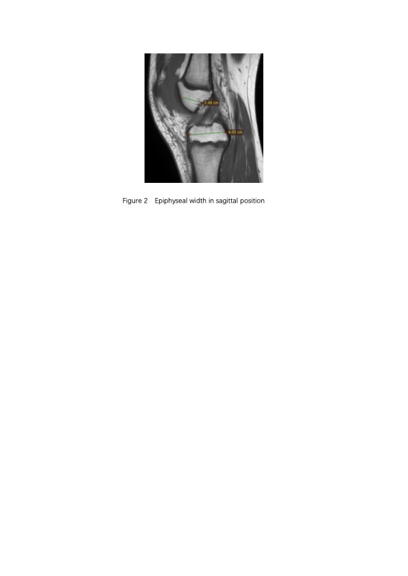
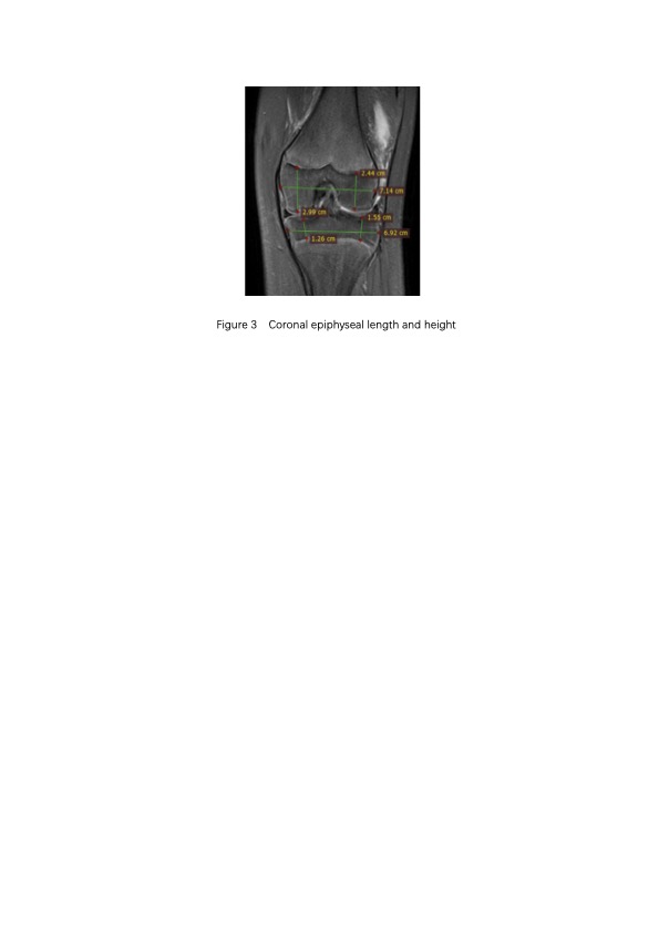
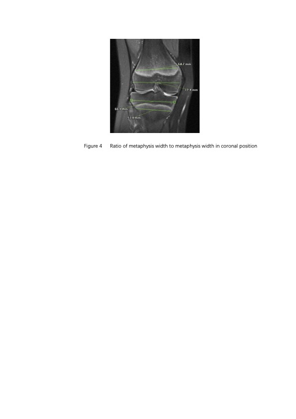
Methods: rom January 2016 to January 2022, a total of 1167 knee MRI patients aged 10 to 20 years old from 5 Grade III hospitals in Shaanxi Province were retrospectively included, including 685 males and 482 females. Indicators representing epiphyseal development at distal femur and proximal tibia were selected for measurement, including cartilage thickness and epiphyseal volume, metaphysis to metaphysis width ratio,To explore the difference of developmental indicators of distal femur and proximal tibial epiphysis between different sex and age groups.
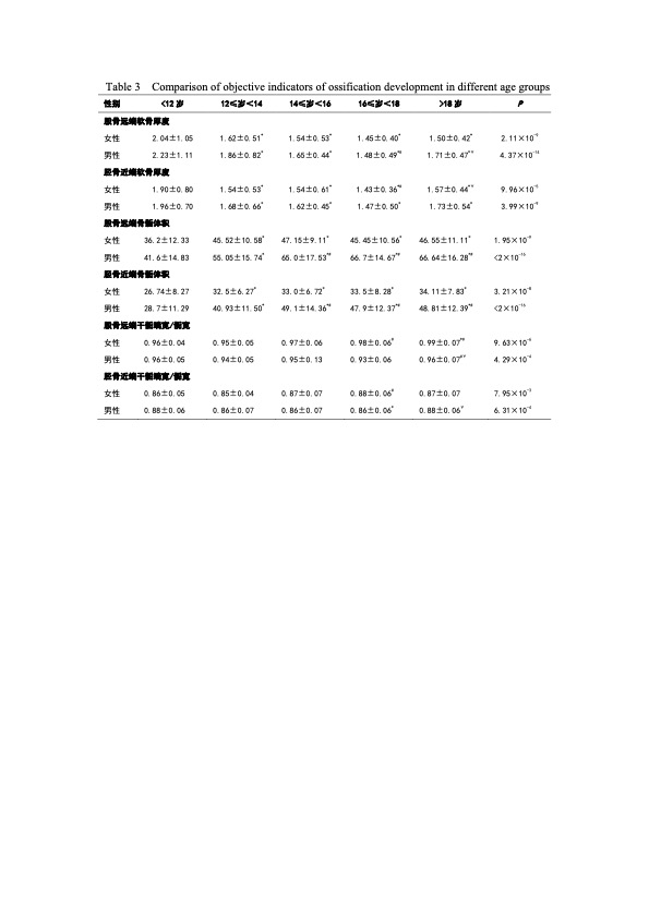
Results: From the results of this study, it can be seen that normal articular cartilage gradually thinned with the increase of bone age before 18 years old. The cartilage thickness of male distal femur was the smallest in 16≤year< 18 age group, and that of female proximal tibia was the smallest in 16≤year< 18 age group;The volume of epiphysis increased with age, male epiphysis volume increased significantly before 14 years old; Female epiphyseal volume increased significantly before 12 years of age;In terms of metaphysis width/epiphysis width, women in the age group 16≤year< 18 developed earlier than men, the metaphyseal width/epiphyseal width of male distal femur 12≤year< 14 age group was significantly lower than that of 16≤year< 18 age group, and that of female proximal tibia 12≤year< 14 age group was significantly lower than that of 16≤year< 18 age group.
Discussion: MRI images have high soft tissue contrast and resolution, and the development of epiphysis is clearly displayed on the images. It is worth noting that we used three scan sequences (T1WI/T2WI/PDWI) and two scan directions (sagittal/coronal) in our study to provide more detailed information about the epiphyseal cartilage plate and to assist in a comprehensive evaluation. It can be seen from the results of this study that normal articular cartilage becomes progressively thinner with bone age before the age of 18, and the volume of epiphyseal increases with age, which is consistent with the results of many studies [9,10]. In terms of metaphyseal width/epiphyseal width, females in 16≤ age < 18 age group developed earlier than males, and the metaphyseal width/epiphyseal width of male distal femur in 12≤ age < 14 age group was significantly lower than that in 16≤ age < 18 age group, and the metaphyseal width/epiphyseal width of female proximal tibia in 12≤ age < 14 age group was significantly lower than that in 16≤ age < 18 age group. It provides a new way to evaluate epiphysis. Studies have shown that males perform better in age prediction of distal femur and females in age prediction of proximal tibia, which may be related to the process of adolescent development [11]. In future knee MRI age estimation studies, it is necessary to evaluate gender separately and consider various factors related to gender and age differences.
Conclusion: The age group of 12≤ years old < 14 years old and 16≤ age < 18 years old have good evaluation, and the age prediction of the distal femur in males and the proximal tibia in females is better, and individual evaluation is needed if necessary, which provides a new idea for epiphyseal evaluation.
Acknowledgements
No acknowledgement found.References
1. Sironi E, Gittelson S, Bozza S, Taroni F .Minor or adult? Introducing decision analysis in forensic age estimation [J]. Sci Justice. 2021;61(1):47–60.
2. Berlin L .Shared decision-making: is it time to obtain informed consent before radiologic examinations utilizing ion- izing radiation? Legal and ethical implications [J]. J Am Coll Radiol.2014; 11(3):246–251.
3. Lu Ting, Fan Fei, Bai Wanjing,et al. Based on magnetic resonance imaging (MRI) concluded that teenagers living research status and prospect of bone age [J]. Chinese journal of forensic science, 2022; (02): 184-188.
4. Xiao‐Dong Deng,Ting Lu, Guang‐Feng Liu, et al. The feasibility of age estimation based on MRI and radiographs of the knee joint in the Chinese population of Han nationality [J]. International Journal of Legal Medicine. 2022;136:841–852
5. Oguzhan Ekizoglu, Ali Er, Mustafa Bozdag, et al. Forensic age estimation via magnetic resonance imaging of kneein the Turkish population: use of T1-TSE sequence[J]. Int J Legal Med. 2021 Mar;135(2):631-637.
6. Ekizoglu O, Hocaoglu E, Inci E, et al. Forensic age estimation via 3-Tmagnetic resonance imaging of ossification of the proximal tibial and distal femoral epiphyses: use of a T2-weighted fast spin-echo technique[J]. Forensic Sci Int.2016 Mar: 260: 102.e1-102.e7
7. Ottow C, Schulz R, Pfeiffer H, et al. Forensic age estimation by magnetic resonance imaging of the knee: the definite relevance in bony fusion of the distal femoral- and the proximal tibial epiphyses using closest-to-bone T1 TSE sequence[J]. Eur Radiol.2016;27:5041–5048.
8. Vieth V, Schulz R, Heindel W, et al. Forensic age assessment by 3.0T MRI of the knee: proposal of a new MRI classification of ossification stages[J]. Eur Radiol.2018; 28(8):3255–3262
9. DEDOUIT F, AURIOL J, ROUSSEAU H, et al. Age assessment by magnetic resonance imaging of the knee:A preliminary study[J]. Forensic Sci Int.2014; 012, 217(1/2/3):232. e1-232. e7.
10. EKIZOGLU O, HOCAOGLU E, INCI E, et al. Forensic age estimation via 3-T magnetic resonance imaging of ossification of the proximal tibial and dis‐ tal femoral epiphyses:Use of a T2-weighted fast spin-echo technique[J]. Forensic Sci Int.2016;260: 102.e1-102.e7.
11. Hao Hongxia, Wang Yahui, Zhou Zhilu, etc. Research progress on MRI living age in the knee joint [J]. Journal of Forensic Medicine, 2023,39(01):66-71+82.
Figures