5135
The Value of Deep Learning Reconstruction In Improving the Image Quality of rectum MRI Images1Department of Diagnostic Radiology, National Clinical Research Center for Cancer/Cancer Hospital, Chinese Academy of Medical Sciences and Peking Union Medical College, Beijing, China, 2MR Research Collaboration, Siemens Healthineers, Beijing, China, 3MR Application Predevelopment,, Siemens Healthineers AG, Erlangen, Germany
Synopsis
Keywords:
Motivation: TSE sequences are crucial for rectum MRI, but have limitations. DL-TSE is expected to improve image quality and reduce acquisition time for rectum MRI.
Goal(s): To assess the viability of employing TSE sequences with deep learning reconstruction for rectal MRI when compared to conventional TSE sequences.
Approach: This study included 16 patients with colorectal cancer confirmed by pathology. SNR and CNR were analyzed by SPSS 22.0 software.A P-value below 0.05 was considered statistically significant.
Results: The results show that the application of deep learning can shorten the scanning time while maintaining high image resolution, and improve the diagnostic efficiency of rectal diseases.
Impact: Deep learning reconstruction of TSE sequence in rectal MRI has the advantages of shortening acquisition time, improving image quality, and improving diagnostic efficiency. DL-TSE may also be extended to MRI examinations of other organs, such as the prostate and pelvis.
Introduction
Rectum cancer, a prevalent malignancy of the digestive tract, presents a complex challenge due to its intricated anatomical relationship with the pelvic cavity. The role of rectum MRI is paramount in understanding lesion localization, anatomical relationships, and accurate preoperative clinical staging, thereby facilitating the development of effective treatment strategies. T1WI and T2WI are essential for rectum MRI examinations, and turbo spin echo (TSE) is the standard sequence owing to its high signal-to-noise ratio (SNR) and spatial resolution. However, the TSE sequence in rectum MRI is susceptible to motion artifacts induced by intestinal peristalsis and susceptibility artifacts near air-tissue interfaces during its extended acquisition time. Several approaches have been used to mitigate these challenges, such as conventional in-plane parallel imaging (PI) method, administration of anticholinergic agents and the application of a saturation band to the subcutaneous fat of the anterior body wall. However, in practice, the conventional PI techniques often have limitations in achieving an acceleration factor of beyond 2 due to the introduction of parallel acquisition artifacts and decrease in SNR, which reduces by the square root of the acceleration factor [3]. Deep learning reconstruction, with its trainable components in the latest technological innovations, holds the promise of achieving higher acceleration factors while simultaneously enhancing SNR [4], and more homogeneity in the images and denoising. Thus, it is hypothesized that TSE combined with deep learning reconstruction (TSE-DL) can produce similar image quality that is comparable to clinically conventional in-plane parallel imaging TSE images. Further, with higher acceleration and reduced averages, the time of acquisition (TA) can also be significantly reduced. Therefore, this study aims to evaluate the feasibility of using the TSE sequence with deep learning reconstruction in rectum MRI compared with conventional TSE sequences.Method
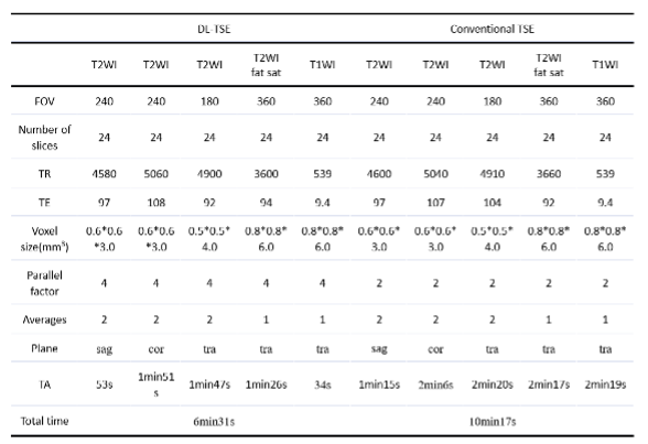
16 patients with rectal cancer underwent MRI scans on a 3T system (MAGNETOM Prisma, Siemens Healthineers, Erlangen, Germany) acquiring routine rectum MR scans and in addition the corresponding research sequences using deep learning image reconstruction. The detailed imaging parameters are listed in Table 1. Two radiologists (reader1, Reader2) with 8 and 20 years of experience independently evaluated MR images in random order. Quantitative assessment was performed on a workstation (Syngo MR, Siemens Healthineers, Erlangen, Germany). Two radiologists independently placed as large an area of interest (ROI) as possible and free of serious artifacts on the rectum and adjacent tissues of all patients. Signal to noise ratio (SNR) and contrast-to-noise ratio (CNR) were calculated by the following equations: SNR=SI/SD and CNR= |SIA – SIB|/√SDA2+SDB2, where SI is signal intensity and SD is standard deviation. Two radiologists independently assessed the image quality in terms of artifacts, image clarity and lesion detectability of both sequences. All imaging datasets are in a blind reading, using the Rickett scale 5-point criteria: 5 = no artifacts, with excellent image quality; 4 = no artifacts, with good image quality; 3 =minor artifacts, with acceptable image quality, 2 = moderate artifacts, with low image quality, and 1 = non-diagnostic, with major streak artifacts. The CNR and SNR of deep learning image reconstruction sequences and conventional sequences are tested by paired T-test for normally distributed data, and the Wilcoxson singned-rank test for non-distribution data.Result
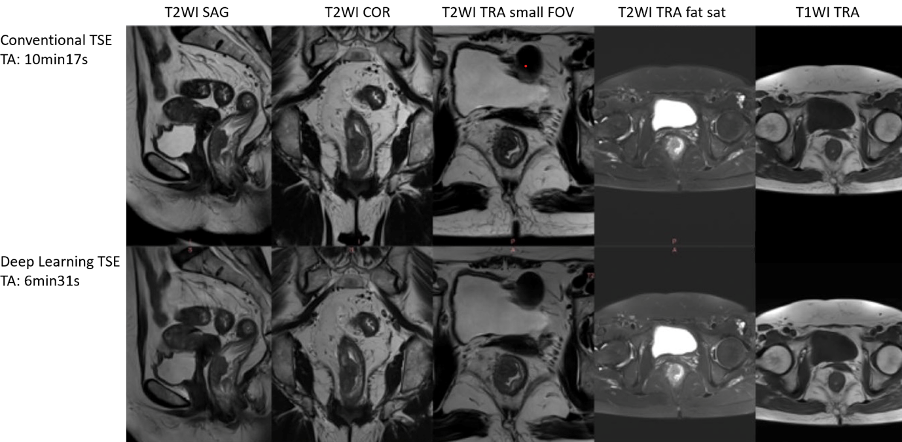
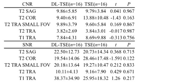
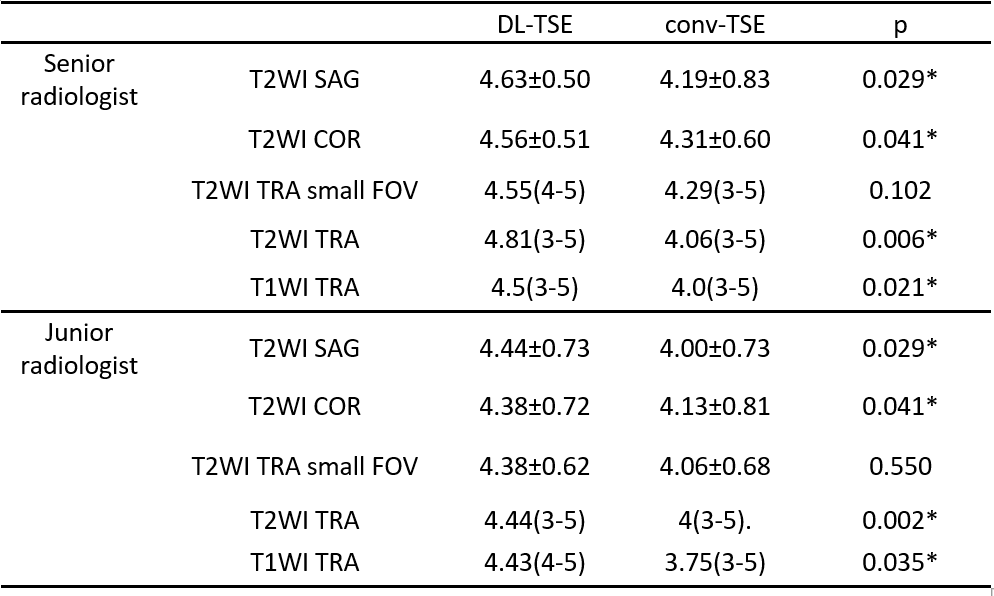
Figure 1 shows routine TSE images and DL-TSE images of a 68-year-old female with moderately differentiated adenocarcinoma of the rectum. The acquisition time is 10min17s in conventional TSE MRI (top row) and 6min31s in DL-TSE MRI scheme (bottom row) were obtained.The quantitative evaluation of DL TSE sequences and TSE sequences is shown in Table 2. The analysis shows that SNR and CNR of DL sequences is not significantly different from that of conventional sequences. This shows that deep learning reconstruction has the advantages of shortening scan time without the loss of SNR and CNR. In quantitative evaluation, the score of DL sequence in image quality is significantly higher than of conventional sequences in both T1WI and T2WI of sagittal, coronal, and axial position except small FOV of T2WI tra (as shown in Table 3). Both diagnostic imaging doctors prefer to the DL sequence, and they believe that the deep learning sequences provides higher image quality, enhance the diagnostic confidence of doctors, and improve the diagnostic efficiency of rectal diseases.Conclusion
The feasibility of deep learning image reconstruction to improve the quality of rectal MRI images was studied. Deep learning reconstructions performed well on T2 and T1-weighted TSE rectal MRI, both acquiring images faster and maintaining high image quality with reduced scanning artifacts compared to traditional TSE. Compared to traditional TSE sequences, the time can be reduced by 36.6% at 3T.Acknowledgements
The Author Yueluan Jiang and Marcel Dominik Nickel are from a commercial company, Siemens Healthineers Ltd., are a MR collaboration scientist doing technical support in this study under Siemens collaboration regulation without any payment and personal concern regarding to this study.References
[1] Klessen C, Rogalla P, Taupitz M. Local staging of rectal cancer: the current role of MRI. Eur Radiol 2007;17:379-389.
[2] Fernandes MC, Gollub MJ, Brown G. The importance of MRI for rectal cancer evaluation. Surg Oncol 2022;43:101739.
[3] Fritz J, Guggenberger R, Del Grande F. Rapid Musculoskeletal MRI in 2021: Clinical Application of Advanced Accelerated Techniques. AJR AmJ Roentgenol 2021;216(3):718–733
[4] Hammernik K, Klatzer T, Kobler E, Recht MP, Sodickson DK, Pock T, Knoll F. Learning a variational network for reconstruction of accelerated MRI data. Magn Reson Med. 2018 Jun;79(6):3055-3071. doi: 10.1002/mrm.26977. Epub 2017 Nov 8. PMID: 29115689; PMCID: PMC5902683.
Figures