cheng tang1, jianjian huang2, peng peng1, shunzu lu1, yiwu lei1, huiting zhang3, and chen zhao3
1THE FIRST AFFILIATED HOSPITAL OF GUANGXI MEDICAL UNIVERSITY, nanning, China, 2Guangdong Sencond Provincial General Hospital, guangzhou, China, 3MR Scientific Marketing, Siemens Healthnieers, wuhan, China
Synopsis
Keywords:
Motivation: Minimize scanning time while ensuring the quality of MRH images.
Goal(s): This study investigated the feasibility of using compressed perception acceleration sequences to evaluate the quality of MRH images.
Approach: We compared data from traditional sequences and CS sequences with different acceleration factors.
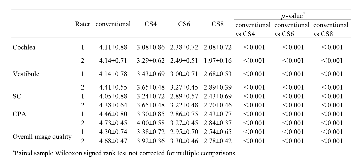
Results: The results showed that CS4 can guarantee diagnostic requirement with 44%time reduction. Conventional images were more commonly used than CS SPACE images for radiologists. However, CS4 SPACE images still get more than 3-point. This suggests that CS4 SPACE images is still considered acceptable.
Impact: This preliminary study showed that for the MRH of inner ear, T2 SPACE sequence with 4-fold CS technique provided similar image quality to conventional T2 SPACE with 44% shorter scan time. Therefore, we can save a lot of examination time.
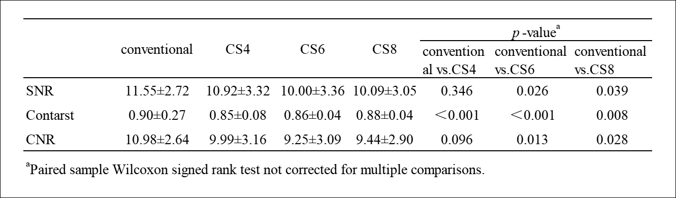
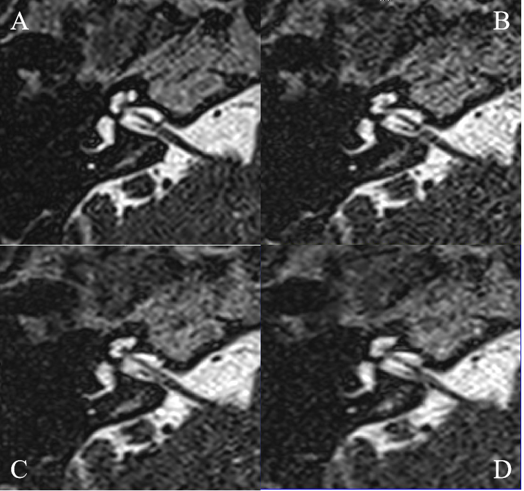
Purpose: To evaluate the image quality and acquisition time of magnetic resonance hydrography (MRH) of inner ear with routine technique and with different fold accelerated compressed sense (CS) technique by T2 SPACE sequence. Materials and Methods:The prospective study was approved by the institutional review board of our hospital. Thirty-seven consecutive volunteers were recruited and signed the informed consent. All volunteers underwent MR scans on a 1.5T MR scanner (MAGNETOM Altea; Siemens Healthcare, Erlangen, Germany) with the conventional T2 SPACE and CS SPACE sequences. The same parameters were as follows: flip angle =150°, turbo factor= 88, matrix = 288 × 288, FOV = 190mm × 190mm, slice thickness=0.7mm. Other parameters were as follows: accelerated factor =2(GRAPPA2), 4 (CS4), 6 (CS6), and 8 (CS8); TR=1400ms, TE=169ms,total scan time =163s /91s/66s /55s. For the CS SPACE, variable-density Poisson disc pattern and compressed sensing reconstruction with a regularization factor of 0.005 were employed. Unilateral images for each sequence were separated, then placed into mixed folders for independent, blinded review by 2 neuroradiologists during 1+ weeks apart. Visualization of cochlea, vestibule, semicircular canal (SC), cerebellopontine angle(CPA), overall image quality were rated using ordinal scales (1-5 points, ≥3 points are considered as meeting the diagnostic requirements). The contrast, signal-to-noise-ratio (SNR), and contrast-noise-ratio (CNR) between the semicircular canal (SC) and peri semicircular canal tissues on MRH of inner ear were evaluated quantitatively.Data analysis was performed using SPSS 22 (IBM Ltd, U.S.A). Paired sample Wilcoxon signed rank test was used to assess the differences in subjective scales and in objective results (SNR and CNR) between conventional SPACE and CS SPACE with different accelerated factors. P < 0.05 was considered statistically significant different.Results:The image acquisition time was 44%, 59%, 66% shorter in 4-fold, 6-fold and 8-fold accelerated CS SPACE sequences than conventional SPACE sequence. There were no significant differences in the SNR, and CNR of the SC between conventional SPACE and CS SPACE sequence with 4-fold CS technique (P>0.05). The contrast of the SC in conventional sequence was significantly higher without CS technique than with it (P < 0.01). The SNR, and CNR of the SC in conventional sequence was significantly higher than with 6-fold and 8-fold accelerated CS sequence (P < 0.01). The detailed results are shown in Table 1.CS SPACE images are 0.71–2.03 lower than conventional images on 5-point scale, P < 0.001, but 4-fold accelerated CS SPACE images acceptable, as shown in Table 2 and Figure 1.Discussion:This preliminary study showed that for the MRH of inner ear, T2 SPACE sequence with 4-fold CS technique provided similar image quality to conventional T2 SPACE with 44% shorter scan time. However, as the acceleration fold increased to 6-fold even 8-fold, the image quality reduced, which had significant difference compared with the conventional SPACE. In the previous internal auditory canal MRI with 5-fold CS SPACE [1], researcher found that the subjective scale had significant difference, but they also supposed the rated CS CSPACE acceptable.Conventional images were superior to accelerated CS SPACE images for radiologists. However, 4-fold accelerated CS SPACE images is still considered acceptable (>3).Conclusion:Using 1.5T MRI for MRH of inner ear with the 4-fold accelerated CS technique preserved diagnostic image quality with a shorter image acquisition time when compared to image of conventional sequence.Acknowledgements
This work was supported by Guangxi Medical and Health Appropriate Technology Development and Promotion Application Project (No.S2022067).References
1. Mikell Yuhasz, Michael J. Hoch, Mari Hagiwara, et al. Accelerated IAC screening MRI protocol with compressed sensing 3D T2-weighted sequence, Invest Radiol. 2018; 53(12): 742–747.