5122
MULTI-ECHO FUNCTIONAL IMAGING ON AN ULTRA HIGH-PERFORMANCE HEAD-ONLY GRADIENT SYSTEM1Radiology, USU/WRNMMC, Bethesda, MD, United States, 2GE Global Research, Niskayuna, NY, United States
Synopsis
Motivation: This abstract focuses on educating on how high-gradient MRI systems can utilize the multi-echo functional MRI (ME-fMRI) techniques.
Goal(s): We explain the blood-oxygen-level-dependent (BOLD) signal technique and how additional echoes can improve the signal fidelity.
Approach: The ME-fMRI, using three or more echoes allow for the pixelwise T2* decay to be modeled, and as BOLD contrast is a function of T2* evolution over time, an experiment sampling the voxel-wise T2* signal decay can be used to separate BOLD from artifact signal constituents.
Results: Gradient systems with ultra-rapid slew rates (> 400 T/m/s) allow ME-fMRI to reduce artefacts from flow, motion, and susceptibility effects.
Impact: MRI systems with ultra-high gradient systems can reduce artefacts from flow, motion, and susceptibility effects in the BOLD contrast technique using an ME-fMRI technique with three or more echoes to improve fidelity of fMRI.
Background:
Scientists have long wondered about how the human brain functions, so when MRI was developed it was only a matter of time before MRI would be used to develop a technique to look at the functional aspects of the brain. The theory by Roy and Sherrington in 1890 postulated that functional activity of the brain is related to blood supply and cerebral metabolism (1). Dr. John Belliveau is credited with the first successful fMRI experiments in 1984 using two gadolinium injections and EPI to collect two cerebral blood volume maps, one resting and one after a stimulation (2,3). Today, fMRI has evolved to assess changes in blood contrast without the use of a contrast agent using changes in cerebral blood flow (CBF) and blood oxygenation to provide temporal resolution on the order of hemodynamic response times (1). The recent high gradient MRI systems are allowing for a new, improved multi-echo fMRI technique to emerge (4).Teaching Point:
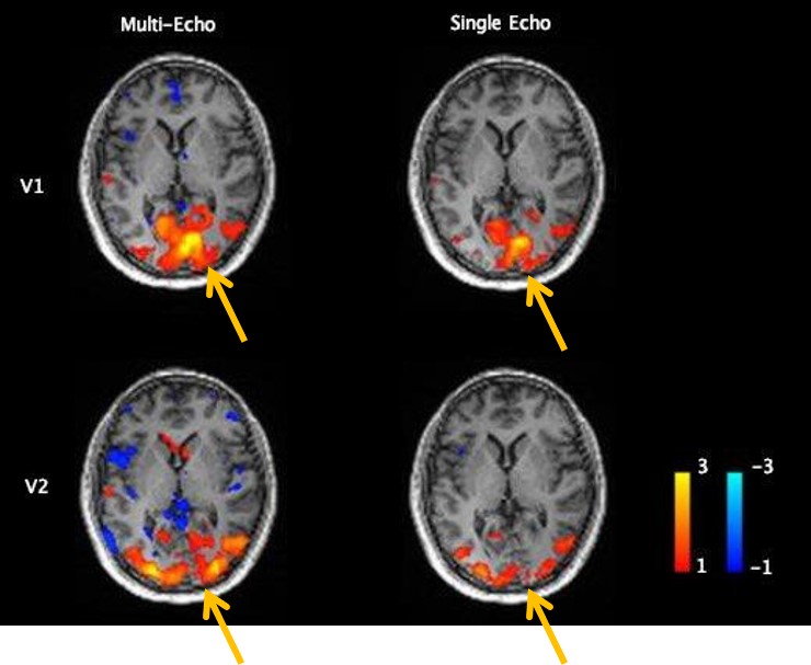
Traditional fMRI is acquired using gradient echo EPI sequences to acquire data at a single TE optimized for detection of blood-oxygen-level-dependent (BOLD) signal. Optimum sensitivity is achieved at an echo time close to the apparent T2* value of the grey matter in the tissue (5). In brain imaging the optimal TE can vary according to the T2* of the underlying susceptibility artifacts resulting in signal indeterminacy (or uncertainty in the precision of the measurement) (6). The interplay of blood flow, blood volume and magnetic susceptibility effects can be influenced by scanner and physiological noise sources, which affect the BOLD signal making traditional fMRI measurements struggle with reliability and repeatability. Multi-echo (ME) fMRI has emerged as an augmented acquisition approach allowing for improved data fidelity of fMRI. MRI systems with strong gradient system are needed to acquire adequate signal for the later echoes (7). By acquiring multiple-echo images per TR, the approach allows the pixelwise T2* decay to be modeled. As BOLD contrast is a function of T2* evolution, an experiment sampling the voxel-wise T2* signal decay can be used to separate BOLD from non-BOLD signal changes (8). ME-fMRI involves relatively small changes from standard fMRI acquisition but can better recover functional activation in regions of signal loss from high magnetic susceptibility and geometric distortion. The use of three or more echoes per TR interval is recommended with the first TE as short as possible (9). It has been determined that the earliest TE has highest signal intensity but lower contrast between gray and white matter and CSF. Longer TE’s have less MR signal but higher BOLD-related differences. The increased echo sampling requirements can compromise temporal resolution, while longer echo spacing constraints due to performance and PNS limits of whole-body scanners results in long ΔTE and late TE. The multiple echoes on a whole-body 3T scanner, are too far out to be of any benefit, thus limiting usage on conventional whole-body scanners (8). Improved gradient performance from high performance head only gradient systems not only reduces distortion in EPI, but the high gradient slew rates further reduce echo spacing and readout durations. For the same 2.5-mm acquisition, on MAGNUS-1 (200 mT/m and 500 T/m/s), the parameter space allows for a ΔTE of 14ms, and the first echo to be <11.8 ms (9). This acquisition space is optimal for R2*(1/T2*) to get the largest benefit in SNR returns from all echoes and minimal loss in signal. Readout duration has a benefit of x3 the number of echoes that can fit in the same scan TR compared to conventional scanners. By acquiring multiple echo images per slice, the ME approach allows T2* decay to be modeled at every voxel at every time point. Using a high-performance gradient system, we are able to acquire 3 echoes readouts in the time usually for a single echo readout. The increased fidelity of the ME-fMRI method (Figure 1) shows the increased sensitivity of the BOLD signal for ME-fMRI versus SE-fMRI in visual cortex area.Summary:
Multi-echo fMRI with a high-performance gradient system yields greater sensitivity to BOLD dynamic contrast in task-based and resting state fMRI. The use of specialized head-only gradient systems with ultra-rapid slew rates (> 400 T/m/s) allow ME-fMRI to reduce artefacts from flow, motion, and susceptibility effects. This also produces higher quality fMRI results in frontal and basal areas of the brain where air-bone-tissue boundary susceptibility distort or mute BOLD contrast.Acknowledgements
The authors would like to thank the MAGNUS MRI team members, Haymanot Yalewayker and Samrawit Yalewayker for their efforts in project management and clinical coordination to recruit subjects for us to learn and develop ME-fMRI techniques.References
- Belliveau JW, Kwong KK, Kennedy DN, et al. Magnetic resonance imaging mapping of brain function. Human visual cortex. Invest Radiol. 1992 Dec;27 Suppl 2(0 2):S59-65.
- Bandettini PA. Twenty years of functional MRI: the science and the stories. Neuroimage. 2012 Aug 15;62(2):575-88.
- Belliveau JW, Kennedy DN Jr, McKinstry RC, et al. Functional mapping of the human visual cortex by magnetic resonance imaging. Science. 1991 Nov 1;254(5032):716-9.
- Posse S, Wiese S, Gembris, et al. Enhancement of BOLD-contrast sensitivity by single-shot multi-echo functional MR imaging. Magn Reason Med. 1999.42:87-97.
- Heunis S, Breeuwer M, Caballero-Gaudes C, et al. The effects of multi-echo fMRI combination and rapid T2*-mapping on offline and real-time BOLD sensitivity. Neuroimage. 2021 Sep;238:118244.
- Kovářová A, Gajdoš M, Rektor I, Mikl M. Contribution of the multi-echo approach in accelerated functional magnetic resonance imaging multiband acquisition. Hum Brain Mapp. 2022 Feb 15;43(3):955-973.
- Kang D, In MH, Jo HJ, et.al. Improved Resting-State Functional MRI Using Multi-Echo Echo-Planar Imaging on a Compact 3T MRI Scanner with High-Performance Gradients. Sensors (Basel). 2023 Apr 27;23(9):4329.
- Kundu P, Voon V, Balchandani P, Lombardo MV, Poser BA, Bandettini PA. “Multi-echo fMRI: Areview of applications in fMRI denoising and analysis of BOLD signals. “Neuroimage. 2017 Jul 1;154:59-80.
- Foo TKF, Tan ET, Vermilyea ME, et.al. Highly efficient head-only magnetic field insert gradient coil for achieving simultaneous high gradient amplitude and slew rate at 3.0T (MAGNUS) for brain microstructure imaging. Magn Reson Med. 2020 Jun;83(6):2356-2369.
Figures