5117
Cortical mapping provide insights into whole-brain tumor burden in diffuse midline glioma1Huaxi MR Research Center (HMRRC), Department of Radiology, West China Hospital of Sichuan University, Chengdu, China, Chengdu, China
Synopsis
Keywords: Tumors (Pre-Treatment), Neuro, diffuse midline glioma; H3K27M altered; whole-brain tumor burden; cortical myelin content; cortical thickness;
Motivation: Diffuse midline glioma (DMG) represents a systemic disease due to its ability to disseminate tumor cells throughout the whole brain. Current imaging techniques, however, provide information only about the main tumor and its immediate surroundings.
Goal(s): We employed comprehensive cortical mapping to gain insights into the individual tumor burden across whole-brain using structural MRI.
Approach: Cortical thickness and myelin content was calculated from participants using Human Connectome Project pipeline.
Results: DMG has the capacity to induce cortical thickness compensation while concurrently leading to cortical demyelination in numerous non-lesional regions. Notably, DMG harboring H3K27M altered exhibited specific cortical myelin and thickness reorganization patterns.
Impact: These findings may open up the possibility of tailoring treatment strategies to the individual disease severity and distribution within the patient's brain, potentially enhancing the effectiveness of both current and future treatment approaches.
Objectives
Systemic infiltration is a hallmark of diffuse midline glioma (DMG) pathogenesis which can trigger distant disturbances in cortical structure1,2. However, the existence and effects of these changes have been underexamined. This study aimed to investigate whole-brain cortical myelin and thickness alternations which may allowing for a holistic assessment of tumor burden induced by DMG.Methods
High-resolution T1-weighted (T1w) and T2-weighted (T2w) structural images were acquired from 90 patients with DMG H3K27M altered (DMG-A) and 64 patients with wild type (DMG-W) and 86 healthy controls (HC). Cortical thickness and myelin contents was calculated using Human Connectome Project pipeline3. Significant differences in cortical thickness and myelin contents were detected between DMG-A, DMG-W and HC groups. Short-term survival prediction model was constructed using automated machine learning approach.Results
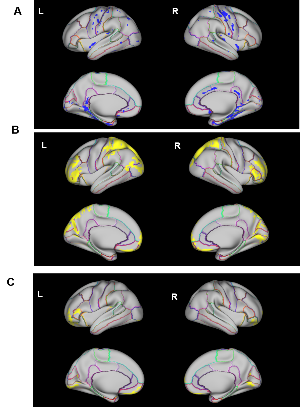
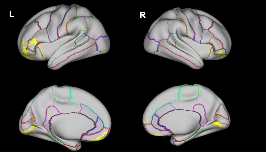
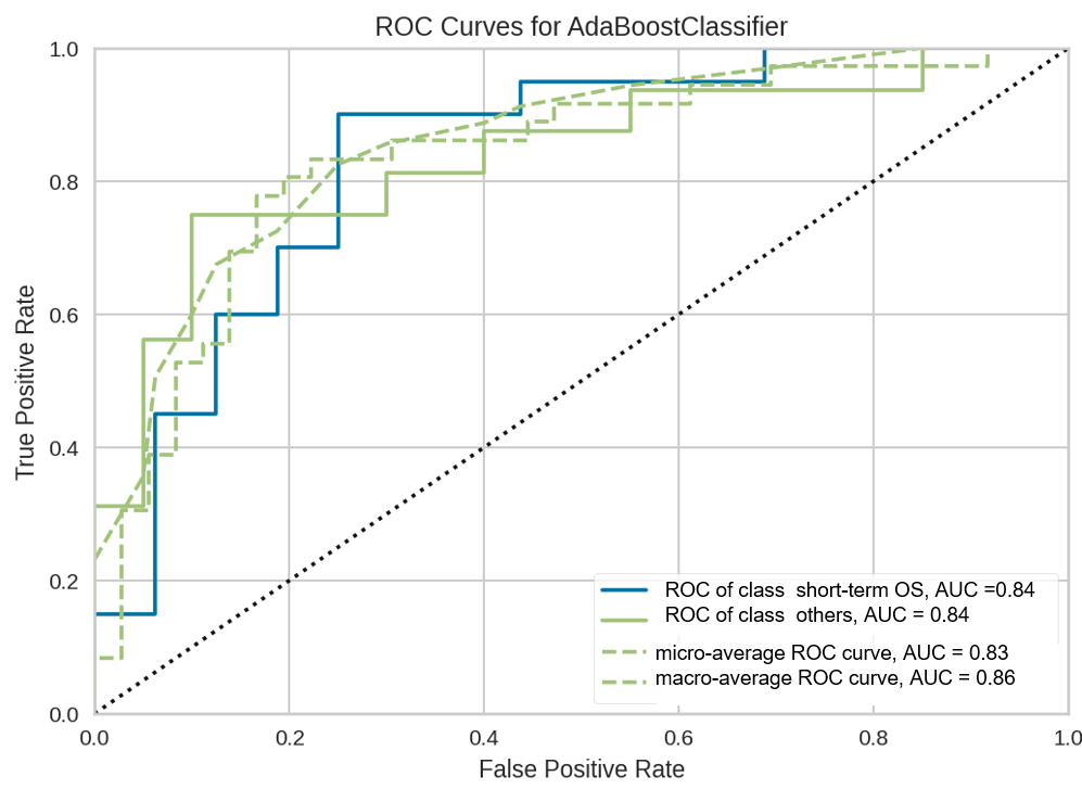
Compared with HC, both DMG-A and DMG-W showed cortical thickening in normal-appearing cortex. As for cortical myelin, DMG-A patients showed significantly lower cortical myelin contents in bilateral precentral gyrus, postcentral gyrus, insular, parahippocampal gyrus, fusiform gyrus, and cingulate gyrus, whereas DMG-W patients exhibited well-preserved myelin contents(Fig.1). Furtherly, when compared DMG-A with DMG-W, the decreased cortical thickness in parietal and occipital regions and additional demyelination in medial orbitofrontal cortex was observed in DMG-A(Fig.2). Besides, cortical features and tumor radiomics allowed short-term survival prediction with accuracy 0.80 and AUC 0.84(Fig.3).Conclusion
Utilizing comprehensive whole-brain cortical mapping, our study revealed that DMG has the capacity to induce cortical thickness compensation while concurrently leading to cortical demyelination in numerous non-lesional regions. These invisible alternations on conventional MRI and cortical features are correlated with patient short-term survival. The exploration of cortical thickness and myelin may provide a valuable tool for precise treatment for patients with DMG.Acknowledgements
The authors declare no potential conflicts of interest.This work was supported by the National Natural Science Foundation of China (Grant Nos. 82271961).References
1.Wang J, Xu S-L, Duan J-J, Yi L, Guo Y-F, Shi Y, Li L, Yang Z-Y, Liao X-M, Cai J et al: Invasion of white matter tracts by glioma stem cells is regulated by a NOTCH1–SOX2 positive-feedback loop. Nature Neuroscience 2019, 22(1):91-105.
2.Giese A, Westphal M: Glioma Invasion in the Central Nervous System. Neurosurgery 1996, 39(2).
3.Glasser MF, Van Essen DC: Mapping human cortical areas in vivo based on myelin content as revealed by T1- and T2-weighted MRI. The Journal of neuroscience : the official journal of the Society for Neuroscience 2011, 31(32):11597-11616.
Figures