5115
Diffusion-Weighted Imaging Using Echo Planar Imaging with Compressed SENSE (EPICS) for Pancreatic Protocol MRI: A Multicenter Study1Department of Radiology, Gifu University, Gifu, Japan, 2Department of Radiology Services, Gifu University Hospital, Gifu, Japan, 3Department of Radiology, Kagoshima University Graduate School of Medical and Dental Sciences, Kagoshima, Japan, 4Center for One Medicine Innovative Translational Research, Institute for Advanced Study, Gifu University, Gifu, Japan, 5Department of Radiology, Kawasaki Medical School, Kurashiki, Japan
Synopsis
Keywords: Diffusion Reconstruction, Pancreas
Motivation: To prove the feasibility of DWI using echo planar imaging with Compressed SENSE (EPICS-DWI).
Goal(s): Are the images and values obtained by EPICS-DWI reliable?
Approach: Taking both conventional DWI using parallel imaging (PI-DWI) and EPICS-DWI images for the same patient with untreated PDAC within the same examination and compare them.
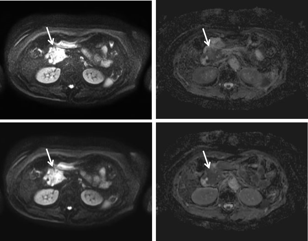
Results: The ADC value of PDAC did not differ between PI-DWI and EPICS-DWI. EPICS-DWI can improve the qualitative overall image quality and PDAC-to-pancreas CNR compared to PI-DWI. The qualitative PDAC conspicuity was comparable between PI-DWI and EPICS-DWI.
Impact: EPICS could improve image quality of high-b value DWI images without any worries about significant changing of ADC values of PDAC.
INTRODUCTION
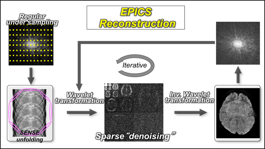
Diffusion-weighted imaging (DWI) is one of the essential sequences for evaluating pancreatic ductal adenocarcinoma (PDAC) in pancreatic protocol MRI1-5. However, the degraded image quality and lack of reproducibility of ADC value measurements are clinically considerable problems in DWI. Recently, the echo planar imaging with Compressed SENSE (EPICS) (Figure 1) has been newly applied to DWI (EPICS-DWI). EPICS-DWI could reduce image noise and significantly improve image quality of high-b value DWI compared with conventional DWI using parallel imaging (PI-DWI)6-10. It is a matter of concern that previous studies reported that the mean ADC values of various normal organs were higher in EPICS-DWI than PI-DWI6-9. The purpose of this study was to evaluate the impact of EPICS-DWI on the evaluation of PDAC.METHODS
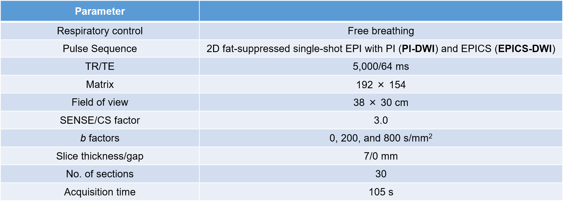
This multicenter prospective study was approved by our and Kagoshima university Institutional Review Boards, and written informed consent was obtained from all participants. We analyzed participants who underwent pancreatic protocol MRI between October 2020 and July 2022, diagnosed as PDAC by pathological examination, and had not received any treatment for PDAC at the time of MRI examination. Both free-breathing two-dimensional fat-suppressed PI-DWI and EPICS-DWI were obtained in all participants using a 3.0T MRI system (Ingenia 3.0T CX; Philips Healthcare) equipped with a 32-channel digital coil. The scanning parameters were presented in Figure 2. As qualitative image analysis, two radiologists independently and randomly reviewed high-b value DWI images and assigned confidence scores for the overall image quality, image noise, pancreas conspicuity, and PDAC conspicuity using a 5-point scale. As quantitative image analysis, one of the aforementioned radiologists measured the signal intensity (SI) of the pancreatic parenchyma (SIPANC) and PDAC (SIPDAC) and those of standard deviations (SDPANC and SDPDAC) on high-b value DWI images, and ADC values of the pancreatic parenchyma (ADCPANC) and PDAC (ADCPDAC) using a region of interest. The PDAC-to-pancreas contrast-to-noise ratio (CNR) was calculated using the following formula;$$CNR = \frac{|SI_{PANC} - SI_{PDAC}|}{ \sqrt{\frac{{SD_{PANC}}^2 + {SD_{PDAC}}^2}{2}}}$$
The Wilcoxon signed-rank test was conducted to compare the qualitative and quantitative items between PI-DWI and EPICS-DWI.
RESULTS
The median confidence scores for the overall image quality (P < .001 in both radiologists) and image noise (P < .001 in both radiologists) were higher in EPICS-DWI than in PI-DWI. The median confidence score for the pancreas conspicuity was higher in EPICS-DWI than in PI-DWI in only radiologist 1 (P = .02 in radiologist 1 and P = .06 in radiologist 2). The median confidence score for the PDAC conspicuity was not different between PI-DWI and EPICS-DWI (P > .99 in both radiologists). The SIPANC (183.0 in PI-DWI and 167.9 in EPICS-DWI; P > .99) and SIPDAC (289.7 in PI-DWI and 286.4 in EPICS-DWI; P = .85) were not different between PI-DWI and EPICS-DWI. The PDAC-to-pancreas CNR was higher in EPICS-DWI than in PI-DWI (5.3 in PI-DWI and 6.4 in EPICS-DWI; P = .02). The ADCPANC was higher in EPICS-DWI than in PI-DWI (1.4 × 10⁻³ mm2/s in PI-DWI and 1.5 × 10⁻³ mm2/s in EPICS-DWI; P = .01). The ADCPDAC was not different between PI-DWI and EPICS-DWI (1.3 × 10⁻³ mm2/s in PI-DWI and 1.3 × 10⁻³ mm2/s in EPICS-DWI; P = .48).DISCUSSION
According to the previous studies6-10, EPICS-DWI can drastically improve the image quality, signal-to-noise-ratio, and CNR by reducing image noise compared to PI-DWI through wavelet transformation framework in Compressed SENSE, and our results of improved overall image quality, pancreas conspicuity, and PDAC-to-pancreas CNR were consistent with them. On the other hand, the PDAC conspicuity was not improved in EPICS-DWI compared to PI-DWI in this study. Regarding this aspect, one possible cause is that PDACs don’t always manifest as a well-defined mass or hyper-intense lesions compared to the pancreatic parenchyma on DWI images. The ADC value of PDAC was not different between PI-DWI and EPICS-DWI contrary to the past studies which have reported that the mean ADC values of various normal organs were higher in EPICS-DWI than in PI-DWI6-9. Although the reason why focal lesions did not exhibit noticeable changes in ADC values has yet been unclear, we thought of the following reasons: the influence of noise reduction did not affect the ADC values of PDAC because of its relatively high signal on DWI images, and the fact that respiratory motion was not negligible in relatively small lesions. This newly adapted EPICS-DWI could be applied in clinical use without any worries about significant changing of ADC valuesCONCLUSION
The PDAC-to-pancreas CNR was higher in EPICS-DWI than PI-DWI. However, PDAC conspicuity and ADC values of PDAC were almost comparable between PI-DWI and EPICS-DWI.Acknowledgements
The authors of this manuscript declare no relationships with any companies whose products or services may be related to the subject matter of the article.References
1. T. Ichikawa, S.M. Erturk, U. Motosugi, et al. High-b value diffusion-weighted MRI for detecting pancreatic adenocarcinoma: preliminary results, AJR Am J Roentgenol 188(2) (2007) 409-14.
2. T. Kamisawa, K. Takuma, H. Anjiki, et al. Differentiation of autoimmune pancreatitis from pancreatic cancer by diffusion-weighted MRI, Am J Gastroenterol 105(8) (2010) 1870-5.
3. A. Kurita, Y. Mori, Y. Someya, et al. High signal intensity on diffusion-weighted magnetic resonance images is a useful finding for detecting early-stage pancreatic cancer, Abdom Radiol (NY) 46(10) (2021) 4817-4827.
4. P. Wiggermann, R. Grützmann, A. Weissenböck, et al. Apparent diffusion coefficient measurements of the pancreas, pancreas carcinoma, and mass-forming focal pancreatitis, Acta Radiol 53(2) (2012) 135-9.
5. Dalah. B. Erickson, K. Oshima, D. Schott, et al. Correlation of ADC With Pathological Treatment Response for Radiation Therapy of Pancreatic Cancer, Transl Oncol 11(2) (2018) 391-398.
6. T. Kaga, Y. Noda, T. Mori, et al. Diffusion-weighted imaging of the abdomen using echo planar imaging with compressed SENSE: Feasibility, image quality, and ADC value evaluation, Eur J Radiol 142 (2021) 109889.
7. Yoneyama, K. Morita, J. Peeters. Noise Reduction in Prostate Single-Shot DW-EPI utilizing Compressed SENSE Framework, Proc. ISMRM (1634) (2019).
8. N. Yoshida, T. Nakaura, K. Morita, et al. Echo planar imaging with compressed sensitivity encoding (EPICS): Usefulness for head and neck diffusion-weighted MRI, Eur J Radiol 155 (2022) 110489.
9. T. Kaga, Y. Noda, M. Asano, et al. Diagnostic ability of diffusion-weighted imaging using echo planar imaging with compressed SENSE (EPICS) for differentiating hepatic hemangioma and liver metastasis, European Journal of Radiology 167 (2023) 111059.
10 T. Tamada, Y. Ueda, A. Kido, et al. Clinical application of single-shot echo-planar diffusion-weighted imaging with compressed SENSE in prostate MRI at 3T: preliminary experience, MAGMA 35(4) (2022) 549-556
Figures