5111
A Novel Ultra-High b-Value Diffusion-Weighted MRI Technique for ALS Diagnosis and Disease Tracking in Mouse Spinal Cords In vivo1Department of Radiology and Imaging Sciences, Emory University, Atlanta, GA, United States, 2Department of Biomedical Engineering, University of Illinois Chicago, Chicago, IL, United States, 3Department of Electrical and Computer Engineering, University of Illinois Chicago, Chicago, IL, United States, 4Research Resources Center, University of Illinois Chicago, Chicago, IL, United States
Synopsis
Keywords: Microstructure, Diffusion/other diffusion imaging techniques
Motivation: Amyotrophic Lateral Sclerosis (ALS) significantly impacts global human health, but its etiology remains unclear.
Goal(s): To develop a novel diffusion-weighted MRI technique to detect early changes in ALS-affected spinal cord in vivo.
Approach: We applied ultra-high b-values by using long diffusion time to examine the restricted diffusion in spinal white matter tracts in SOD1G93A mice at ages of 75 and 90 days.
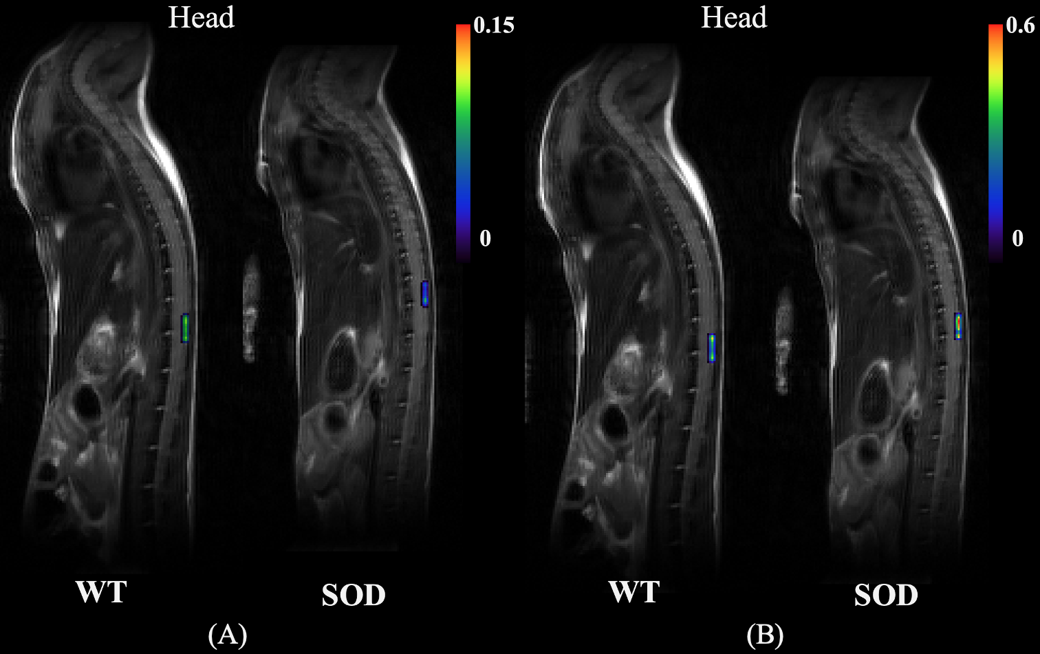
Results: Significant differences were found in diffusion of ventral roots between SOD1G93A mice and control at ages of 75 and 90 days. A shift of diffusion distribution was observed in SOD1G93A mice between 75 and 90 days.
Impact: This in vivo study potentially presents a novel view in non-invasive evaluating alterations in spinal cord tissue associated with ALS pathology, thus benefiting investigations related to drug delivery and therapeutic response monitoring of ALS.
Introduction
Amyotrophic Lateral Sclerosis (ALS) has a global impact on human health, leading to profound consequences[1, 2]. However, the precise underlying cause of ALS remains incompletely understood [3], which needs innovative methodologies for further investigation. Diffusion-weighted MRI (dMRI) plays a vital role in ALS research due to its non-invasive nature. Specifically, while multi-direction diffusion techniques have been extensively explored, their wealth of directional information is largely redundant in spinal cord investigations ascribed to highly organized neural fiber structure. This characteristic can facilitate the application of specific diffusion gradient directions, such as those aligned with the radial diffusivity (RD) plane, to sensitize minimal changes in the confined diffusion space. As such, we developed a novel dMRI technique using ultra-high b-values on a RD direction to specifically focus on detecting subtle ALS-induced alterations in mouse spinal cords in vivo.Methods
Seven wild type mice and seven SOD1G93A mice were scanned at postnatal 75 days (P75, pre-symptomatic stage) and postnatal 90 days (P90, early symptomatic stage). The experimental protocol was approved by the local IACUC. MR images were acquired on a 9.4 T Agilent MRI scanner (Santa Clara, CA). The diffusion-weighted imaging protocol includes 8 b-values ranging from 0 to 1× 10⁵ s/mm² achieved by a stimulated echo sequence with the following parameters: TR/TE = 2000/21.69 ms, mixing time = 197.02 ms, diffusion time = 210 ms, diffusion gradient duration = 6.5 ms, slice thickness = 1.5 mm, FOV = 43 mm ×25.6 mm, matrix = 128 × 64, average = 8. The total scan time is 2 hours 16 minutes. A rectal temperature of 29°C was sustained (SA Instruments, NY).The obtained images underwent analysis through both manual drawing of lumbar level ROIs and voxel-wise assessment using the multicomponent diffusion analysis approach, employing the L1-norm regularized non-negative least squares (NNLS) method with 𝛼=1.05 and 𝛾=0.7 [4]. To quantify the fitted weight sum within the range of diffusion coefficients (Ds), the sum of weights on the averaged ROI data from three sections (a, b, and c) was computed, yielding an index denoted as SaDw. Furthermore, the sum of Ds weights (SDw) in each voxel and SDw maps for individual subintervals were also generated.
Results
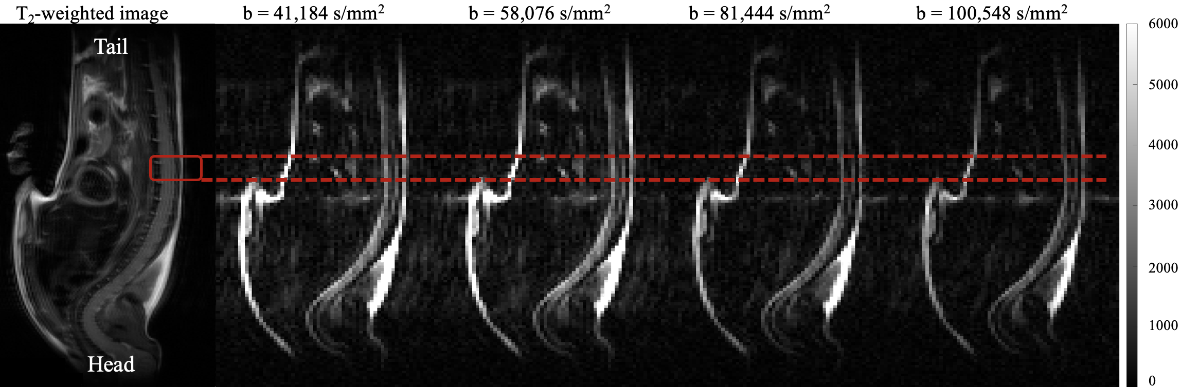
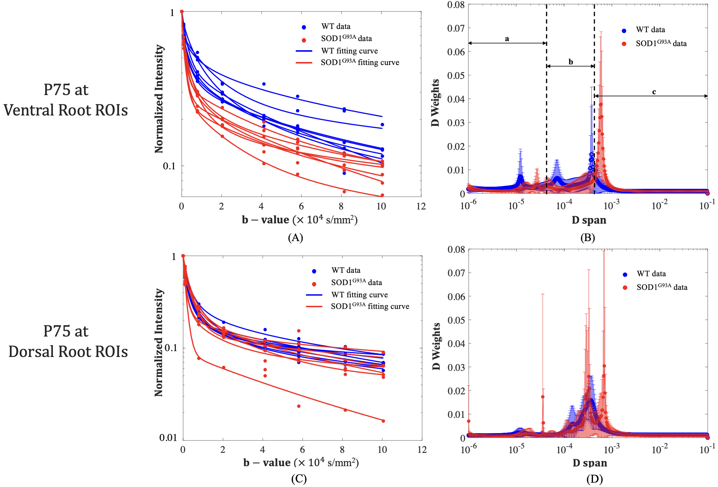
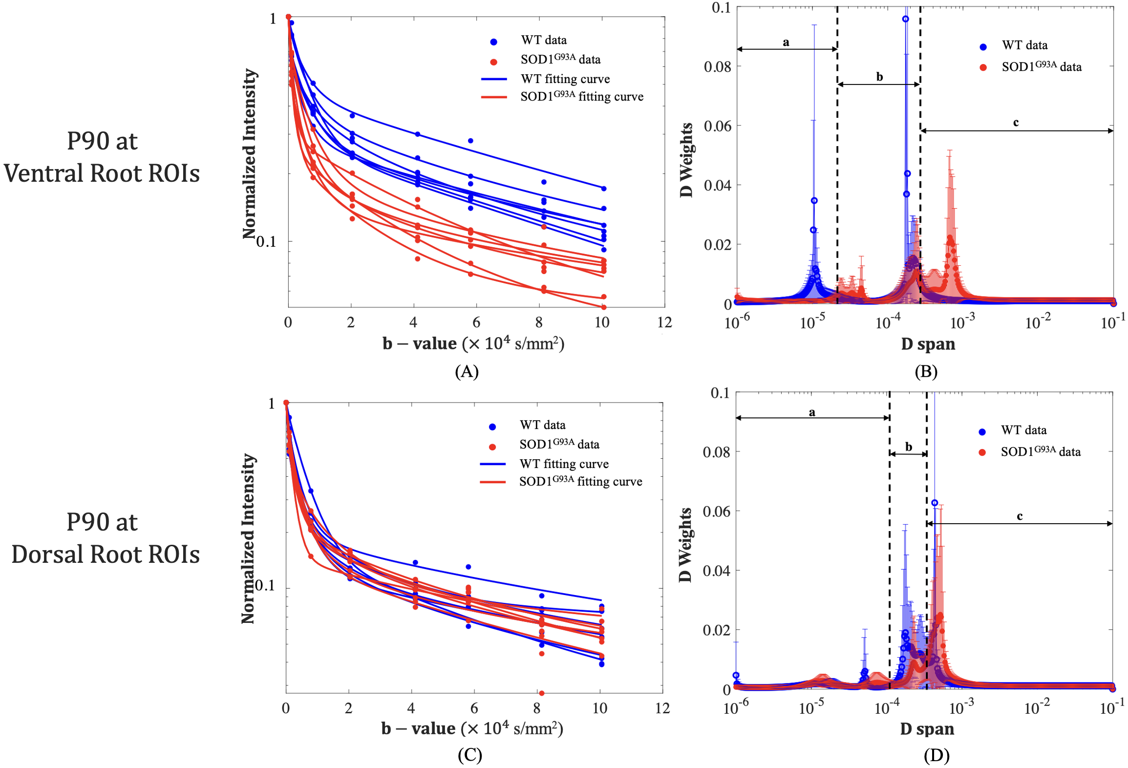
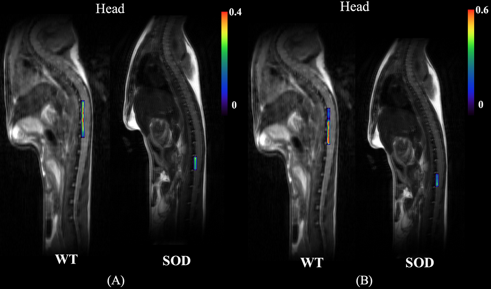
As the b-value increases, the signal-to-noise ratio (SNR) decreases. At the maximum b-value of 1×10⁵ s/mm², the SNR decreases to a value of 12 at the lumbar level. Notably, as shown in Fig. 1, ventral roots display stronger signal intensities than dorsal roots do at each b-value. Fig. 2A shows the NNLS fitting curves to the measured ventral ROIs. Wide type (WT) control (blue) exhibits higher SNRs for the ventral root when comparing to those of SOD1G93A mice (red) at P75. Fig. 2B displays the fitted D weights with error bars for the ventral ROIs. The weight distribution was identified as three distinct intervals characterized by peak grouping: region (a) [1×10⁻⁶, 4.34×10⁻⁵] mm²/s, region (b) [4.35×10⁻⁵, 4.34×10⁻⁴] mm²/s, and region (c) [4.35×10⁻⁴, 0.1] mm²/s. Significant differences were found in region (a) (𝑃= 0.001) and region (b) (𝑃= 0.048). For the dorsal ROIs (Fig. 2(C)), the signal intensities at each b-value for both groups are indistinguishable. The fitted D weights (Fig. 2(D) confirmed no identifiable intervals relating to peak grouping.At P90, the differences in signal intensities between WT and SOD1G93A mice in the ventral roots were more pronounced (shown in Fig. 3(A)). Furthermore, the weight distributions displayed a notable shift towards the high diffusion coefficient range, as shown in Fig. 3(B). Three distinct intervals were identified: region (a) [1×10⁻⁶, 2.11×10⁻⁵] mm²/s, region (b) [2.12×10⁻⁵, 2.99×10⁻⁴] mm²/s, and region (c) [3×10⁻⁴, 0.1] mm²/s. Significant differences were found in region (a) (𝑃= 3.84×10⁻⁵) and region (c) (𝑃= 0.002). The SDw maps of P75 and P90 are shown in Fig. 4 and Fig. 5 respectively.
Discussion
This study showed the suitability of a wide range of b-values (up to 1× 10⁵ s/mm²) for in vivo SOD1G93A mouse spinal cord applications. Notably, the post-processing results obtained using the L1-norm NNLS showed potential to detect microenvironmental changes in ventral roots of the spinal cord attributed to ALS in both pre-symptomatic stage (P75) and early symptomatic stage (P90). Despite challenges, in vivo animal research is critical to evaluating a new technique. To gauge effectiveness of the proposed ultra-high b-value dMRI technique, this study involves live SOD1G93A mice and their wild-type counterparts indispensably addressing gaps in previous study [5]. Consequently, this in vivo study holds substantial potential for advancing physiology research of ALS, as well as investigations related to drug delivery and response monitoring in this specific genetic mouse model.Acknowledgements
No acknowledgement found.References
[1] A. Chiò et al., "Global Epidemiology of Amyotrophic Lateral Sclerosis: A Systematic Review of the Published Literature," Neuroepidemiology, vol. 41, no. 2, pp. 118-130, 2013, doi: 10.1159/000351153.
[2] W. Robberecht and T. Philips, "The changing scene of amyotrophic lateral sclerosis," Nature Reviews Neuroscience, vol. 14, no. 4, pp. 248-264, 2013/04/01 2013, doi: 10.1038/nrn3430.
[3] B. R. Foerster, R. C. Welsh, and E. L. Feldman, "25 years of neuroimaging in amyotrophic lateral sclerosis," (in eng), Nat Rev Neurol, vol. 9, no. 9, pp. 513-24, Sep 2013, doi: 10.1038/nrneurol.2013.153.
[4] W. L. Jin Gao, Richard Magin, and D Accurate andRobust Detection of Alternations in Spinal Cord," in Proceedings of the 29th Annual Meeting of the ISMRM, virtual conference, 2021, pp. 3440.
[5] J. Gao et al., "Multicomponent diffusion analysis reveals microstructural alterations in spinal cord of a mouse model of amyotrophic lateral sclerosis ex vivo," PLoS One, vol. 15, no. 4, p. e0231598doi: 10.1371/journal.pone.0231598.
Figures