5110
Enabling 1-minute High-resolution Clinical Diffusion-weighted Imaging at 7 Tesla via Improved Non-local-PCA Denoising1Tiantan Neuroimaging Center of Excellence, Beijing Tiantan Hospital, Capital Medical University, Beijing, China, 2Wellcome Centre for Integrative Neuroimaging, FMRIB, Nuffield Department of Clinical Neurosciences, University of Oxford, Oxford, United Kingdom, 3Department of Radiology and Imaging Sciences, University of Utah, Salt Lake City, UT, United States, 4MR Research Collaboration Team, Siemens Healthineers, Beijing, China, 5Department of Neurology, Beijing Tiantan Hospital, Capital Medical University, Beijing, China, 6Center for Biomedical Imaging Research, Department of Biomedical Engineering, School of Medicine, Tsinghua University, Beijing, China, 7Center for Magnetic Resonance Research, Radiology, Medical School, University of Minnesota, Minneapolis, MN, United States
Synopsis
Keywords: Diffusion Acquisition, Diffusion/other diffusion imaging techniques
Motivation: High-resolution diffusion-weighted imaging (DWI) is crucial for diagnosing neurological pathologies, but traditionally requires long scan times due to low SNR, hindering its application in clinical settings.
Goal(s): To evaluate how our new non-local principal-component-analysis (PCA)-based denoising method can help achieve high-resolution DWI at 7 Tesla within a clinically viable timeframe.
Approach: We compared our method to two existing local-PCA-based approaches by collecting whole-brain DWI at 1.2-mm isotropic resolutions from a healthy volunteer and a patient with multiple sclerosis.
Results: Our non-local PCA method provided improved denoising performances, producing quality DWI where the lesion was identifiable even with 1-minute acquisition.
Impact: Demonstrated capable of enabling high-resolution DWI under 1-minute scan at 7 Tesla, our non-local PCA method is believed to promote the utility of DWI in clinical settings while having the potential to improve many other neuroimaging applications.
Introduction
Diffusion-weighted imaging (DWI) is essential in the clinical screening and diagnosis of neurological pathologies, including ischemic stroke. High-resolution DWI has been demonstrated valuable in identifying small ischemic infarcts(1,2). However, even at ultra-high fields with potential SNR gains, the intrinsic low SNR of high-resolution DWI necessitates multiple averages for acceptable image quality for diagnostics, resulting in clinically impractical scan durations.Previously, we proposed a new two-step non-local principal-component-analysis (PCA) denoising method(3) and demonstrated its effectiveness for improving SNR in high-resolution whole-brain diffusion tensor imaging at 7 Tesla (7T). In this study, we explore the potential of our method to improve fast high-resolution clinical DWI, in comparison to two existing local-PCA-based denoising approaches(4,5). Our results, obtained with 7T high-resolution DWI scans of both health and disease, indicate that our method enables 1-minute whole-brain clinical DWI scans at 1.2-mm isotropic resolutions.
Method
Human scans were performed on a 7T MR system (MAGNETOM Terra, Siemens Healthcare, Erlangen, Germany) equipped with a body gradient (200 T/m/s slew rate and 80 mT/m Gmax).We started by acquiring DWI data from one healthy volunteer using the commercial Nova 8-channel transmit 32-channel receive coil (operated in its circularly polarized mode for excitation). The multi-band diffusion sequence used in the Human-Connectome Project(6) was utilized with relevant parameters: resolution=1.2-mm isotropic, slice gap=0, slice number=70, TE/TR=51/5500 ms, in-plane acceleration=4, diffusion encoding with 3 orthogonal directions with b=1000 s/mm2, averages=12 (corresponding to 12 b=0 and 12 repetitions of 3 b=1000 s/mm2 volumes), and total scan time=4 min 57 sec.
Both magnitude and phase images saved in DICOM format were exported for complex-valued denoising.
Image processing and evaluation (Figure 1) were conducted as follows. Data were retrospectively extracted to mimic scans with one through six averages (corresponding to scan times ranging from 55 sec to 2 min 45 sec) and were subsequently denoised using our non-local denoising method(3).
In each case, apparent diffusion coefficient (ADC) maps were calculated.
Normalized root-mean-square-error (nRMSE) relative to the 12 averages (serving as the reference) was evaluated in the white-matter mask generated using the SynthSeg(7) toolbox, of both DWI and ADC domains.
Results were compared with those obtained using existing alternatives, including MPPCA (applied in the magnitude domain as originally proposed, using the implementation in MRTrix(8)) and NORDIC(5).
We then collected data from one multiple sclerosis (MS) patient using the same protocol as for the healthy volunteer and denoised the data using our non-local method.
The study was approved by the local Ethical Standards Committee and informed consents were obtained.
Results
Our non-local method outperformed both MPPCA and NORDIC (Figure 2) for scans under 1.5 minutes, improving image quality with reduced noise levels.Correspondingly, our non-local denoising method led to the lowest nRMSE (Figure 3) in both image and ADC domains, especially when reducing scan time to under 2 min (≤4 averages).
A close inspection further revealed that our denoising method effectively improved the image quality even with scan durations of under 1 minute and began to preserve finer brain structures when extending the scan time to ~ 1.5 minutes (Figure 4).
Encouragingly, our denoising method effectively enhanced image quality in the MS patient (Figure 5), visualizing the lesion even with a ~1-minute scan.
Discussion
We have demonstrated the utility of our non-local denoising method for rapid acquisition of high-resolution whole-brain clinical DWI at 7T in both health and disease.The results show that our method outperforms existing local-PCA approaches in improving SNR in DWI with even four images/volumes, thanks to its ability to promote low rankness by assembling similar non-local patches.
Part of our future work is to include more participants, study other diseases (e.g., ischemic stroke) and incorporate radiologists’ assessments.
We will also seek to optimize diffusion acquisition (e.g., by using FLEET(9) to speed up calibration and a new reconstruction(10) to enable slice acceleration without increasing calibration time).
Conclusion
Our improved non-local PCA denoising method can be used to enhance SNR for diffusion MRI with a few images, enabling ~1-minute high-resolution whole-brain DWI scans at ultra-high field with a potential for clinical diagnostics.Acknowledgements
XW was supported in part by USA NIH grants (NIBIB P41 EB027061 and U01 EB025144).References
1. Misquitta K, Daou M, Conklin J, Liao C, Setsompop K, Poublanc J, Shirzadi Z, MacIntosh BJ, Tomlinson G, Cohn M, Aviv RI, Silver FL, Mandell DM. Detecting Silent Acute Microinfarcts in Cerebral Small Vessel Disease Using Submillimeter Diffusion-Weighted Magnetic Resonance Imaging: Preliminary Results. Stroke 2022;53(7):e251-e252.
2. Hou Z, Jing J, Yan L, Zhang Z, Fu W, Liu J, Yu Y, Jiang L, Yang J, Wang Y, Miao Z, Lou X, Ma N. New Diffusion Abnormalities Following Endovascular Treatment for Intracranial Atherosclerosis. Radiology 2023;307(4):e221499.
3. Ye X, Ma X, Pan Z, Moller S, Auerbach E, Ugurbil K, Wu X, Guo H. Non-local low rank denoising method for complex-valued DWI. Proc. Intl. Soc. Mag. Reson. Med. 30 (2022). 2022. p 3041.
4. Veraart J, Novikov DS, Christiaens D, Ades-Aron B, Sijbers J, Fieremans E. Denoising of diffusion MRI using random matrix theory. Neuroimage 2016;142:394-406.
5. Moeller S, Pisharady PK, Ramanna S, Lenglet C, Wu X, Dowdle L, Yacoub E, Ugurbil K, Akcakaya M. NOise reduction with DIstribution Corrected (NORDIC) PCA in dMRI with complex-valued parameter-free locally low-rank processing. Neuroimage 2021;226:117539.
6. Vu AT, Auerbach E, Lenglet C, Moeller S, Sotiropoulos SN, Jbabdi S, Andersson J, Yacoub E, Ugurbil K. High resolution whole brain diffusion imaging at 7T for the Human Connectome Project. Neuroimage 2015;122:318-331.
7. Billot B, Greve DN, Puonti O, Thielscher A, Van Leemput K, Fischl B, Dalca AV, Iglesias JE, Adni. SynthSeg: Segmentation of brain MRI scans of any contrast and resolution without retraining. Med Image Anal 2023;86:102789.
8. Tournier JD, Smith R, Raffelt D, Tabbara R, Dhollander T, Pietsch M, Christiaens D, Jeurissen B, Yeh CH, Connelly A. MRtrix3: A fast, flexible and open software framework for medical image processing and visualisation. Neuroimage 2019;202:116137.
9. Polimeni JR, Bhat H, Witzel T, Benner T, Feiweier T, Inati SJ, Renvall V, Heberlein K, Wald LL. Reducing sensitivity losses due to respiration and motion in accelerated echo planar imaging by reordering the autocalibration data acquisition. Magn Reson Med 2016;75(2):665-679.
10. Pan Z, Ma X, Dai E, Auerbach EJ, Guo H, Ugurbil K, Wu X. Reconstruction for 7T high-resolution whole-brain diffusion MRI using two-stage N/2 ghost correction and L1-SPIRiT without single-band reference. Magn Reson Med 2023;89(5):1915-1930.
Figures
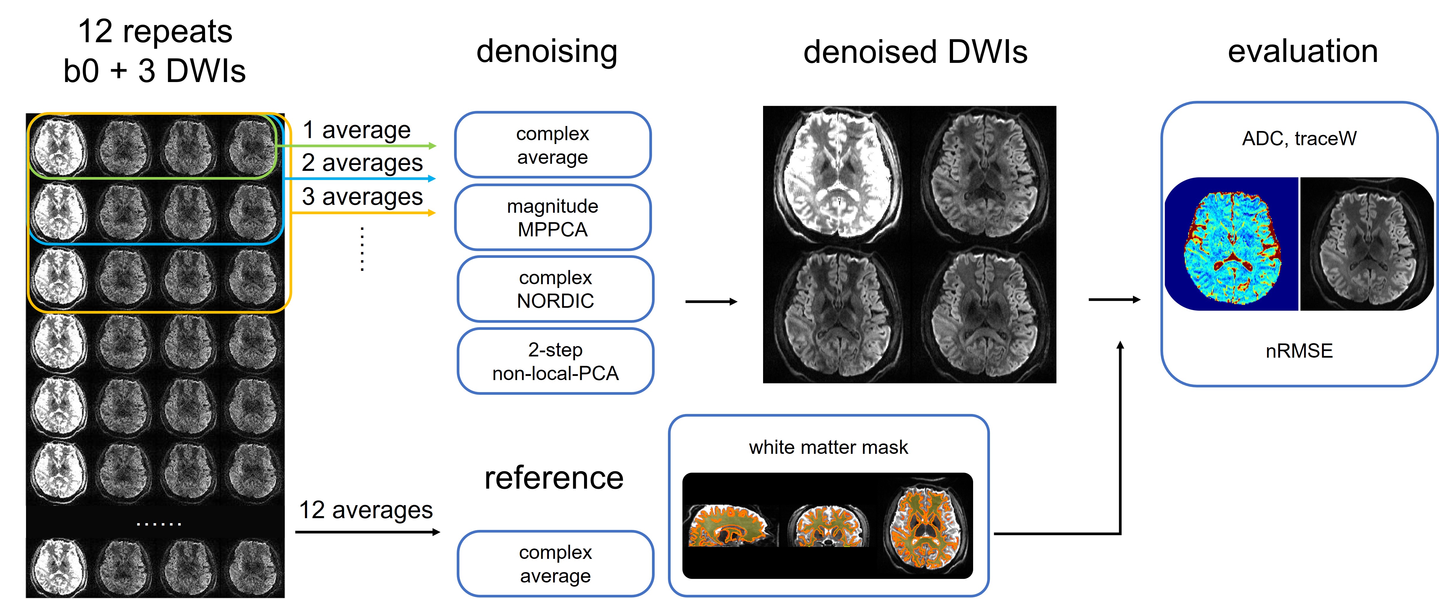
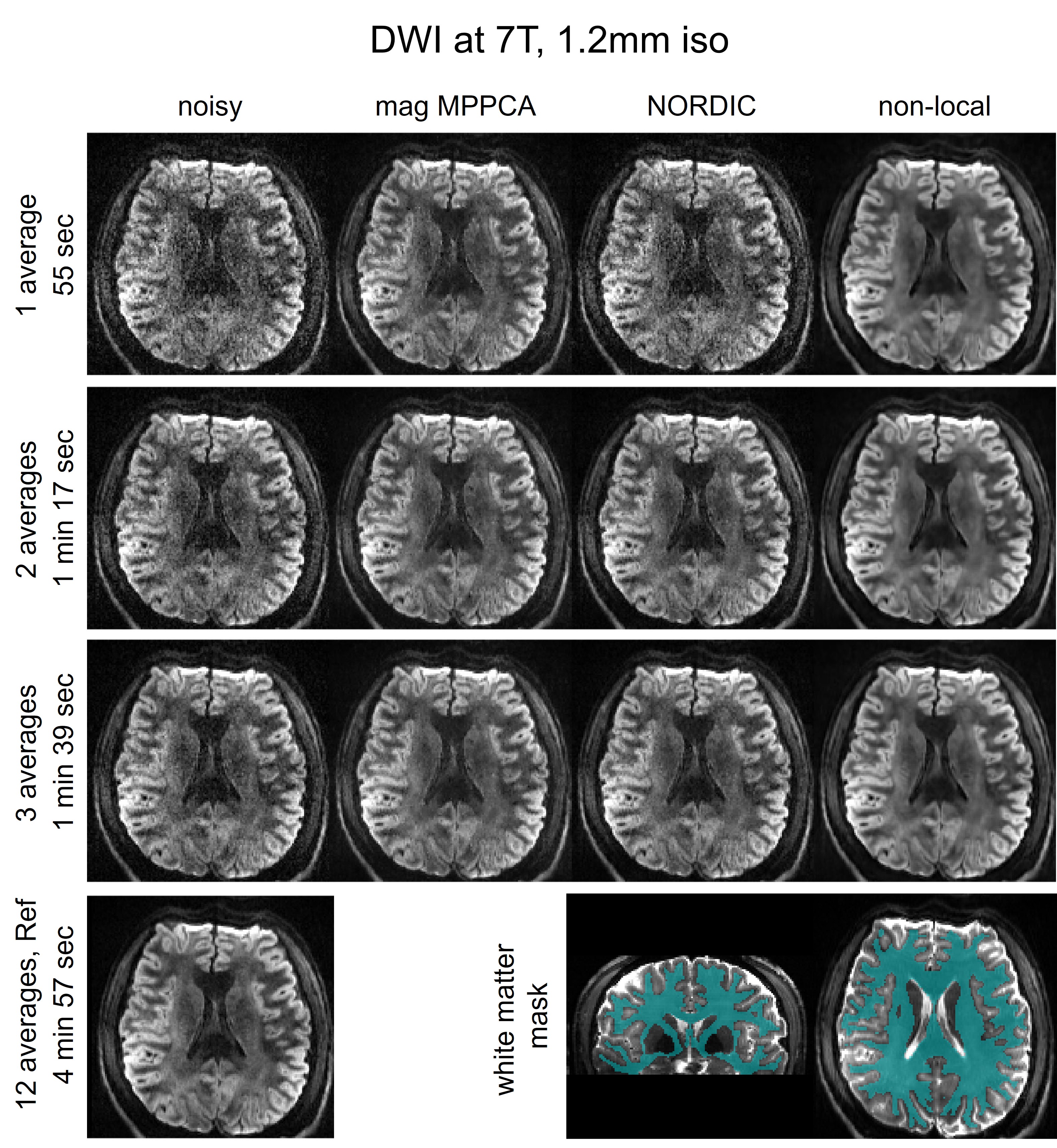
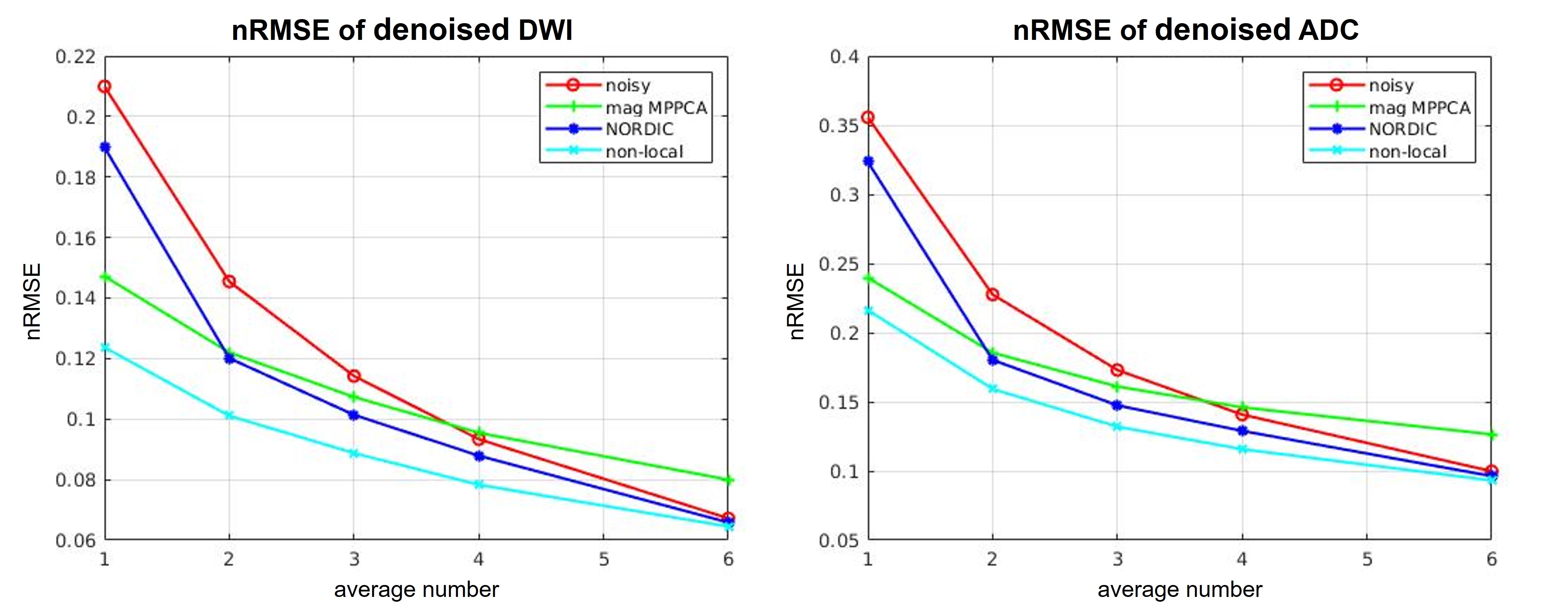
Figure 3. Comparing normalized root-mean-square-error (nRMSE) of noisy vs. denoised using MPPCA (applied in the magnitude domain), NORDIC and our non-local denoising method, in DWI and ADC domain. Results are shown for different data averages, corresponding to scans under 3 min. Note that our non-local denoising method outperformed both MPPCA and NORDIC, leading to the lowest nRMSE for both DWI and ADC, especially when pushing scan time under 2 min (≤4 averages).
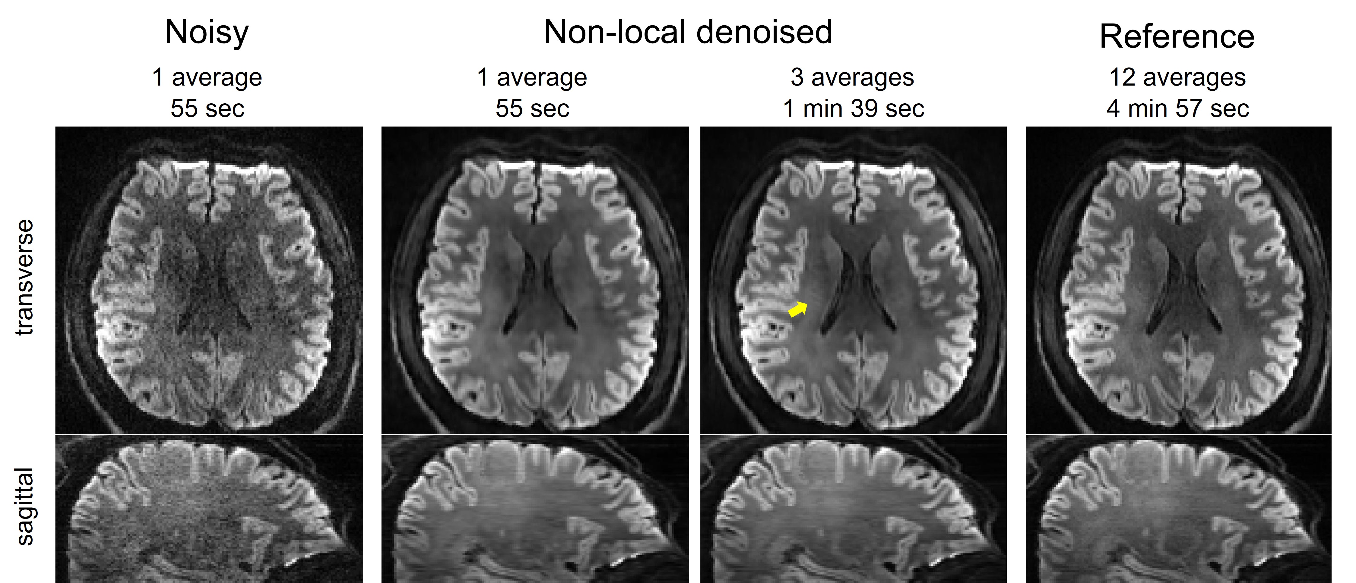
Figure 4. Demonstrating the utility of our non-local denoising method in health. Shown are trace-weighted DWIs in two representative views for unprocessed noisy images and denoised ones using our method, alongside the reference (i.e., 12 averages). Note that our denoising method effectively improved the image quality even with under 1-minute scan and started to preserve fine brain structures (e.g., indicated by the yellow arrow) when increasing the scan time to ~1.5 minutes.
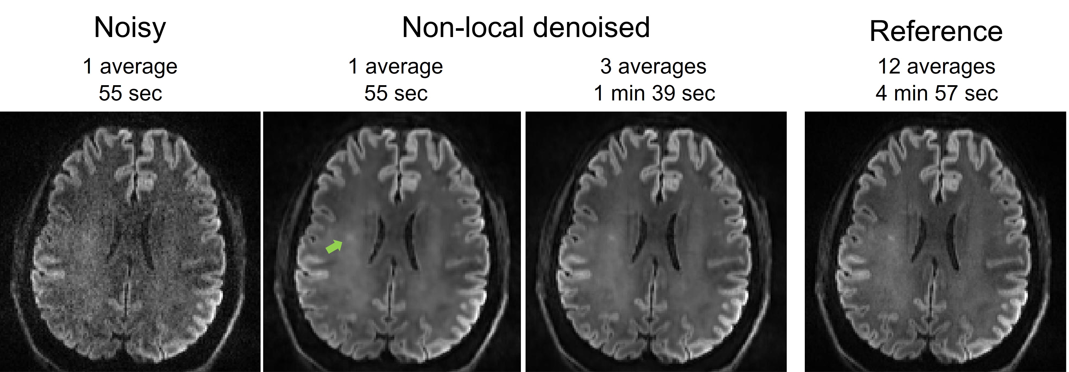
Figure 5. Demonstrating the utility of our non-local denoising method in disease. Shown are trace-weighted DWIs in a representative axial slice for unprocessed noisy images and denoised ones using our method, alongside the reference (i.e., 12 averages). Note that our denoising method effectively enhanced image quality, by visualizing the lesion (pointed by the green arrow) even with a ~1-minute scan and further improving lesion sharpness when increasing scan time to ~1.5 minutes.