5107
Improved visualization of high b-value prostate diffusion weighted imaging using LIPO-only multi-shot EPI with 2D navigator (LION-IRIS)1Radiology, Eastern Chiba Medical Center, Chiba, Japan, 2Philips Japan, Tokyo, Japan, 3Eastern Chiba Medical Center, Chiba, Japan, 4Philips Australia & New Zealand, North Ryde, Australia
Synopsis
Keywords: Diffusion Acquisition, Diffusion/other diffusion imaging techniques, fat suppresstion
Motivation: One of the drawbacks of IRIS is its low SNR compared with ss-DWI. High b-value DWI improves the ability of prostate cancer detection by increased cancer conspicuity. However, such high b value images may also suffer from decreased SNR.
Goal(s): We hypothesized that using only slice selection gradient reversal (SSGR, LIPO) technique without fat suppression pre-pulse (LIpo-ONly :LION) could improve the SNR of high b-value IRIS DWI.
Approach: SNR,CR and ADC values were calculated with ROIs placed in internal obturator muscle and prostate region in peripheral zone.
Results: LION IRIS-DWI could provide high quality distortion-free high b-value prostate DWI sufficient SNR.
Impact: The present findings suggest that LION IRISs-DWI sequence demonstrated improved image distortion and blurring compared to ss-DWI, with having sufficient SNR and CR.It may help to further assess the prostate cancer pathology.
Purpose
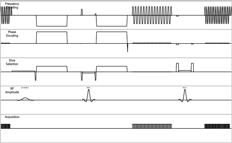
Prostate multiparametric MRI commonly consists of T2-weighted imaging (T2WI), diffusion-weighted imaging (DWI), and dynamic contrast-enhanced MRI (DCE-MRI). Multiparametric MRI, especially for DWI, is the most promising technique for investigating regarding the detection and localization of significant primary prostate cancer and local recurrence, assessment of tumor aggressiveness local staging. [1] Therefore, prostate DWI requires high lesion detectability and stable image quality with minimal artifacts. [2] DWI with single-shot echo-planar imaging (ss-DWI) is the most used in routine clinical practice due to its high signal-to-noise ratio (SNR), rapid acquisition, and insensitivity to motion. However, it suffers from geometry distortion and signal loss in the case of susceptibility differences. [3] Multi-shot EPI DWI (ms-DWI), on the other hand, can reduce distortion and image blurring because the effective echo spacing and echo train length are shorter. However, ms-DWI is sensitive to motion among each shot, causing phase variations that results in signal loss. Image reconstruction using the image-space sampling function (IRIS) [4] can minimize motion effects among shots by implementing two-dimensional (2D) navigator echo, it enables a reduction in image distortion and blurring effectively compared with conventional ss-DWI. One of the drawbacks of IRIS is its low SNR compared with ss-DWI.High b-value DWI improves the ability of prostate cancer detection by increased cancer conspicuity. However, such high b value images may also suffer from decreased SNR. [5] Recently, it has been introduced that using only slice selection gradient reversal (SSGR, LIPO) technique without use of fat suppression pre-pulse (LIpo-ONly: LION) DWI can improve the SNR while maintain the sufficient fat suppression. [6,7] We hypothesized that the use of LION could improve the SNR of high b-value IRIS DWI. In this study, we examined the feasibility of combining IRIS-DWI and LION (LION IRIS) (Fig.1) to achieve sufficient SNR without distortion in high b-value imaging.Methods
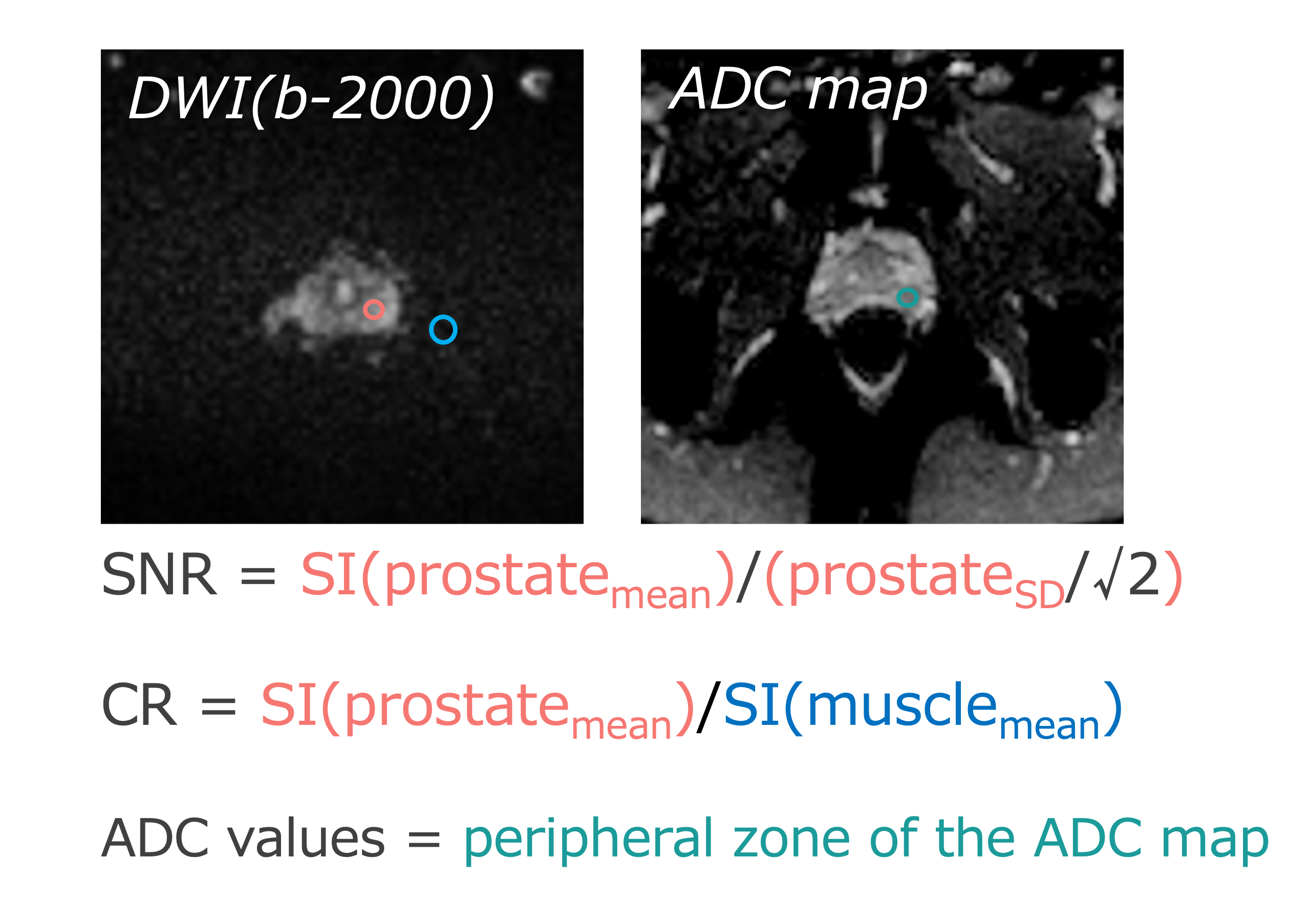
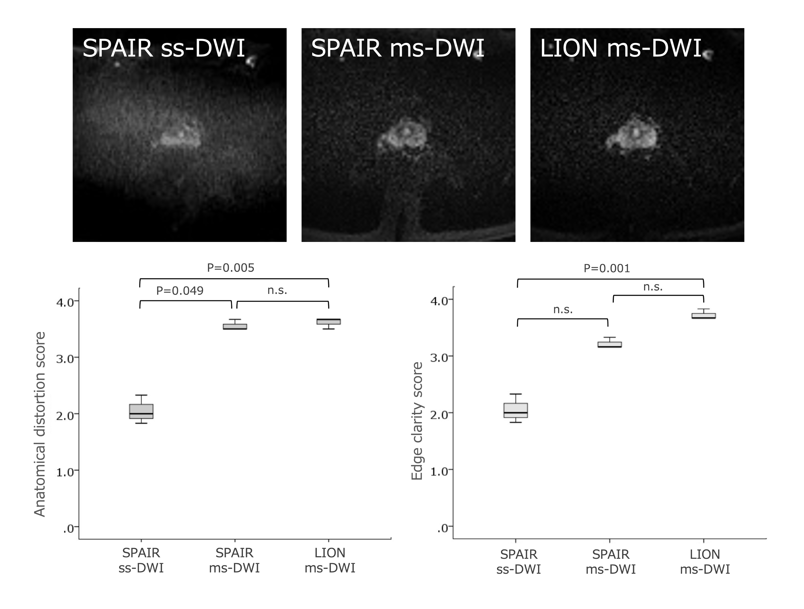
Volunteers were examined on 3.0T whole-body clinical systems (Ingenia CX, Philips Healthcare, Best, the Netherlands) with a 32ch flexible torso coil. The study was approved by the local IRB, and written informed consent was obtained from all subjects.A total of 6 volunteers underwent three DWI scans, including conventional SPAIR ss-DWI, SPAIR IRIS-DWI, and LION IRIS-DWI. All DW images in prostate were acquired with the transverse direction with the following parameters: 1) SPAIR ss-DWI: TR = 5500 ms, TE = 68 ms, SENSE factor = 2, NSA = 5, scan time = 4:49; 2) SPAIR ms-DWI (IRIS) : TR = 5500 ms, TE= 77 ms, SENSE factor = 2, NSA= 1, scan time = 4:59 ; 3) LION ms-DWI (IRIS): TR = 5500ms, TE = 80 ms, SENSE factor = 2, NSA = 1, scan time = 4:57 ; and the other parameters of these four protocols are identical: FOV = 220*220 mm2 , Matrix = 112*110 mm2, 20 slices, slice thickness = 3 mm, b-value = 2000 s/mm2 .For LION IRIS, LIPO technique was optimized to maximize the effect of fat suppression for large FOV, based on previous studies [6,7].SNR and contrast ratio (CR) were calculated with ROIs placed in internal obturator muscle and prostate region in peripheral zone. ADC values of prostate region in peripheral zone were measured from the ADC-map.(Fig.2) In qualitative analysis, we used two types of indices: anatomical distortion and edge clarity score of prostate for 6 volunteers. 3 radiological technologists/radiologists assessed qualitative indices in the DW images visually using a 4-point grading scale.Results and Discussion
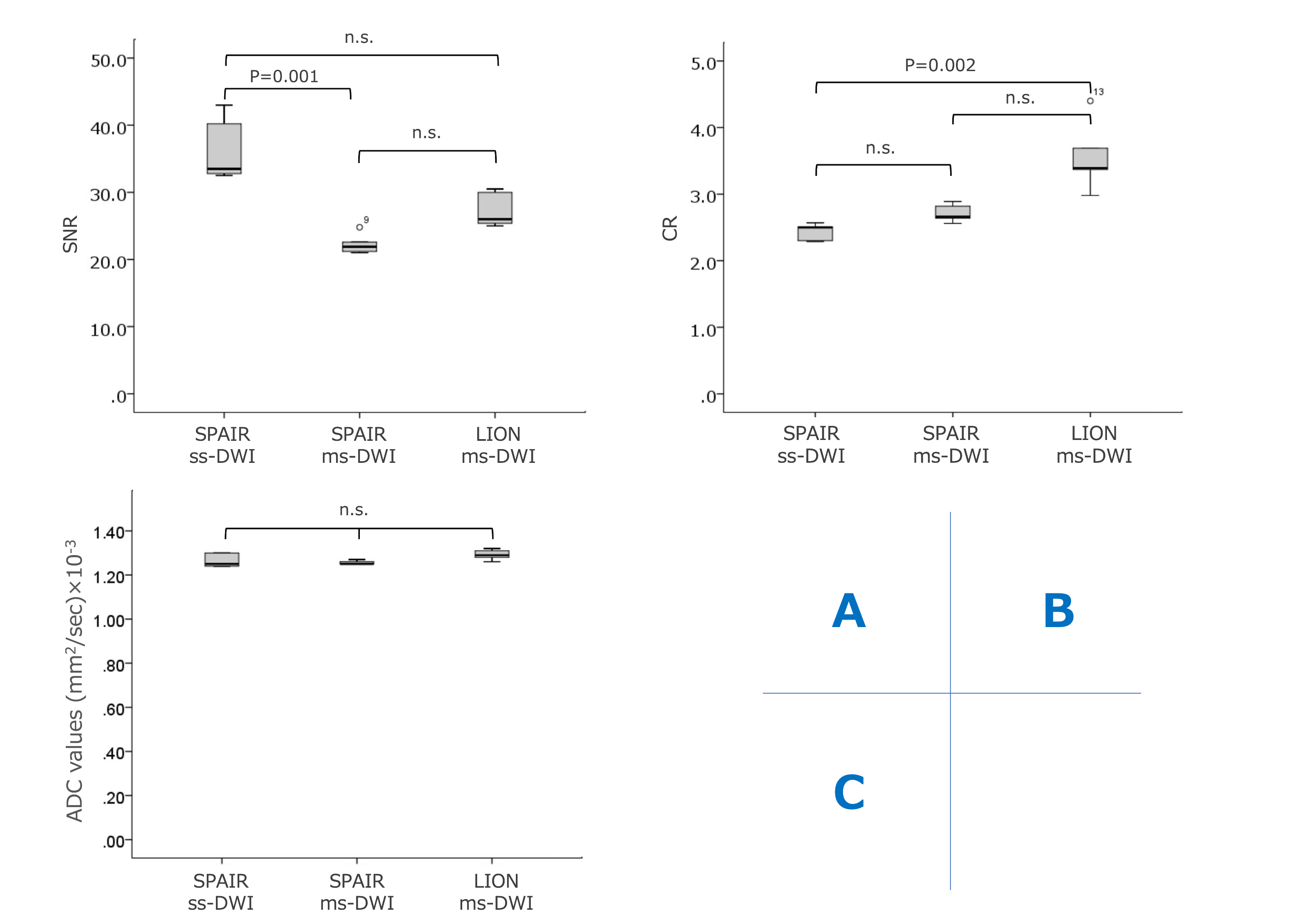
The SNR of LION IRIS-DWI was similar to that of SPAIR ss-DWI, significantly higher than that of SPAIR IRIS-DWI. LION IRIS-DWI showed significantly higher CR compared with SPAIR ss-DWI, there were no significant differences in CR between the SPAIR ss-DWI and SPAIR IRIS-DWI. There were no significant differences in ADC values among the SPAIR ss-DWI and SPAIR IRIS-DWI, LION IRIS-DWI.(Fig.3) In the qualitative analysis, anatomical distortion and prostate edge clarity were significantly higher in LION IRIS-DWI than in SPAIR ss-DWI.(Fig.4) The present findings suggest that LION IRISs-DWI sequence demonstrated improved image distortion and blurring compared to ss-DWI, with having sufficient SNR and CR. It may contribute to improve the diagnostic performance.Conclusion
LION IRIS-DWI could provide high quality distortion-free high b-value prostate DWI sufficient SNR. It may help to further assess the prostate cancer pathology.Acknowledgements
No acknowledgement found.References
1. Fütterer JJ. et al. Can clinically significant prostate cancer be detected with multiparametric magnetic resonance imaging? A systematic review of the literature. Eur Urol. 2015; 68: 1045-53.
2. Schieda N. et al. Quantitative prostate MRI. J Magn Reson Imaging. 2021; 53:1632–1645.
3.Le Bihan D. et. al. Artifacts and pitfalls in diffusion MRI. J Magn Reson Imaging. 2006; 24:478-488.
4. T Tamada. et al .Comparison of single‑shot EPI and multi‑shot EPI in prostate DWI at 3.0 T. Scientific Reports. 2022; 12:16070.
5. Kitajima K. et al. Clinical utility of apparent diffusion coefficient values obtained using high b value when diagnosing prostate cancer using 3 tesla MRI: comparison between ultra-high b value (2000 s/mm(2)) and standard high b value (1000 s/mm(2)). J Magn Reson Imaging. 2012;36:198-205.
6.Murayama D. et al. Reduced acquisition time of female pelvis diffusion weighted imaging (DWI) using LIPO-only (LION) DWI. ISMRM2023; 2960.
7. Sakai T. et al. Volume isotropic thin-slice high-quality brain DWI with no fat-suppression pre-pulse. ISMRM2023; 3970.
Figures