5106
Improved readout-segmented EPI using deep learning reconstruction1Siemens Healthineers AG, Erlangen, Germany
Synopsis
Keywords: Diffusion Reconstruction, Image Reconstruction, Diffusion
Motivation: We explore the potential of deep learning reconstruction (DLR) to overcome challenges for readout-segmented EPI (rs-EPI) , ultimately leading to more efficient and high-quality diffusion-weighted imaging (DWI).
Goal(s): We evaluate DLR's applicability for rs-EPI, aiming to improve image quality, reduce scan durations, and expand rs-EPI's clinical utility.
Approach: We adapted the successful DLR method used in single-shot EPI (ss-EPI) to rs-EPI, conducting experiments for head and prostate diffusion imaging.
Results: Our study demonstrates that DLR can improve image quality and reduce scan times in rs-EPI DWI, promising more efficient clinical imaging and potential applications in diverse diffusion imaging scenarios.
Impact: The successful implementation of DLR in readout-segmented EPI DWI promises accelerated, high-quality diagnostics, directly benefiting clinicians and patients. Furthermore, DLR's potential for diverse diffusion imaging applications opens new research horizons, enhancing the field of MR imaging.
INTRODUCTION
Diffusion-weighted imaging (DWI) stands as a cornerstone in modern medical imaging, playing a pivotal role in lesion detection and disease diagnosis. Traditionally, single-shot echo-planar imaging (ss-EPI) has been the go-to choice for DWI due to its rapid image acquisition. Nevertheless, ss-EPI is prone to susceptibility artifacts and T2*-related blurring, restricting its clinical applications. To address these limitations, readout-segmented echo-planar imaging (rs-EPI) with 2D navigation1 has emerged as a well-established technique, offering reduced distortion and T2*-related blurring in high-resolution DW images for various clinical applications. However, it's worth noting that rs-EPI entails longer scan times, primarily due to multiple excitations and navigator acquisitions. Recent advances in deep learning (DL) have presented a promising solution for image reconstruction, enhancing imaging resolution and signal-to-noise ratios (SNR) for high acceleration factors or specific regions. While deep learning reconstruction (DLR) has been successfully applied to ss-EPI diffusion and extended to multi-shot EPI diffusion2-5, it's noteworthy that, to the best of our knowledge, no prior report exists concerning k-space-based DLR in the context of rs-EPI. In this study, we set out to assess the feasibility of implementing rs-EPI with the same k-space-based DLR method used in ss-EPI diffusion and compare it to the conventional reconstruction technique.METHODS
We have adapted the k-space DLR method from ss-EPI for use in a research application based on the commercial RESOLVE rs-EPI sequence. This integration required adoptions to implement phase correction among segments and subsequent segment splicing, enabling compatibility with the network architecture designed for ss-EPI. All data collection was performed on a 3 Tesla scanner (MAGNETOM Vida, Siemens Healthineers AG, Erlangen, Germany). Experimental data were acquired from a healthy volunteer using both a product sequence and the newly developed research application. Imaging parameters for head imaging with a 20-channel head-neck coil were as follows: Field of View (FOV) = 220x220 mm², 56 slices with 2mm slice thickness and a 30% slice gap, utilizing a 4-scan trace diffusion mode, in-plane acceleration of 3, slice acceleration of 2, matrix size of 192x192, TE/TR = 57/5460 ms, echo spacing = 0.36 ms, and b-values of 0/1000 s/mm² with 1 average for both b-values, resulting in a total scan time of 3 minutes and 28 seconds. For prostate imaging, a 32-channel spine coil and an 18-channel body coil were employed with the following parameters: FOV = 200x200 mm², 22 slices with 3mm slice thickness and no slice gap. The diffusion mode was 4-scan trace, with in-plane acceleration of 2. The matrix size was 118x118, TE/TR = 53/4200 ms, echo spacing = 0.36 ms. Two b-values were used: 50 s/mm² with 1 average and 800 s/mm² with 2 averages in the product sequence and 1 average in the research application. In the product sequence, this resulted in a scan time of 4 minutes and 26 seconds, while in the research application, the scan time was reduced to 3 minutes and 2 seconds.RESULTS
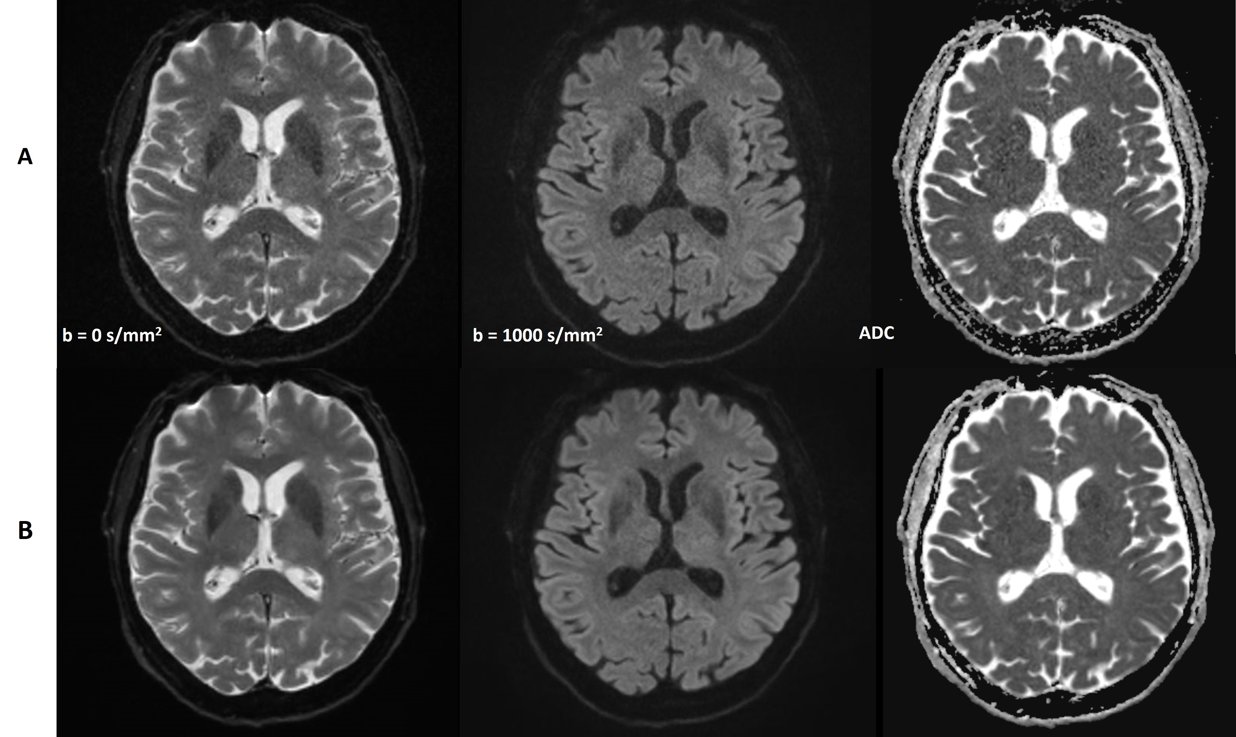
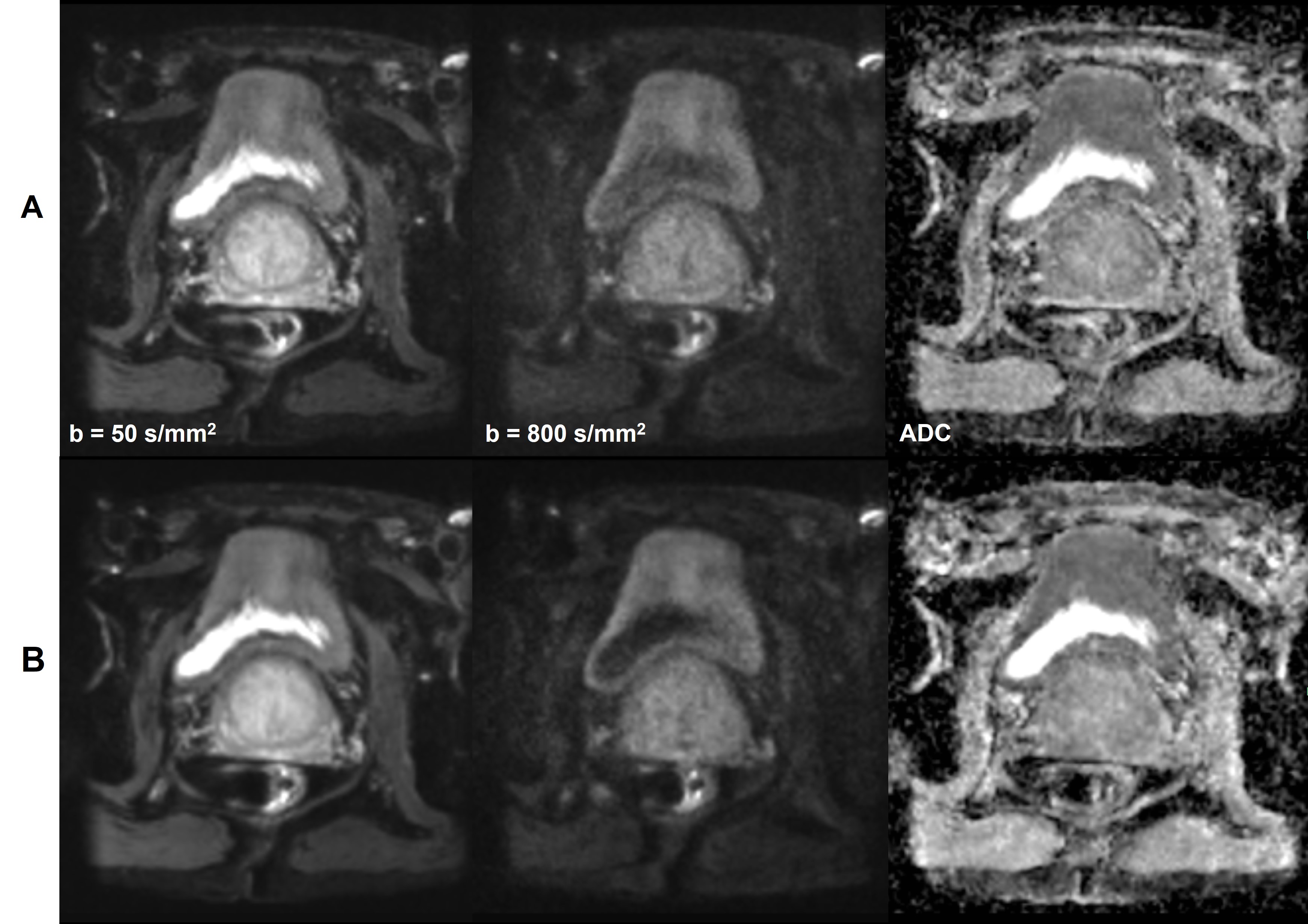
In Figs 1A and 1B, we present rs-EPI images of the head with conventional reconstruction and DLR. As anticipated, the application of DLR noticeably enhances image quality in such a highly accelerated case. Moving to Figs 2A and 2B, these images illustrate the results for rs-EPI prostate images with conventional reconstruction and DLR. Fig 2 highlights the difference between conventional reconstruction with 2 averages and DLR with only 1 average. As evident in Fig 2, the implementation of DLR significantly reduces scan times in rs-EPI while preserving a high standard of image quality. These findings underline the potential of DLR to improve image quality and efficiency in rs-EPI, offering valuable insights for its clinical applications.DISCUSSION AND CONCLUSION
Our study showcases the feasibility of applying DLR to rs-EPI DWI. This approach not only improves image quality but also reduces the need for multiple averages in rs-EPI compared to conventional reconstruction. Furthermore, it opens doors for enhanced image resolution and acceleration factors without compromising diagnostic image quality. The potential applications of DLR in various regions hold promise for further advancements in clinical imaging, offering a pathway for optimized, efficient, and high-quality DWI MRI solutions.Acknowledgements
No acknowledgement found.References
1. Porter DA, Heidemann RM. High resolution diffusion-weighted imaging using readout-segmented echo-planar imaging, parallel imaging and a two-dimensional navigator based reacquisition. Magn Reson Med. 2009; 62(2):468-475.
2. Bae SH, Hwang J, Hong SS, Lee EJ, Jeong J, Benkert T, Sung J, Arberet S. Clinical feasibility of accelerated diffusion weighted imaging of the abdomen with deep learning reconstruction: Comparison with conventional diffusion weighted imaging. Eur. J. Radiol. 2022; 154:110428.
3. Lee EJ, Chang YW, Sung JK, Thomas B. Feasibility of deep learning k-space-to-image reconstruction for diffusion weighted imaging in patients with breast cancers: Focus on image quality and reduced scan time. Eur. J. Radiol. 2022; 157, 110608.
4. Hu Y, Xu Y, Tian Q, Chen F, Shi X, Moran CJ, et al.. RUN-UP: Accelerated multishot diffusion-weighted MRI reconstruction using an unrolled network with U-Net as priors. Magn Reson Med. 2021; 85:709-20.
5. Aggarwal HK, Mani MP, Jacob M. MoDL-MUSSELS: model-based deep learning for multishot Sensitivity-Encoded diffusion MRI. IEEE Trans. Med. Imaging. 2019, 39(4):1268-1277.
Figures