5103
High b-value in vivo Whole-brain Diffusion MRI at 7T with a High-performance Gradient System1Department of Biomedical Magnetic Resonance, Institute for Physics, Otto von Guericke University Magdeburg, Magdeburg, Germany, 2German Center for Neurodegenerative Diseases (DZNE), Magdeburg, Germany, 3Center for Behavioral Brain Sciences (CBBS), Magdeburg, Germany, 4Leibniz Institute for Neurobiology (LIN), Magdeburg, Germany
Synopsis
Keywords: Diffusion Acquisition, Challenges
Motivation: Probing brain microstructure with high-b-value brain diffusion imaging was mainly performed at 3T. At higher field strength, the long echo times prohibited a significant SNR boost over 3T. At 7T, a high-performance gradient allows much shorter echo times and harvesting of the field-related signal gain.
Goal(s): Determine SNR and explore feasibility of high b-value DWI.
Approach: The SNR was measured in high b-value DWI at whole-body and high-performance 7T scanners.
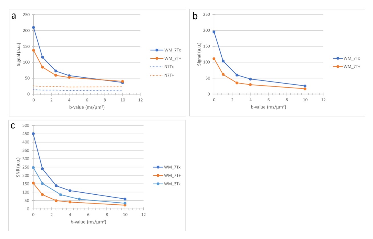
Results: The SNR improvement on the high-performance 7T is 2.76 ±0.12 and 1.73 ±0.05 compared to the whole-body 7T and high-performance Connectome 3T respectively, allowing even higher spatial resolution and higher b-value imaging.
Impact: The shortcoming of 7T for high b-value diffusion MRI can be overcome when leveraging a high-performance gradient system, effectively reducing scan time 7.6-fold compared to a whole-body 7T scanner. This enables the next generation of diffusion imaging.
Background
7T MRI enabled higher SNR in many applications, but diffusion MRI remains challenging1,2. The reason is a reduced apparent transversal relaxation time (T2) and increased geometric distortion compared to 3T. Due to the required diffusion encoding time in whole-body 7T systems, the reasonably applicable b-value is lower than at 3T. b-values larger than 3000 s/mm2 with a spatial resolution higher than 2 mm isotropic have rarely been applied. A high-performance gradient system can significantly reduce encoding and readout duration to compensate for the accelerated signal decay. We compare high b-value DWI at 7T with whole-body and high-performance gradient scanners and on a 3T Connectome scanner.Methods
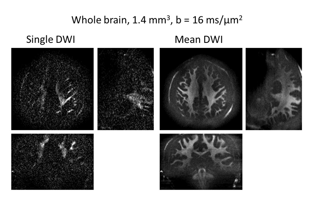
Experiments are performed on two 7T human scanners (Siemens Healthineers, Erlangen, Germany). The first scanner (Magnetom 7T Plus) is equipped with a conventional whole-body gradient system (70 mT/m, 200 T/m/s) and an 8 TX/32 RX head coil, referred to as 7T Plus, while the Magnetom Terra.X Impulse Edition has a high-performance gradient (200 mT/m, 900 T/m/s) and an 8 TX/64 RX head coil, referred to as Terra.X. Diffusion MRI data of one healthy subject (scanned after giving written, informed consent, study approved by local institutional review board) were acquired with the vendor-supplied spin-echo EPI sequence in the latest software versions (VE12U for 7T Plus and XA70 for Terra.X). The first imaging protocol was adapted from the MGH Adult Diffusion Data protocol3 (1.5 mm3, iPAT 3×1, whole-brain coverage, maximum b-value 10 ms/μm2) with the changes of iPAT 3×2, b-value (1, 2.5, 4, 10 ms/μm2) and three orthogonal diffusion directions for each b-value. The TR was 8000 ms for both scanners, and the minimum TE is 110 ms for 7T Plus and 59 ms for Terra.X. The data was corrected by FSL topup and eddy4. An SNR comparison with 3T Connectome data (300 mT/m, 200 T/m/s) was also performed. The second experiment was performed on the Terra.X with a b-value of 16 ms/μm2, 1.4 mm3, iPAT 3×2, 128 diffusion directions, TR/TE 9000/67 ms; the total scan time was 21 minutes.Results
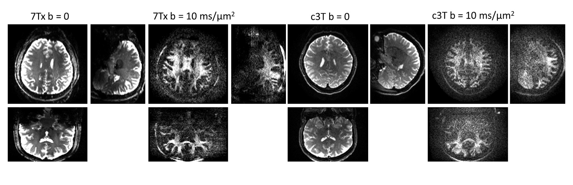
Figure 1 shows the minimum TE for different b-values on both 7T systems. On the 7T Plus, TE increases roughly linearly with b-value. On the Terra.X, TE remains unchanged from b = 0 to b = 4 ms/μm2, and only then increases linearly to the b-value of 16 ms/μm2. The TE-slope for Terra.X is only 20% of the 7T Plus. In Figure 2, the b = 0 images and DW images from both 7T scanners are shown. The white matter structure is barely detectable on the 7T Plus for b > 2.5 ms/μm2, while the white matter remains clear up to b = 10 ms/μm2 on the Terra.X. In Figure 3, the DW images at b = 10 ms/μm2 are compared between Terra.X and the 3T Connectome. The 7T images show a visually higher SNR and lower eddy-current related Nyquist ghost. Figure 4 displays the SNR evaluation of all data. The noise floor of the 7T Plus data is 1.95 ± 0.06 times higher than Terra.X (Fig. 4a). The noise floor-subtracted SNR (Fig. 4b) is 2.76 ± 0.12 times higher for the Terra.X compared to the 7T Plus. The SNR of the 7T Plus is 0.63 ± 0.05 times the SNR of the Connectome 3T and Terra.X shows 1.73 ± 0.11 time the SNR of the 3T Connectome (Fig. 4c). The achievable high quality of the Terra.X for high resolution, high b-value diffusion imaging is shown in Figure 5 (whole brain DWI with 1.4 mm3 resolution and b = 16 ms/μm2).Conclusions
7T in combination with a high-performance gradient system allows to realize the envisioned field strength related SNR gain also for high b-value DWI. The SNR is 2.76 times that of a whole-body 7T scanner and 1.73 times that of the 3T Connectome scanner. Imaging parameters were kept as similar as possible in this study. The high-performance gradient system enables further increase of the readout bandwidth allowing further reduction of TE, SNR gain, and higher resolution single-shot EPI.Acknowledgements
Data collection and sharing for the Connectome 3T data was provided by the Human Connectome Project (HCP). HCP funding was provided by the National Institute of Dental and Craniofacial Research (NIDCR), the National Institute of Mental Health (NIMH), and the National Institute of Neurological Disorders and Stroke (NINDS).References
- Pohmann R, Speck O, Scheffler K. Signal-to-noise ratio and MR tissue parameters in human brain imaging at 3, 7, and 9.4 tesla using current receive coil arrays. Magn. Reson. Med. 2016; 75(2), 801-809.
- Ladd M, Bachert P, Meyerspeer M, et al. Prog Nucl Magn Reson Spectrosc. 2018; Dec:109:1-50.
- MGH Adult Diffusion Data Acquisition Details. https://www.humanconnectome.org/study/hcp-young-adult/document/mgh-adult-diffusion-data-acquisition-details.
- Andersson J, Sotiropoulos S. An integrated approach to correction for off-resonance effects and subject movement in diffusion MR imaging. NeuroImage. 2015; 125, 1063-1078.
Figures
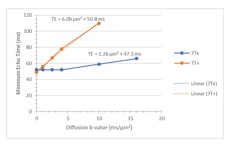
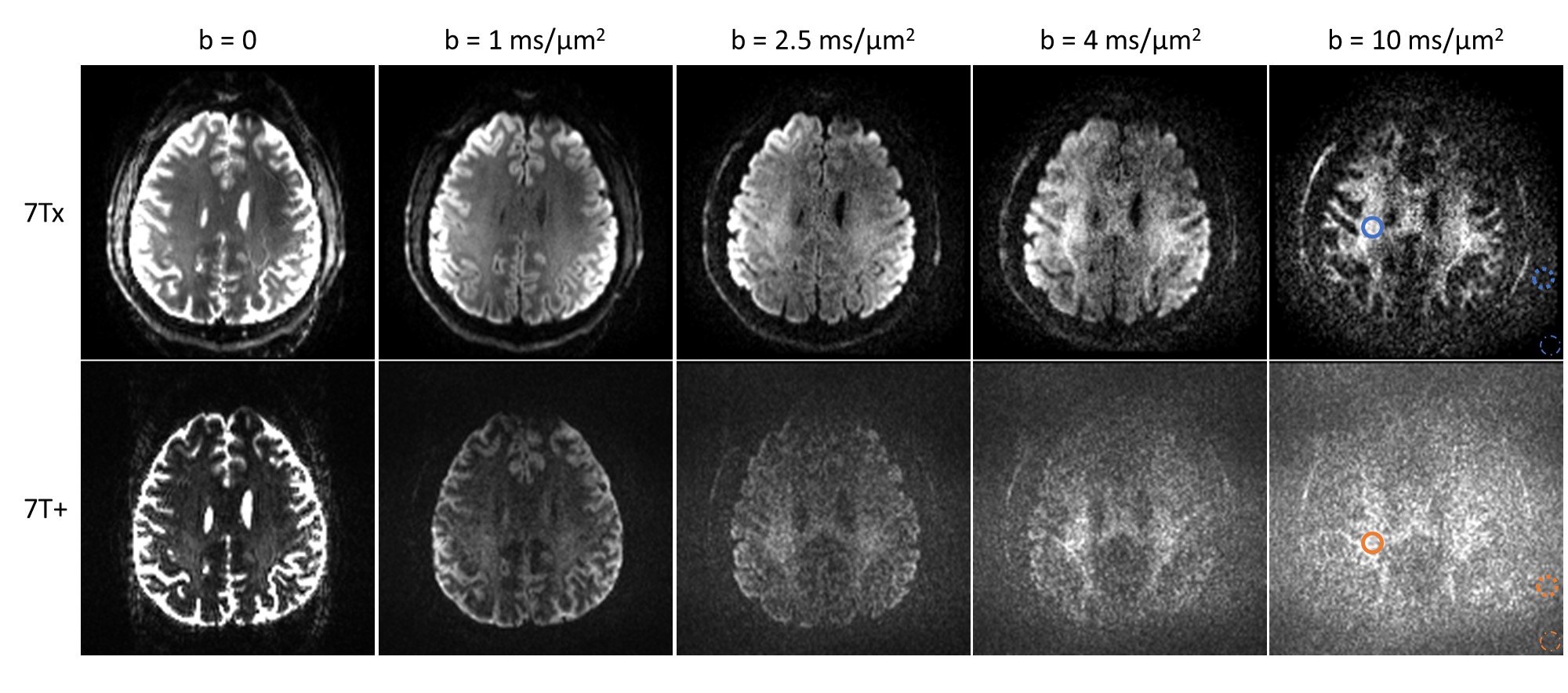
Fig. 2 High b-value imaging comparison on the 7T Plus and Terra.X scanners. The imaging protocol was adapted from the 3T Connectome high b-value protocol (bmax = 10 ms/μm2, 1.5 mm3). The Terra.X system enables considerably shorter TEs and provides sufficient white matter signal even at b-values of up to 10 ms/µm2. In contrast, white matter signal of 7T plus from b > 2.5 ms/μm2 is barely detectable. 7T+: 7T Plus. 7Tx: Terra.X.