5102
Studying topographic organization of the brain with directional derivatives of connectivity1University of Eastern Finland, Kuopio, Finland, 2University of Helsinki, Helsinki, Finland, 3Aalto University, Espoo, Finland
Synopsis
Keywords: Tractography, Tractography & Fibre Modelling
Motivation: The currently available tools to describe changes in structural connectivity preclude an in-depth topography study of the brain’s white matter. We sought to quantify the degree of change in structural connectivity through the mathematical notion of directional derivatives.
Goal(s): To define and compute the directional derivatives of tractograms.
Approach: We defined a measure of connectivity at a point in the brain, that is expressed on the brain's surface. By using numerical differentiation, we computed directional derivatives of connectivity.
Results: Our directional derivative method allows a comprehensive topographic study of the brain and highlights potential topographic patterns in structural organization.
Impact: Directional derivatives quantify connectivity changes, enabling systematic topographic study of the brain. The computational neuroimaging tool developed may aid in neurosurgical planning, precise brain stimulation, and biomarkers identification. This versatility may contribute significantly to both neuroscience research and clinical practice.
INTRODUCTION
A key feature of the brain is its topographic organization1. This principle also applies to structural connectivity2,3,4, as evidenced by tractography-based studies of sensory and motor systems5,6.Gradients have been used to describe spatial changes in connectivity patterns7,8. While informative, since these methods ultimately rely on dimensionality reduction, they may not fully capture the complexity of structural organization.
Here we propose a novel approach, based on the mathematical definition of directional derivatives, that quantifies changes in structural connectivity along many directions, thus providing a valuable tool for the topographical study of the brain.
METHODS
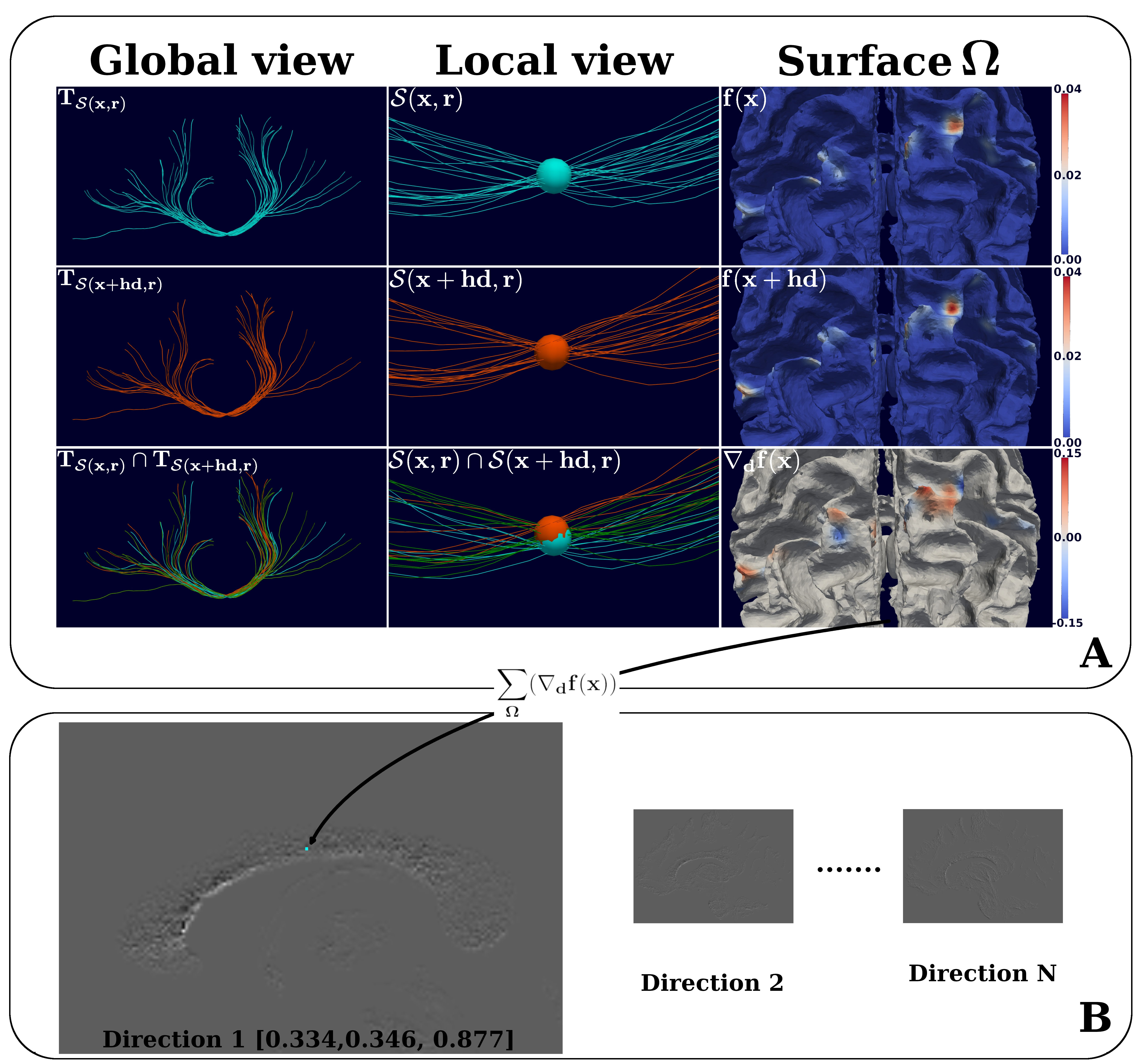
We propose to employ the mathematical notion of directional derivatives to study the change in structural connections derived from dMRI-based tractography, as illustrated in Figure 1.Let the set, $$$T=\{s_1,s_2,\dots\}$$$ be the input tractogram. Here $$$s_i$$$ is a streamline, which is a sequence of points $$$(p_i^1,p_i^2,\dots)$$$. Let $$$ \overline{p_i^j} $$$ represent the segment between the points $$$ p_i^j $$$ and $$$ p_i^{j+1} $$$, of length $$$ |\overline{p_i^j}| $$$. Let $$$ T_{\mathcal{S}(x,r)} $$$ be the subset of streamlines in $$$ T $$$ which cross $$$ \mathcal{S}(x,r) $$$, that is the sphere with radius, $$$ r $$$, centered at $$$ x \in \mathbb{R}^3 $$$. Each streamline's portion contributes to the structural connectivity with a weight $$ w_{x,i} = \sum_{p_i^j \in s_i \cap \mathcal{S}(x,r)} G(p_i^j)|\overline{p_i^j}|, $$ where $$ G(\mathbf{p_j}) = \frac{1}{(2\pi)^{3/2} \sigma^3} \exp\left(-\frac{\|\mathbf{p_j} - x\|^2}{2\sigma^2}\right) $$ is the 3D Gaussian function with variance $$$ \sigma^2 $$$ that is used to scale each segment with respect to its distance to the point, $$$x$$$. In our implementation, we set $$$ \sigma = r$$$. Using the above definitions, and $$$\Omega $$$ as the brain surface mesh, we can define and compute the structural connectivity of a given point $$$x \in \mathbb{R}^3$$$ with the following function, $$$f(x): \mathbb{R}^3 \rightarrow \mathbb{R}^\Omega$$$, as follows: $$ f(x)=\sum_{s_i \in T_{\mathcal{S}(x,r)}}\sum_{p_i^j \in s_i \cap \mathcal{S}(x,r)} w_{x,i}\mathcal{C}(s_i) $$ where $$$ \mathcal{C}(s_i) : T \rightarrow \mathbb{R}^\Omega $$$, denotes the connectivity of a streamline with the brain surface. In our work, $$$ \mathcal{C}(s_i) $$$ is precomputed as a scalar field over the surface mesh $$$ \Omega $$$ that assigns values to the vertices around the intersection points of $$$ s_i $$$ and $$$ \Omega $$$ such that $$$\sum_{\Omega} \mathcal{C}(s_i) = 1 $$$. Finally, the directional derivative along direction $$$\mathbf{d} \in \mathbb{R}^{3} $$$ can be estimated using a finite difference, e.g. the forward difference as shown below: $$ \nabla_\mathbf{d} f(x)= \frac{f(x+h\mathbf{d}) - f(x)}{h}. $$ $$$ \sum_{\Omega}(\nabla_\mathbf{d} f(x)) $$$ is the scalar stored in the output image at position $$$x$$$.
RESULTS
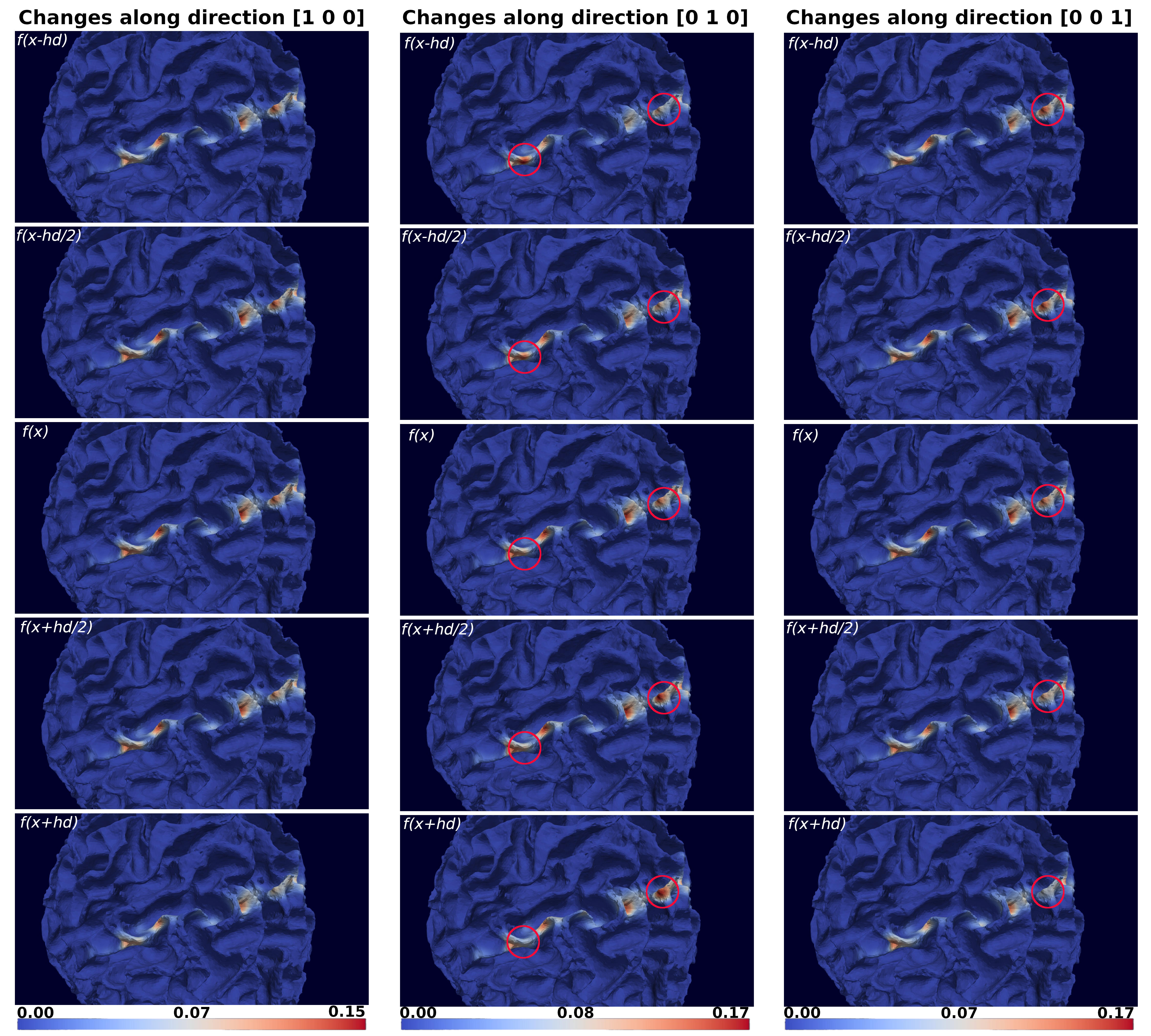
To demonstrate our method, we ran experiments using the Human Connectome Project9 subject 100307. The whole brain tractogram contained 100 million streamlines, generated using parallel transport tractography10 with anatomically constraint tractography11, ensuring consistent streamline termination on $$$\Omega$$$ obtained with FreeSurfer12.Figure 2 shows the gradual change in connectivity and how $$$f(x)$$$ informs about the topographic organization along different directions.
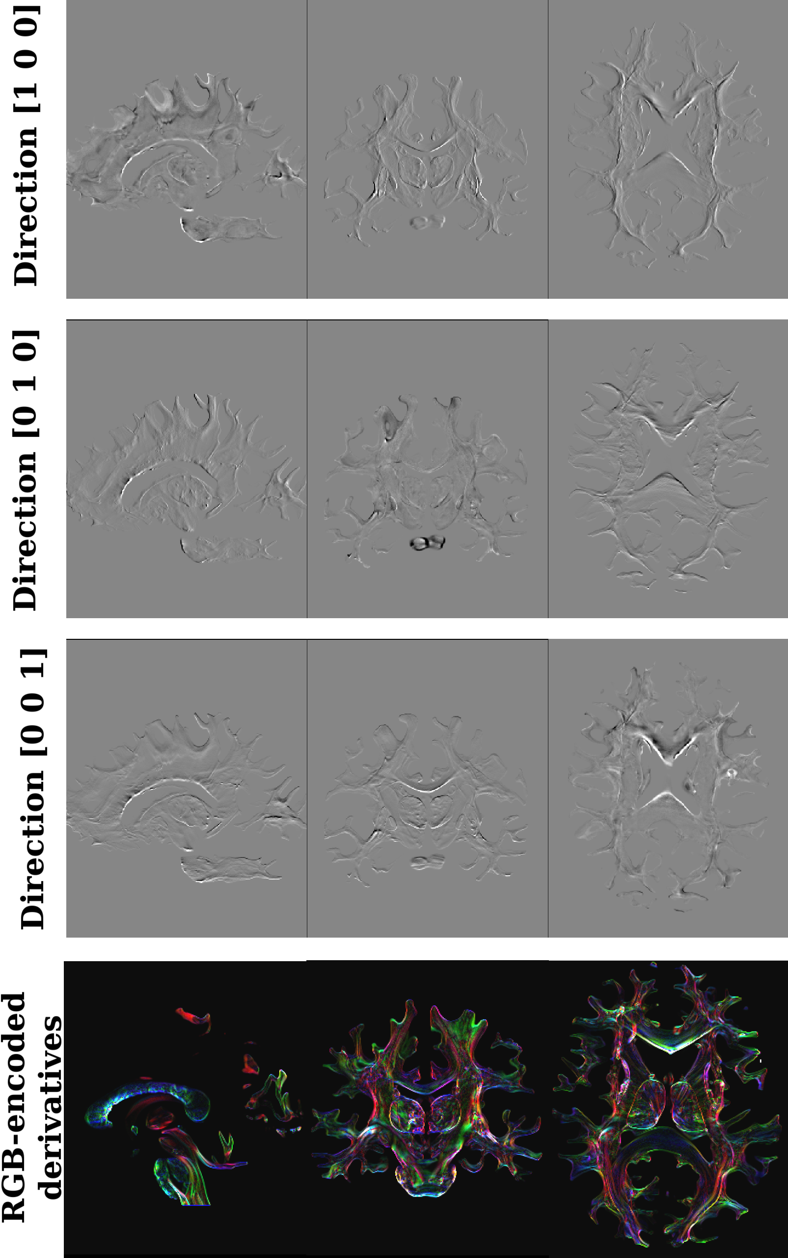
Figure 3 shows the directional derivative images evaluated along three directions, as well as a combined RGB image, highlighting the rich novel contrast that our technique offers.
DISCUSSION
Figure 2 well exemplifies why directional derivatives are powerful tools to study the topographic organization of the brain. As we gradually move in the brain along a direction, we observe a shift in the connectivity pattern. Thus small displacements lead to measurable changes in connectivity, and indeed $$$f(x)$$$ captures information about the topographic organization of structural connectivity. Since the change in connectivity is different in different directions, it is evident that capturing information about the change along many directions is key to understanding the complex organization of brain’s wiring.In Figure 3 we combine information about the derivative along three orthogonal directions. The novel contrast of the RGB image highlights boundaries of brain regions sharing similar connectivity patterns. This new information could advance our understanding of the brain's topographic organization and enhance the segmentation of small nuclei.
Despite the computational challenge of obtaining the derivatives on large tractograms for small voxel sizes, our implementation can evaluate derivatives on feasible computational times, e.g. few minutes for a few millions of streamlines for 1 mm voxel size along a single direction.
CONCLUSIONS
In this work, we proposed a novel computational tool to study the brain. While our current focus was only on structural connectivity, the concept of directional derivatives of connectivity can be extended to functional measurements such as MRI and EEG. Apart from advancing our knowledge regarding the brain’s organization, we believe our method will be relevant in all contexts where precision and personalization matter, such as surgical planning, or brain stimulation studies.Acknowledgements
This project was funded through grant agreements #348631 and #353798, Research Council of Finland.References
1. Wedeen, Van J., et al. "The geometric structure of the brain fiber pathways." Science 335.6076 (2012): 1628-1634.
2. Thivierge, Jean-Philippe, and Gary F. Marcus. "The topographic brain: from neural connectivity to cognition." Trends in neurosciences 30.6 (2007): 251-259.
3. Jbabdi, Saad, Stamatios N. Sotiropoulos, and Timothy E. Behrens. "The topographic connectome." Current opinion in neurobiology 23.2 (2013): 207-215.
4. Patel, Gaurav H., David M. Kaplan, and Lawrence H. Snyder. "Topographic organization in the brain: searching for general principles." Trends in cognitive sciences 18.7 (2014): 351-363.
5. Aydogan, Dogu Baran, and Yonggang Shi. "Tracking and validation techniques for topographically organized tractography." NeuroImage 181 (2018): 64-84.
6. Lee, Dong-Hoon, Do-Wan Lee, and Bong-Soo Han. "Topographic organization of motor fibre tracts in the human brain: findings in multiple locations using magnetic resonance diffusion tensor tractography." European radiology 26 (2016): 1751-1759.
7. Margulies, Daniel S., et al. "Situating the default-mode network along a principal gradient of macroscale cortical organization." Proceedings of the National Academy of Sciences 113.44 (2016): 12574-12579.
8. Bajada, Claude J., et al. "A tutorial and tool for exploring feature similarity gradients with MRI data." NeuroImage 221 (2020): 117140.
9. Van Essen, David C., et al. "The Human Connectome Project: a data acquisition perspective." Neuroimage 62.4 (2012): 2222-2231.
10. Aydogan, Dogu Baran, and Yonggang Shi. "Parallel transport tractography." IEEE transactions on medical imaging 40.2 (2020): 635-647.
11. Smith, Robert E., et al. "Anatomically-constrained tractography: improved diffusion MRI streamlines tractography through effective use of anatomical information." Neuroimage 62.3 (2012): 1924-1938.
12. Fischl, Bruce. "FreeSurfer." Neuroimage 62.2 (2012): 774-781.
Figures