5098
Spherical tensor diffusion encoding with spectral specificity and isotropy1Institute for Biomedical Engineering, ETH Zurich and University of Zurich, Zurich, Switzerland
Synopsis
Keywords: Diffusion Acquisition, New Signal Preparation Schemes, Spherical b-tensor encoding, oscillating gradient diffusion encoding
Motivation: Lack of spectral selectivity in spherical tensor diffusion encoding impairs the specificity of the measurement.
Goal(s): The goal of this work was to design diffusion gradient waveforms yielding a spherical b-tensor with frequency-specific and isotropic spectral projections.
Approach: Gradient waveform designs derived from oscillating gradient diffusion methodology were conceived by shifting and overlapping cosine-modulated trapezoidal oscillations along the three coordinate axes to achieve orthonormality of the b-tensor.
Results: The proposed gradient waveforms achieved frequency-specific and isotropic diffusion encoding, permitting frequency-dependent diffusion measurements in the in vivo human brain in the same manner as is conventionally performed using multidirectional linear tensor diffusion encoding.
Impact: The proposed diffusion gradient waveforms for spectrally specific and isotropic spherical tensor encoding offer selective frequency measurements that can isolate the signatures of time-dependent diffusion, thereby enabling the study of diffusion dispersion in the framework of tensor-valued encoding.
Introduction
In the presence of time-dependent diffusion, tensor-valued diffusion encoding measurements depend on the spectral content of the employed diffusion-sensitizing gradient waveforms. However, such gradient waveforms are typically optimized for diffusion encoding efficiency1-3 and disregard spectral properties, leading to directionally mismatched spectral encoding (i.e., spectral anisotropy)4 and, consequently, diminished specificity of the measurement. Directionally consistent spectral encoding in spherical tensor encoding (STE) acquisitions can counteract this shortcoming, but such experiments remain frequency-unspecific.By contrast, frequency specificity can be attained using pulsed (PG) and oscillating gradient (OG) linear tensor encoding (LTE), enabling characterization of microstructural motional restrictions and the dependence of diffusivity on frequency (i.e., diffusion dispersion).5 The goal of this work was to extend these capabilities to STE by designing diffusion-sensitizing gradient waveforms achieving spectrally isotropic and spherical b-tensors using both PG and OG for selectivity of zero and non-zero frequencies.
Methods
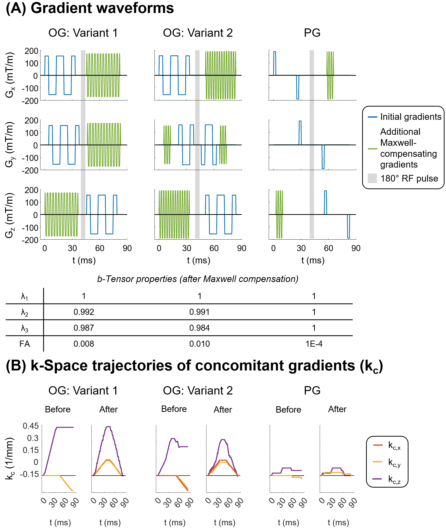
Gradient waveform designOrthogonality of q-space trajectories along two axes using OG can be achieved by using a quarter-wave shift between the simultaneous oscillations on said axes6 or by avoiding temporal overlap of the gradients on the two axes. Combining these criteria for mutually orthonormal diffusion encoding along three axes (i.e., a spherical b-tensor), two variants of OG STE using trapezoidal oscillations were devised: (1) two channels are offset from one another by a quarter wave and the third is non-overlapping with the first two; (2) one channel achieves quarter-wave shifts with respect to the other two, which do not overlap with one another. PG STE was achieved using non-overlapping bipolar gradient pulses along all axes.7 The three STE gradient waveforms are shown in blue in Figure 1A.
To additionally mitigate concomitant gradients (i.e., Maxwell compensation), rapidly oscillating gradient pulses (between 300-400 Hz) were added to each shape along the necessary axes with optimal placement in time to retain orthogonality. These gradients are shown in green in Figure 1A, along with b-tensor properties of each STE design after compensation, indicating near-perfect sphericity. The resulting concomitant gradient terms8 with and without the additional rapid oscillations are shown in Figure 1B for each case.
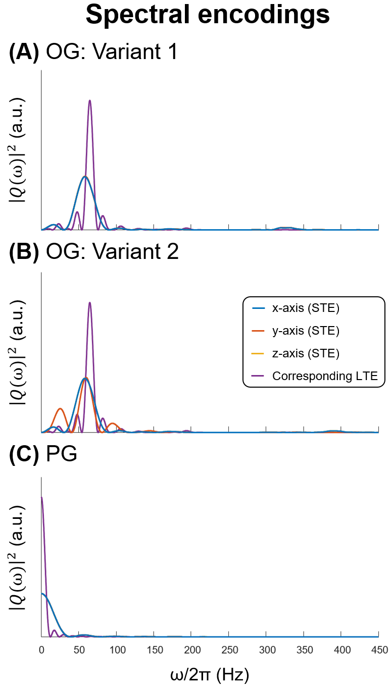
Figure 2 illustrates isotropic spectral encoding along the principal axes for each STE shape. Because one channel of OG STE variant 2 is split by the 180° RF pulse, its spectral specificity is worse than that of the other two channels. As such, variant 1 was used for experimentation.
Data acquisition
Scanning was performed on one healthy volunteer using a Philips 3T Achieva system (Philips Healthcare, Best, the Netherlands) equipped with a high-performance gradient insert (Gmax = 200 mT/m).9 PG and OG STE (variant 1) and PG and OG LTE DTI sequences were performed at b = 1000 s/mm2. LTE sequences (spectra also shown in Figure 2) used the same diffusion encoding duration as STE: PG had the longest possible diffusion time and OG had the closest possible centroid frequency to that of OG STE. Other scan parameters were 20 slices, 2×2×3 mm3 spatial resolution, 2 mm interslice gap, TR/TE = 6000/94 ms, 4 b = 0 acquisitions and 15 diffusion directions (LTE) / repetitions of unrotated diffusion encoding (STE), and 2 averages each.
Reconstructed images were denoised10 and unringed.11 Diffusion tensors were fitted to the LTE data from which mean diffusivity (MD) was computed, whereas MD was directly computed from the STE data using the average tensor-trace–weighted image.
Results
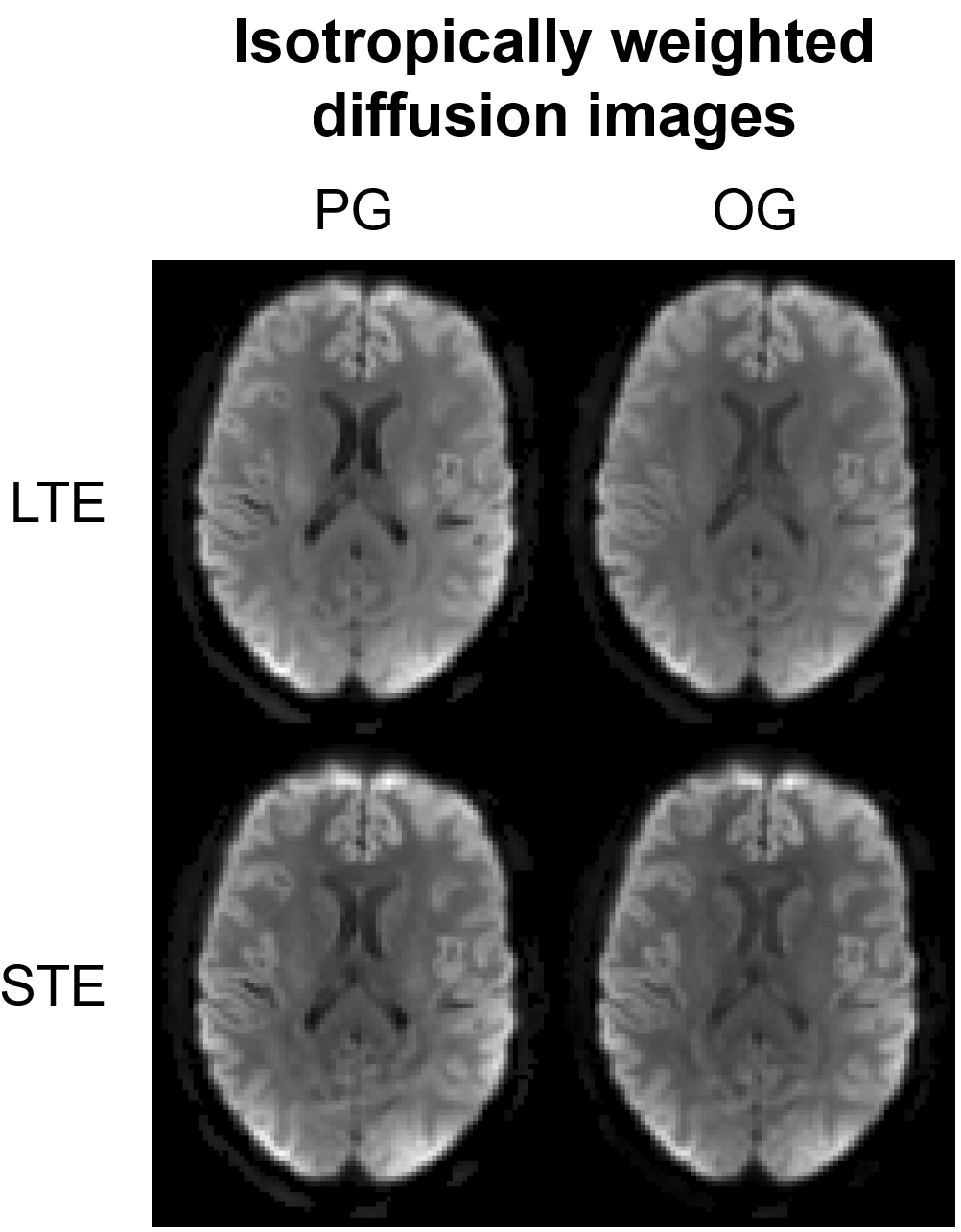
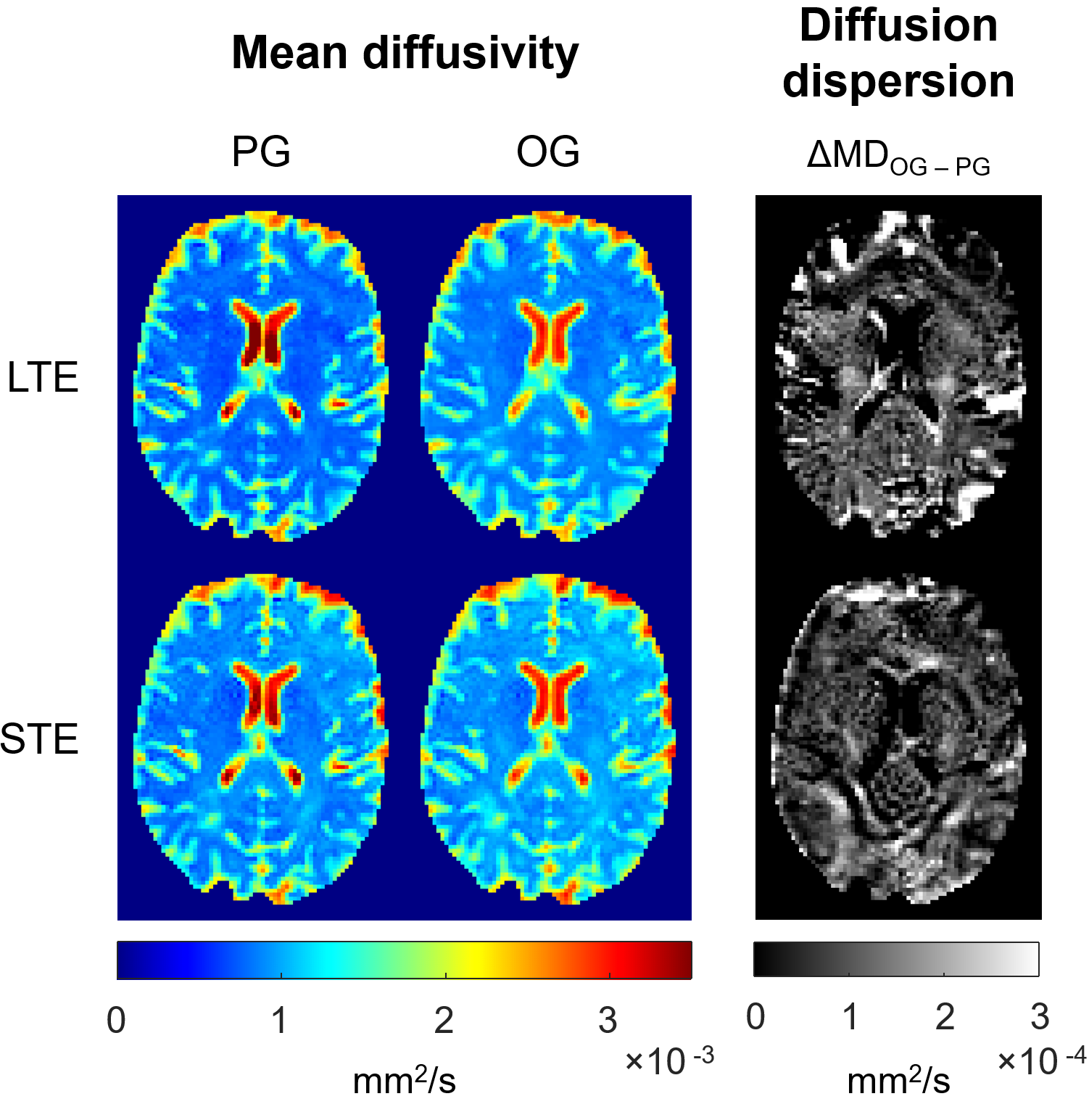
Figure 3 shows average diffusion-weighted images (LTE: powder-averaged; STE: repetition-wise averaged) for a representative slice for the four sequences.Figure 4 shows maps of MD as well as ΔMD (MDOG–MDPG) using LTE and STE.
Discussion and Conclusion
Spectrally isotropic and selective diffusion encodings of PG and OG STE (Figure 2) permit a level of frequency specificity previously limited to linear5 and planar6 b-tensors. DW image contrast for STE resembles that of powder-averaged LTE (Figure 3), devoid of the anisotropy characteristically revealed by individual directional LTE images. Moreover, PG images exhibit better white matter contrast than OG images for both LTE and STE.Positive MD dispersion is seen for both STE and LTE (Figure 4), and ΔMD is greater for LTE because of the superior zero-frequency selectivity brought about by the longer effective diffusion time in PG LTE than in PG STE. Moreover, STE diffusivities are elevated with respect to those of LTE because of the weakened impact of kurtosis induced by orientation dispersion in STE experiments.12
The STE schemes developed here offer isotropic congruity to typical frequency-selective multidirectional LTE experiments and could be used in broader contexts, for instance, to disentangle contributions from orientation dispersion and compartmental heterogeneity in frequency-dependent kurtosis measurements.
Acknowledgements
No acknowledgement found.References
1.Topgaard D. Isotropic diffusion weighting in PGSE NMR: Numerical optimization of the q-MAS PGSE sequence. Microporous Mesoporous Mater. 2013;178:60-63.
2. Sjölund J, Szczepankiewicz F, Nilsson M, Topgaard D, Westin C-F, Knutsson H. Constrained optimization of gradient waveforms for generalized diffusion encoding. J Magn Reson. 2015;261:157-168.
3. Szczepankiewicz F, Westin C-F, Nilsson M. Gradient waveform design for tensor-valued encoding in diffusion MRI. J Neurosci Methods. 2021;348:109007.
4. Lundell H, Nilsson M, Dyrby TB, et al. Multidimensional diffusion MRI with spectrally modulated gradients reveals unprecedented microstructural detail. Sci Rep. 2019;9(1):9026.
5. Does MD, Parsons EC, Gore JC. Oscillating gradient measurements of water diffusion in normal and globally ischemic rat brain. Magn Reson Med. 2003;49(2):206-215.
6. Lundell H, Sønderby CK, Dyrby TB. Diffusion weighted imaging with circularly polarized oscillating gradients. Magn Reson Med. 2015;73(3):1171-1176.
7. Mori S, van Zijl PCM. Diffusion weighting by the trace of the diffusion tensor within a single scan. Magn Reson Med. 1995;33(1):41-52.
8. Szczepankiewicz F, Westin C-F, Nilsson M. Maxwell-compensated design of asymmetric gradient waveforms for tensor-valued diffusion encoding. Magn Reson Med. 2019;82(4):1424-1437.
9. Weiger M, Overweg J, Rösler MB, et al. A high-performance gradient insert for rapid and short-T2 imaging at full duty cycle. Magn Reson Med. 2018;79(6):3256-3266.
10. Veraart J, Novikov DS, Christiaens D, Ades-Aron B, Sijbers J, Fieremans E. Denoising of diffusion MRI using random matrix theory. Neuroimage. 2016;142:384-396.
11. Kellner E, Dhital B, Kiselev VG, Reisert M. Gibbs-ringing artifact removal based on local subvoxel-shifts. Magn Reson Med. 2016;76(5):1574-1581.
12. Westin C-F, Knutsson H, Pasternak O, et al. Q-space trajectory imaging for multidimensional diffusion MRI of the human brain. Neuroimage. 2016;135:345-362.
Figures