5096
Joint estimation of compartment-specific T2 relaxation and tumor microstructure using multi-echo-time IMPULSED MRI1Vanderbilt University Medical Center, Nashville, TN, United States, 2Vanderbilt University Medical Center, nashville, TN, United States
Synopsis
Keywords: Diffusion Modeling, Diffusion/other diffusion imaging techniques, cancer
Motivation: The heterogeneity of T2 in tumors and its influences on estimates of tissue microstructure using diffusion MRI are poorly understood.
Goal(s): Assessing how T2 heterogeneity biases IMPULSED-derived metrics of tumor microstructure and evaluating the potential of estimating multi-compartmental T2 and microstructural parameters simultaneously.
Approach: This study quantifies the impact of T2 relaxation on IMPULSED-derived microstructural parameters using simulations and in vivo animal MRI in five tumor models, including brain, breast, prostate, melanoma, and colon cancer.
Results: TE has a negligible impact on IMPULSED-derived cell sizes, and the TE-dependence of IMPULSED-derived intracellular volume fractions can be used to estimate the compartmental T2 values.
Impact: Findings in this study contribute to the ongoing development and refinement of practical, non-invasive MRI techniques for characterizing tissue microstructure.
Introduction
Cell size and density variations in tissues reflect pathology but are typically invasive to measure, limiting knowledge of their spatial distributions and repeatability. The IMPULSED (imaging microstructural parameters using limited spectrally edited diffusion) method [1] has been shown to be accurate for measuring cell size and density non-invasively in a series of studies in silico, in vitro, and in vivo [2, 3]. More recently, the IMPULSED method has been adapted for imaging human subjects with breast cancer [3, 4], prostate cancer [5], brain cancer [6, 7] and other diseases in the liver [8]. The IMPULSED method assumes transverse relaxation rates are homogeneous, consistent with other contemporary multi-compartment diffusion MRI models [9, 10]. This simplification enables IMPULSED to be acquired with only one echo time (TE), resulting in a total scan time e.g., ~5-7 minutes for clinical studies of the breast. However, it remains questionable if such an approximation is valid. This study quantifies the influence of T2 relaxation heterogeneity on results obtained by the IMPULSED model. It assesses the impact of T2 on IMPULSED-derived microstructural parameters using computer simulations and in vivo animal MRI. A new T2+IMPULSED method was developed which allows simultaneous estimates of multi-compartmental T2 and microstructural parameters. The study compares IMPULSED and T2+IMPULSED and uses voxel-wise model selection to determine the need to consider T2 heterogeneity in IMPULSED MRI in cancer.Methods
Theory: dMRI signals can be considered arising from two compartments, intra- and extracellular spaces, as$$\frac{S\left(b,t_{diff}\middle| T E\right)}{S(b=0|TE)}=v_{in}^0\exp{\left(-\frac{TE}{T_2^{in}}\right)}S_{in}(b,t_{diff})+(1-v_{in}^0)\exp{\left(-\frac{TE}{T_2^{ex}}\right)}S_{ex}(b,t_{diff})$$ [1]
where $$$t_{diff}$$$ is the diffusion time, $$$v_{in}^0$$$ is the intracellular signal fraction at b = 0 and TE = 0, $$$T_2^{in}$$$ and $$$T_2^{ex}$$$ are intra- and extracellular T2 relaxation times, respectively, $$$S_{in}$$$ and $$$S_{ex}$$$ are intra- and extracellular dMRI signal decay functions. $$$S_{in}$$$ can be analytically derived for specific diffusion pulse sequences and typical geometrical shapes. $$$S_{ex}=\exp{\left[-b\left({\rm ADC}_{ex}+\beta_{ex}\cdot f\right)\right]}$$$ where $$$ADC_{ex}$$$ is the apparent extracellular diffusivity with long $$$t_{diff}$$$ (f->0). If T2 relaxation is assumed homogeneous, i.e., , Eq.[1] simplifies to
$$\frac{S(b,t_{diff})}{S_0}=v_{in}S_{in}(b,t_{diff})+(1-v_{in})S_{ex}(b,t_{diff})$$ [2]
where $$$S_0$$$ is the T2-weighted signal at a TE and $$$v_{in}$$$ is the apparent intracellular signal fraction at b = 0. Eq.[2] is the main signal equation of the IMPULSED method used previously. Note that, if $$$T_2^{in}\neq T_2^{ex}$$$ in general cases, IMPULSED-derived $$$v_{in}$$$ is indeed a T2-weighted intracellular signal fraction as
$$v_{in}=\frac{v_{in}^0\exp{\left(-\frac{TE}{T_2^{in}}\right)}}{v_{in}^0\exp{\left(-\frac{TE}{T_2^{in}}\right)}+(1-v_{in}^0)\exp{\left(-\frac{TE}{T_2^{ex}}\right)}}$$ [3]
Therefore, if T2 is heterogeneous, IMPULSED-derived $$$v_{in}$$$ is biased from the true intracellular water fraction $$$v_{in}^0$$$. In the rest of the text, we refer to Eq.[1] as the T2+IMPULSED method and Eq.[2] as the conventional IMPULSED method.
Simulation: The dMRI signals were synthesized using Eq.[1] with parameters (minimum, maximum, step size): $$$T_2^{in}$$$ and $$$T_{ex}^2$$$ (40, 120, 5) ms, d (8, 16, 2) μm, $$$v_{in}$$$ (20, 80, 20) %, $$$D_{ex0}$$$ = 2 μm2/ms, and $$$\beta_{ex}$$$ = 3 μm2.
In vivo animal experiments: Five distinct animal tumor models were utilized, including 9L (brain cancer), MC38 (colon cancer), MDA-MB-231 (breast cancer), B16 (melanoma), and Myc-CaP (prostate cancer). MR imaging in vivo was conducted following tumor growth to approximately 100-300 mm3. PGSE experiments utilized δ = 3 ms and Δ = 46 ms. OGSE acquisitions were performed at 50, 100, and 150 Hz with δ = 20 ms and Δ = 25 ms. Each diffusion acquisition was repeated three times with TE values of 60, 80, and 100 ms.
Results and discussion
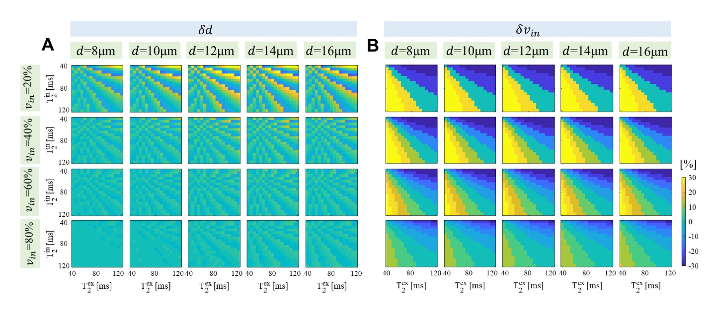
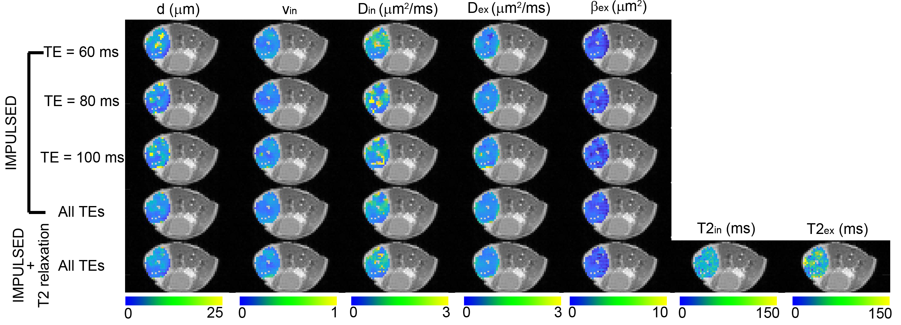
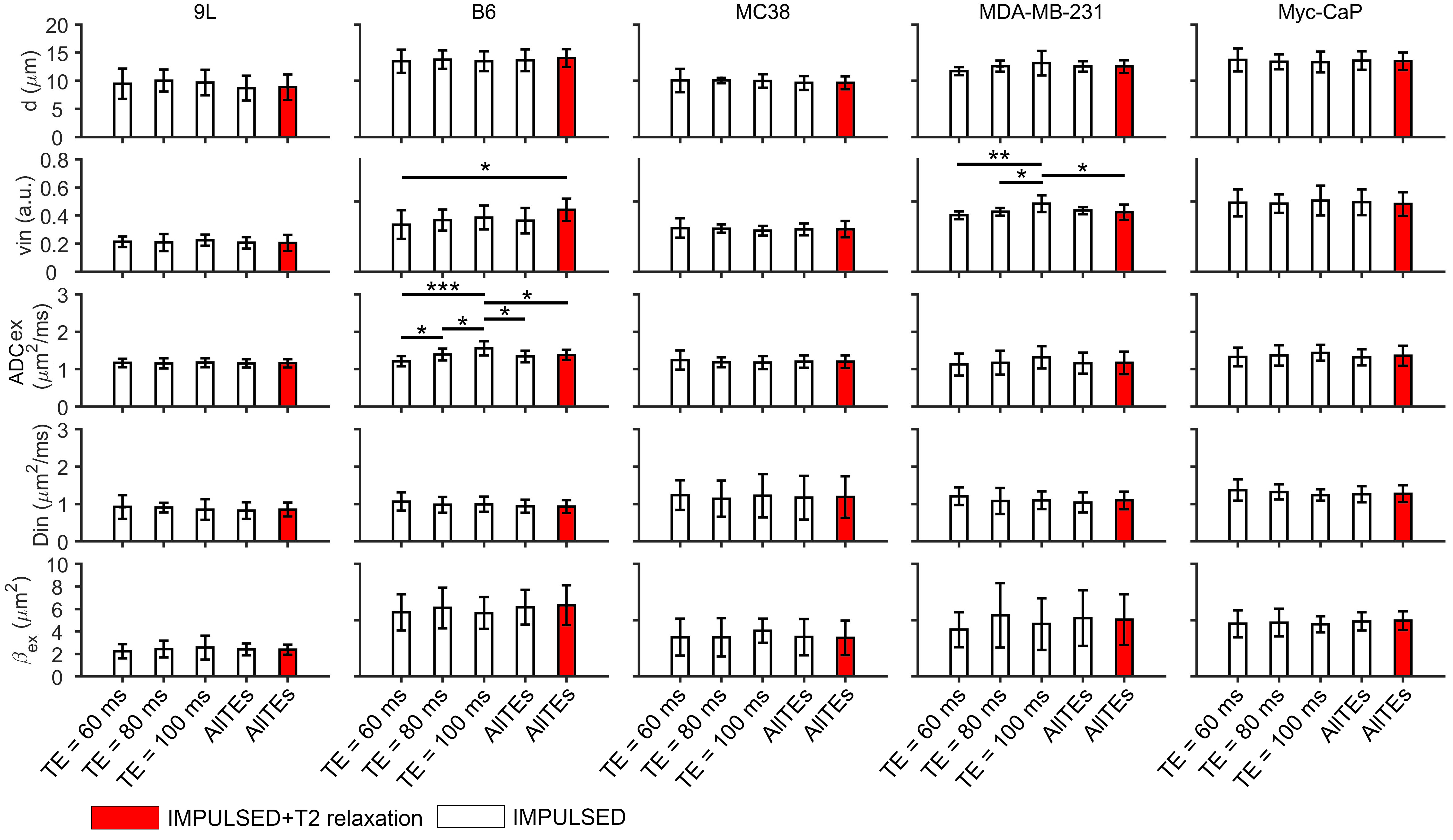
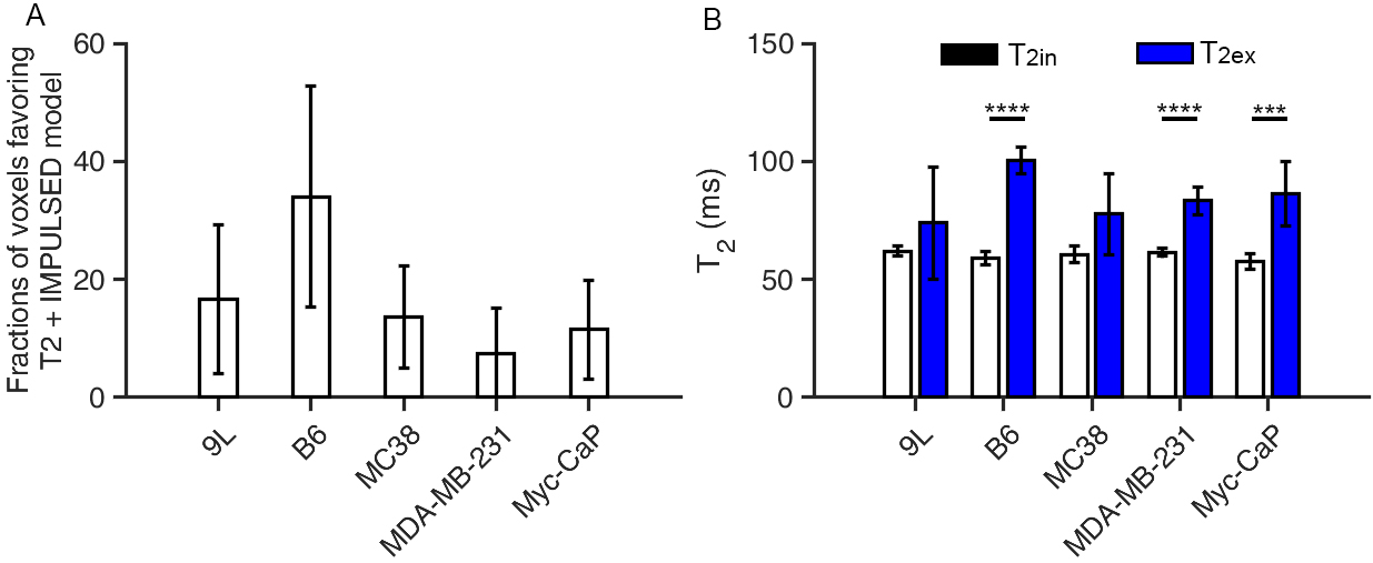
Simulations suggest that T2 heterogeneity has a minor effect on the estimation of d in tissues with intermediate or high cell density, but significantly biases the estimation of $$$v_{in}$$$ with low cell density (Figure 1). An illustrative example is presented in Figure 2 with parametric maps overlaid on corresponding T2-weighted images for a single imaging slice from an MC38 tumor. Figure 3 depicts the group variations in parameters derived from IMPULSED and T2+IMPULSED methods, and the dependency of such variations on TE and tumor type. Notably, no statistically significant differences were observed in all fitted parameters including $$$v_{in}$$$ in 9L, MC38, and Myc-CaP tumors. Fractions of voxels favoring T2+IMPULSED model vary with different types of cancer (Figure 4A). The T2+IMPULSED-derived $$$T_2^{in}$$$ and $$$T_2^{ex}$$$ values of all five tumor models are shown in Figure 4B. $$$T_2^{in}$$$ is consistently smaller than $$$T_2^{ex}$$$, consistent with previous study that combines T2 relaxation and dMRI biophysical model [11]. This is expected since intracellular space contains more macromolecules and components that lead to shorter T2 [12].Conclusion
The findings from this study highlight two key observations: i) TE has a negligible impact on IMPULSED-derived cell sizes, and ii) the TE-dependence of IMPULSED-derived intracellular volume fractions used in T2+IMPULSED modelling to estimate $$$T_2^{in}$$$ and $$$T_2^{ex}$$$.Acknowledgements
No acknowledgement found.References
1. Jiang, X., et al., MR cell size imaging with temporal diffusion spectroscopy. Magn Reson Imaging, 2021. 77: p. 109-123.
2. Jiang, X., et al., Quantification of cell size using temporal diffusion spectroscopy. Magn Reson Med, 2016. 75(3): p. 1076-85.
3. Xu, J., et al., Magnetic resonance imaging of mean cell size in human breast tumors. Magn Reson Med, 2020. 83(6): p. 2002-2014.
4. Ba, R., et al., Diffusion-time dependent diffusion MRI: effect of diffusion-time on microstructural mapping and prediction of prognostic features in breast cancer. Eur Radiol, 2023. 33(9): p. 6226-6237. 5. Wu, D., et al., Time-Dependent Diffusion MRI for Quantitative Microstructural Mapping of Prostate Cancer. Radiology, 2022: p. 211180.
6. Wu, J., et al., IMPULSED model based cytological feature estimation with U-Net: Application to human brain tumor at 3T. Magn Reson Med, 2023. 89(1): p. 411-422.
7. Zhang, H., et al., Histological and molecular classifications of pediatric glioma with time-dependent diffusion MRI-based microstructural mapping. Neuro Oncol, 2023. 25(6): p. 1146-1156. 8. Jiang, X., J. Xu, and J.C. Gore, Mapping hepatocyte size in vivo using temporal diffusion spectroscopy MRI. Magn Reson Med, 2020.
9. Panagiotaki, E., et al., Noninvasive quantification of solid tumor microstructure using VERDICT MRI. Cancer Res, 2014. 74(7): p. 1902-12.
10. Reynaud, O., et al., Pulsed and oscillating gradient MRI for assessment of cell size and extracellular space (POMACE) in mouse gliomas. NMR Biomed, 2016. 29(10): p. 1350-63.
11. Palombo, M., et al., Joint estimation of relaxation and diffusion tissue parameters for prostate cancer with relaxation-VERDICT MRI. Sci Rep, 2023. 13(1): p. 2999.
12. Beall, P.T., C.F. Hazlewood, and P.N. Rao, Nuclear magnetic-resonance patterns of intracellular water as a function of Hela-cell cycle. Science, 1976. 192(4242): p. 904-907.
Figures