5094
Cervical Cancer and Paracancerous Tissue ATPw combined with DWI to predict Parametrial infiltration: An Preliminary Study1The Second Hospital of Jilin University, Changchun, China
Synopsis
Keywords: fMRI Analysis, fMRI
Motivation: Cervical cancer(CC) is one of the most common gynecological malignancies. Parametrial infiltration (PMI) is considered a decisive factor in the treatment of patients with CC.
Goal(s): This study aimed to investigate the potential of Amide proton transfer-weighted (APTw) in predicting PMI of CC, and to further evaluate whether the APTw parameters of paracancerous tissue can add diagnostic value to diffusion-weighted MRI of CC.
Approach: 81 participants underwent pelvic MRI , including APTw MRI ,and takes measurements.
Results: The results showed that the APTw values ,especially in paracancerous tissues, could be used to efficiently distinguish PMI of cervical cancer.
Impact: APTw values ,especially in paracancerous tissues, could be used to efficiently distinguish PMI of cervical cancer. which can provide additional information to improve the results of diffusion-weighted MRI, thereby assisting further clinical decision-making and improving patient prognosis.
Introduction:
As one of the most common gynecological malignancies, cervical cancer(CC) has the fourth highest incidence rate and mortality among female cancers[1]. Parametrial infiltration (PMI) is considered a decisive factor in the treatment of patients with CC[2]. An accurate assessment of the preoperative state of the patient's parauterine tissue is crucial, because it is not only related to the prognosis but also affects the clinical decision-making. DWI, as a commonly used MRI sequence, can reflect the anatomical structure and functional information of the corresponding tissues[3]. Based on this, the obtained ADC sequence, which describes the speed of water molecule diffusion in tissues, has been widely studied in the diagnosis and treatment of cervical cancer[4-5]. Amide proton transfer-weighted (APTw) is a type of non-invasive imaging, and has shown great potential in the diagnosis of cervical cancer (CC), but quantitative research of paracancerous tissues has yet not been well studied in diagnosing parametrial infiltration (PMI). This study aimed to investigate the potential of APTw in predicting PMI of CC, and to further evaluate whether the APTw parameters of paracancerous tissue can add diagnostic value to diffusion-weighted MRI of CC.Methods:
In this single-center retrospective study, 81 patients with pathologic analysis–confirmed cervical cancer with no previous treatment, lesions larger than 10 mm, and adequate MRI quality were enrolled in a university teaching hospital. All patients were examined using a 3.0T MRI system (Ingenia 3.0 T CX; Philips Healthcare, Best, the Netherlands), including APTw MRI and DWI.Detailed MRI protocols are shown in Table 1. The APTw values of tumor(APTw-tu), APTw values of paracancerous tissues (APTw-pa) and apparent diffusion coefficient (ADC) values were independently reviewed by two radiologists experienced in diagnostic gynecologic imaging without knowledge of the patient's clinical and pathologic information to map the regions of interest and measure the corresponding values. Receiver operating characteristic curves were generated to evaluate the diagnostic performance of these quantitative parameters. SPSS25.0, MedCalc 20.0 and Delong test were used to verify whether there were significant differences in AUC of different parameters between the two groups. P < 0.05 was considered to indicate statistically significant differences.Results:
All patients were divided into the PMI group (n = 22) and non-PMI group (n = 59) according to the pathological results (Figure 1). PMI group had higher APTw values (%) than non-PMI group (3.71 [IQR, 3.60–3.98] and 2.75 [IQR, 2.68–2.77] vs 3.33 [IQR, 3.24–3.60] and 1.98 [IQR, 1.82–2.36]; P <0.001). The ADC values of PMI group were lower than those of non-PMI group (0.88 [IQR, 0.83–0.94] ×10-3 mm2/sec vs 0.95 [IQR, 0.88–1.04]×10-3 mm2/sec; P <0.001). (Table 2, Figure 2).The AUCs of APTw-tu, APTw-pa and ADC value for PMI diagnosis were 0.810, 0.831 and 0.806 respectively. In addition, the combination of APTw and DWI had improved diagnostic performance compared with individual analysis of either technique, among them, the AUC value ( 0.918 ) of APTw-pa + ADC was optimal, with a sensitivity and specificity of 91.20% and 87.20% respectively(Table 3, Figure 3). There were statistically differences in AUC among multiple groups(all P < 0.001).Conclusion:
APTw is a promising imaging marker for preoperative clinical prediction of cervical cancer, which can provide additional information to improve the results of diffusion-weighted MRI, This study found that the APTw values, especially in paracancerous tissues has certain potential and value in the evaluation of PMI.Discussion:
Accurate preoperative evaluation of PMI is important as there are different clinical treatments and prognosis for the presence or absence of PMI. In this study, the value of APTw and ADC in the evaluation of cervical cancer PMI was discussed, and the results showed that the APTw and ADC were significant difference in PMI group and non-PMI group. Previous reserch found APTw and dynamic contrast-enhanced magnetic resonance imaging (DCE-MRI) quantitative parameters can help preoperatively predict the occurrence of deep stromalinvasion (DSI) or/and lymphovascular vascular space invasion (LVSI) in cervical cancer[6]. We were considering in the paracancerous tissue with PMI, a large number of lymphocytes are seen, and such infiltrated lymphocytes are rich in cytoplasm and have a high degree of tumor heterogeneity, which was of great significance in diagnosing early PMI. In the future, the sample size should be expanded for further verification, and further molecular experimental research is still needed to validate our findings. APTw imaging has some potential and value in the assessment and diagnosis of PMI,thereby assisting further clinical decision-making and improving patient prognosis.Acknowledgements
No acknowledgement found.References
[1] Sung, H., et al., Global cancer statistics 2020: GLOBOCAN estimates of incidence and mortality worldwide for 36 cancers in 185 countries. Ca-a Cancer Journal for Clinicians, 2021. 71(3): p. 209-249.
[2] Patel-Lippmann, K., et al., MR Imaging of Cervical Cancer. Magnetic Resonance Imaging Clinics of North America, 2017. 25(3): p. 635-+.
[3] Bourgioti, C., et al., Incremental prognostic value of MRI in the staging of early cervical cancer: a prospective study and review of the literature. Clinical Imaging, 2016. 40(1): p. 72-78.
[4] Zhang, B.Q., et al. Whole-lesion Histogram Analysis of Apparent Diffusion Coefficient for Distinguishing Cervical Cancers with Different Differentiation. in 10th IEEE International Conference on Nano/Molecular Medicine and Engineering (NANOMED). 2016. Macau, PEOPLES R CHINA.
[5] Chen, J.J., et al., The Value of Diffusion-Weighted Magnetic Resonance Imaging in Predicting the Efficacy of Radiation and Chemotherapy in Cervical Cancer. Open Life Sciences, 2018. 13(1): p. 305-311.
[6] Song, Q., et al., Amide proton transfer weighted imaging combined with dynamic contrast-enhanced MRI in predicting lymphovascular space invasion and deep stromal invasion of IB1-IIA1 cervical cancer. Frontiers in Oncology, 2022. 12.
Figures
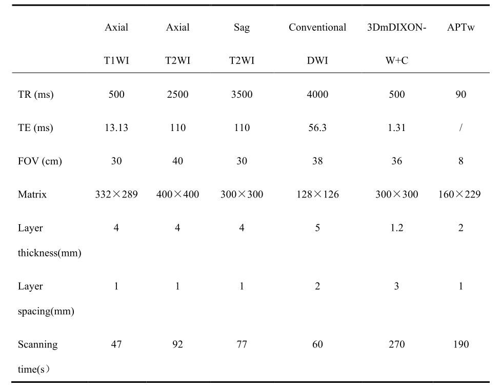
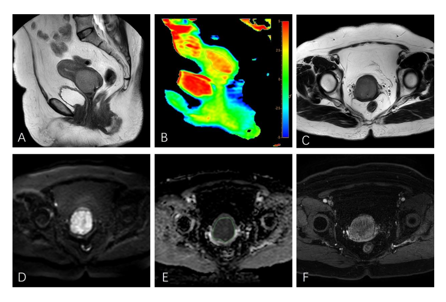
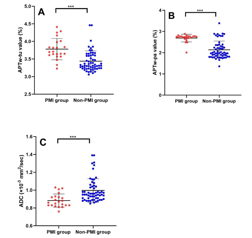
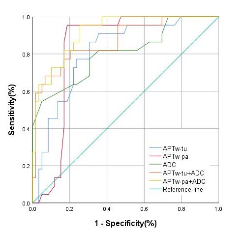
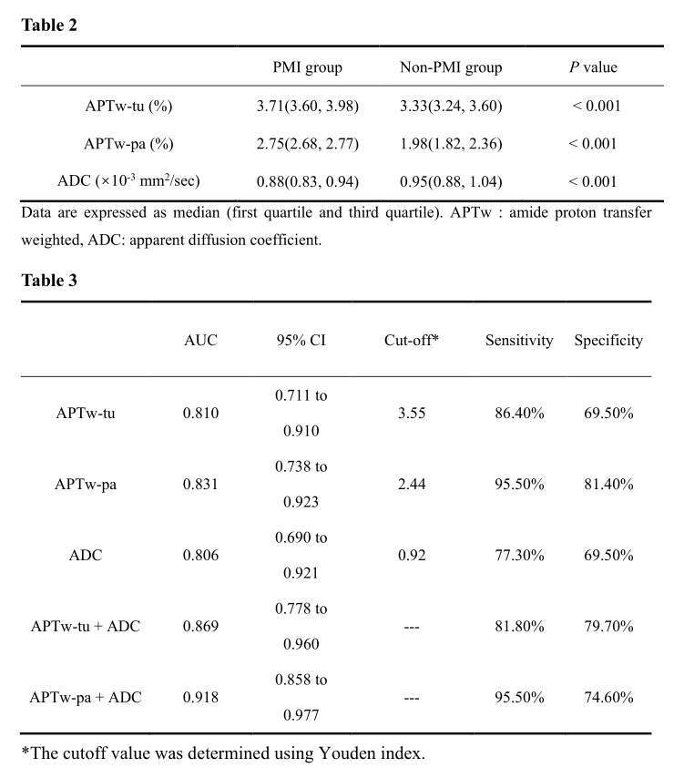
Table2 Comparisons of the Amide Proton Transfer-weighted and Apparent Diffusion Coefficient Values in PMI group and non-PMI group
Table3 Diagnostic Performance of the Amide Proton Transfer-weighted and Apparent Diffusion Coefficient Values in Differentiating PMI group and non-PMI group