5091
Restoration of Bi-contrast Whole Body MRI for Discontinuous Intensity Nonuniformities1Institute for Radiology, South Harz Hospital Nordhausen, Nordhausen, Germany, 2Institute for Diagnostic and Interventional Radiology, University Medicine of Jena, Jena, Germany, 3Department of Information Technologies, University of Limassol, Limassol, Cyprus
Synopsis
Keywords: Screening, Artifacts, bone metastases, signal restoration, cancer screening
Motivation: Whole-body MRI, a screening tool for bone metastatic disease, uses parallel coil imaging to cover a large Field of View. Signal inhomogeneities at the coil junction points create jump discontinuities in the signal intensity with a negative effect on serial image stitching.
Goal(s): To correct for signal jump discontinuities between coils using computer vision.
Approach: We engaged piecewise smooth intensity nonuniformities fields using anisotropic diffusion and quantified for improvement in image entropy (H).
Results: Our algorithm smoothens the signal intensity between parallel coils by 8% based on entropy metrics.
Impact: We implement a novel non-parametric methodology with piecewise smoothness to improve intensity non-uniformities between parallel coil images in whole-body MRI (WB-MRI). Optimized whole-body stitched images render WB-MRI into a one-stop-shop staging method.
INTRODUCTION
Whole-body MRI (WB-MRI) is an imaging tool for the entire body. In clinical oncology, WB-MRI is the future first-line diagnosis in cancer staging and follow-up, currently approved for detecting bone metastasis [1]. WB-MRI images are multi-coil parallel acquisitions over a large Field of View (FoV) that inevitably suffer from extensive intensity nonuniformities due to differences in gains, as well as jump discontinuities between coil junctions. Some of those artifacts cannot be addressed by conventional intensity correction methodologies [2,3,4]. The proposed method addresses this problem with the novelty of estimating piecewise smooth intensity nonuniformities fields using anisotropic diffusion [5] between joint image pairs, as published in previous work by the authors [6]. The methodology has been validated by comparing it with a restoration using conventional isotropic smoothing in an in-domo dataset.METHODS
Forty breast and prostate cancer patients were examined with WB-MRI for possible bone metastases using T1w Turbo Spin Echo (TSE) and Short-TI Recovery (STIR) T1w+T2w. The datasets were screened for not suffering from misregistration and resampled to have the exact spatial resolution, the same size, and two bytes per pixel. The images are first denoised with median filtering. The sum of the two images is processed with Otsu's method. A low-pass and high-pass filter exclude noise and artifacts. The restoration with statistical co-occurrence statistics was based on a Gaussian Point Spread smoothing function linearly increasing in proportion to the intensity of spatial non-uniformities. Spatial smoothing was achieved by back-projection with indexing the restoration matrices using an axial Gaussian filter. The restoration of the images is repeated iteratively for ten iterations. The spatial restoration fields are accumulated multiplicatively along the iterations, and the cumulative fields are smoothed anisotropically along iterations. The optimal iteration is selected retrospectively as the one which minimizes the joint entropy of the statistics.RESULTS
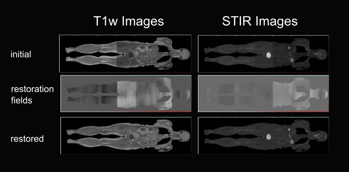
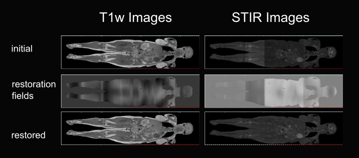
Two examples of initial and restored images of both contrasts are shown in Figure 1 (patient 1) and Figure 2 (patient 2), respectively. In the left figure panels are the T1w TSE images, and in the right are the STIR images. The top rows show the initial images, the middle rows show the cumulative restoration fields and the bottom rows show the restored images. The proposed method was compared to conventional restoration, where the spatial smoothing filter was isotropic. The validation used the entropy of the joint histogram (H). The restoration sharpens the statistics, so it decreases the entropy. A relative improvement of the anisotropic diffusion compared to the isotropic restoration corresponds to a positive value for the measure H-ratio. The H-ratio values were 8.5±9 (mean/SD) and 8.9/-8.9/30.4 (median/min/man). The improvement with the anisotropic method is around 8%. Another conventional intensity correction method, implemented by the module "N4ITK MRI Bias correction" of Slicer3D, was used for experimentation [7]. It was apparent by the observation that N4ITK failed to remove the nonuniformities, and led to an extensive loss of contrast in both the images and in the statistics. This is mainly because N3/N4 is unable to represent field discontinuities.DISCUSSION
The proposed method removes the nonuniformity of the shading together with its discontinuities for bi-contrast WB-MRI data. It improves the stitching performance on average by 8% when considering the spatial nonuniformity compared to when ignoring it. The proposed methodology addresses this problem by first performing deconvolution and restoration of the statistics using the co-occurrence statistics with a lower variance for the dominant distributions. It provides a Bayesian posterior estimate for the restored intensities. These are back-projected to space to give rough estimates of the spatial restorations. They are then processed with piecewise smoothness formulated with MDL, or equivalently MAP, that accommodates spatial nonuniformities discontinuities. Conventional methodologies assume a uniformly smooth nonuniformity. The spatial piecewise smoothness for the nonuniformity shading is novel not only for WB-MRI but more generally for medical imaging and even for computer vision. The method is non-parametric both in the statistics and in space and provides the joint intensity uniformity restorations of two anatomic WB images simultaneously.Major weaknesses of this study, which the authors currently address, are the low data sample and the lack of quantitative comparison to conventional methods.
CONCLUSION
This work presents a novel non-parametric methodology with piecewise smoothness for the nonuniformities for effective and simultaneous joint intensity uniformity restoration of two anatomic WB images. The proposed method is novel and valuable. The current methodology can be enhanced by registering multimodal datasets and is integrable into automated detection pipelines.Acknowledgements
No disclosures or acknowledgmentsReferences
[1] Papageorgiou, I., Dvorak, J., Cosma, I., Pfeil, A., Teichgraeber, U., Malich, A., "Whole-Body MRI: a Powerful Alternative to Bone Scan for Bone Marrow Staging Without Radiation and Gadolinium Enhancer," Clin. Transl. Oncol., 22 (8), (2020) [2] Tustison, N.J., Avants, B.B., Cook, P.A., Zheng, Y., Egan, A., Yushkevich, P.A., Gee, J.C., "N4ITK: Improved N3 Bias Correction," IEEE Trans Med Imaging, 29 (6), (2010) [3] Hadjidemetriou, S., Studholme, C., Mueller, S., Weiner, M., Schuff, N., "Restoration of MRI Data for Intensity Non-uniformities Using Local High Order Intensity Statistics," Medical Image Analysis, 13 (1), (2009) [4] Hadjidemetriou, S., Psychogios, M.N., Lingor, P., Eckardstein, K.V., Papageorgiou, I., "Restoration of Bi-Contrast MRI Data for Intensity Uniformity with Bayesian Coring of Co-Occurrence Statistics," Journal of Imaging, MDPI, 3 (4), (2017) [5] Leclerc, Y.G.: "Constructing Simple Stable Descriptions for Image Partitioning," Int. J. Comput. Vis. 3(1): 73-102 (1989) [6] Hadjidemetriou S., Malich A., Rossknecht LD., Ferrarini L., Papageorgiou IE., "Restoration for Intensity Nonuniformities with Discontinuities in Whole-Body MRI," Signals. 2023; 4(4):725-745. https://doi.org/10.3390/signals4040040 [7] Tustison N., Gee J. "N4ITK: Nick's N3 ITK Implementation For MRI Bias Field Correction," The Insight Journal. 2010 Dec. http://hdl.handle.net/10380/3053Figures