5090
Virtual gadolinium contrast enhancement MRI using deep learning GAN in rectal cancer: a proof-of-concept two-center study1Department of Radiology, Zhejiang Cancer Hospital, Hangzhou, China, 2Department of Colorectal Surgery, Zhejiang Cancer Hospital, Hangzhou, China
Synopsis
Keywords: Pelvis, Safety, rectal cancer, GBCAs, deep learning
Motivation: It would be clinically beneficial if GBCAs enhancement could be accurately synthesized without any GBCAs administration though AI.
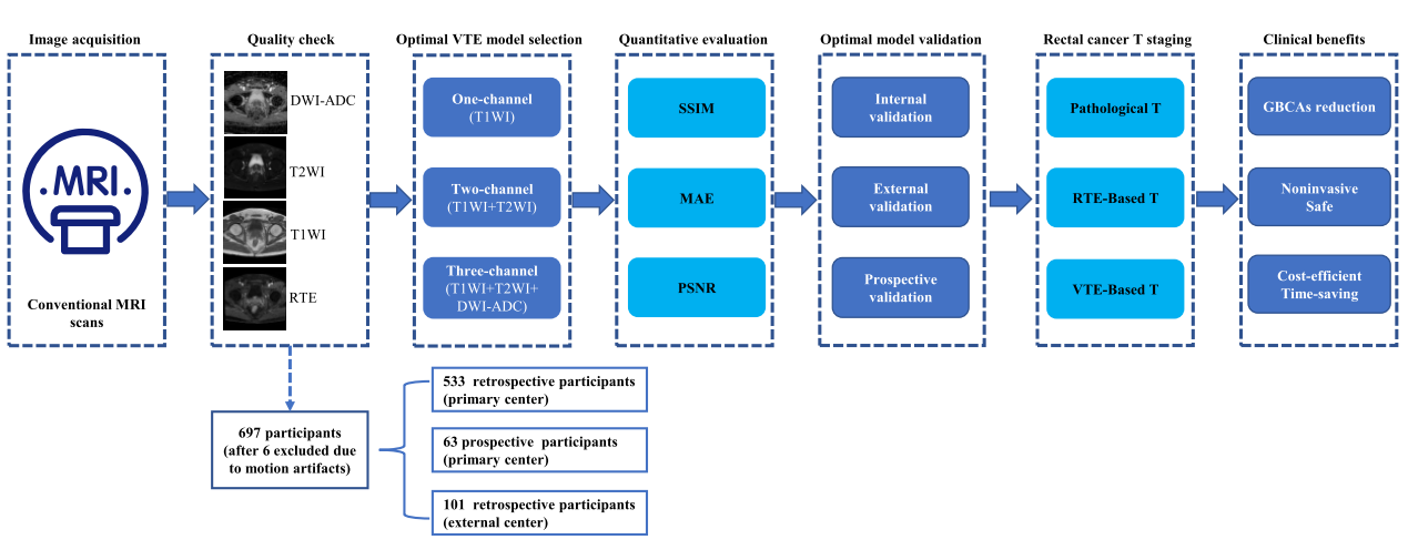
Goal(s): To evaluate the feasibility of deep learning in synthesizing VTE based on noncontrast rectal cancer MRIs obtained without the use of gadolinium.
Approach: Deep learning networks were trained and validated on nonenhanced conventional pelvic MRI (T1WI, T2WI, DWI-ADC) using GAN. MRI scans included 697 rectal cancer patients from two hospitals.
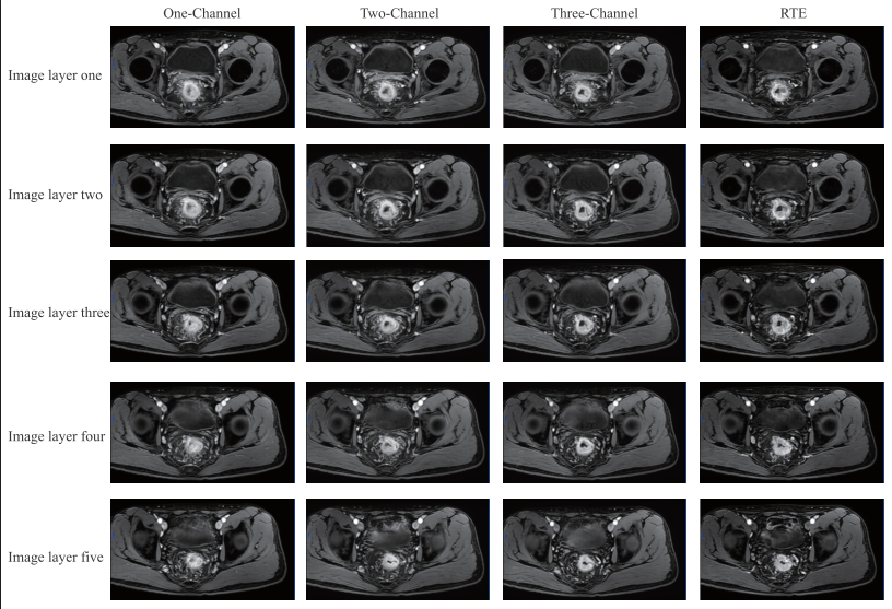
Results: Quantitative and qualitative evaluation of three-channel VTE was significantly better than that of two-channel and one-channel (P<0.001). The T staging accuracy of VTE was comparable with that of RTE.
Impact: VTE synthesized by deep learning based on noncontrast MRI can overcome the limitations of RTE and aid in the clinical diagnosis and management of rectal cancer as a noninvasive, save, affordable and time-saving method that does not require GBCAs.
Introduction
Gadolinium-based contrast agents MRI assists the diagnosis and surveillance of rectal cancer, whereas safety concerns have been raised recently. We aim to explore whether deep learning generative adversarial network (GAN) can synthesize clinically applicable virtual T1WI enhancement (VTE) of rectal cancer based on non-contrast pelvic MRI scans.Methods
Deep learning networks were trained and validated on nonenhanced conventional pelvic MRI (T1WI, T2WI, DWI-ADC) using GAN. MRI scans included 697 rectal cancer patients (634 retrospective, 63 prospective) from two hospitals from June 2020 to October 2022. The real T1WI enhancement (RTE) images served as the ground truth. The similarity between VTE and RTE were quantified using Mean Absolute Error (MAE), Peak Signal Noise Ratio (PSNR), Structural Similarity (SSIM) and qualitatively evaluated by two radiologists. The image quality of input was quantitatively evaluated by Natural Image Quality Evaluator (NIQE). Ablation experiment was conducted to explore the best model. T staging was evaluated on VTE and RTE by two radiologists and compared with pathology.Results
Participants included 493 men and 204 women (average age, 60.87 ± 12.08 years) were enrolled. Quantitative and qualitative evaluation of three-channel VTE was significantly better than that of two-channel and one-channel (P<0.001). In internal validation (IV), external validation (EV) and prospective validation (PV): MAE was 0.03 ± 0.01, 0.04 ± 0.01 and 0.03 ± 0.01; PSNR was 25.86 ± 1.63, 22.34 ± 1.21 and 25.68 ± 1.28; SSIM was 0.87 ± 0.55, 0.66 ± 0.06 and 0.88 ± 0.03, respectively. The T staging accuracy of VTE was comparable with that of RTE in IV, EV, and PV (VTE vs RTE, 71.72% vs 70.71%, 69.31% vs 69.31%, and 73.02% vs 71.43%, respectively). The correlation analysis between NIQE and MAE or SSIM or PSNR was statistically significant (P < 0.001).Discussion
As a technical development and proof-of-concept study, this study underlines the potential of deep learning GAN for synthesizing VTE from clinical routine multiparametric nonenhanced MRIs of rectal cancer obtained without GBCAs injection. Overall, the quantitative and qualitative assessment on two-center dataset showed good consistency between VTE and RTE. The diagnostic ability of VTE in rectal cancer T staging was comparable to that of RTE. This study demonstrated the current frontier in image contrast generation, which has the potential to be harnessed in the future for rectal cancer patient care.Conclusion
The deep learning GAN could synthesize enhancement of rectal cancer from nonenhanced conventional pelvic MRI scans with good image quality quantitatively and qualitatively and allows accurate assessment of rectal cancer patients’ T staging. As a technical development and proof of concept study, this research demonstrates the current frontier in image contrast generation, which has the potential to be harnessed in the future for rectal cancer patient care.Acknowledgements
None.References
1. Cao W, Zou Q, Zhao Y, et al. Application of liver acquisition with volume acceleration enhanced sequence in improving the accuracy of reassessing organ-invasive rectal mucinous adenocarcinoma after chemoradiation. Eur J Radiol 2020; 133: 109368. 2. Lu QY, Guan Z, Zhang XY, et al. Contrast-enhanced MRI for T Restaging of Locally Advanced Rectal Cancer Following Neoadjuvant Chemotherapy and Radiation Therapy. Radiology 2022: 212905. 3. Collidge TA, Thomson PC, Mark PB, et al. Gadolinium-enhanced MR imaging and nephrogenic systemic fibrosis: retrospective study of a renal replacement therapy cohort. Radiology 2007; 245(1): 168-75. 4. McDonald RJ, McDonald JS, Kallmes DF, et al. Intracranial Gadolinium Deposition after Contrast-enhanced MR Imaging. Radiology 2015; 275(3): 772-82. 5. Radbruch A, Weberling LD, Kieslich PJ, et al. Gadolinium retention in the dentate nucleus and globus pallidus is dependent on the class of contrast agent. Radiology 2015; 275(3): 783-91.Figures