5089
Unsupervised Super Resolution of Diffusion Weighted Imaging Guided by High-Resolution Cross-Modality Prior1Biomedical Imaging Research Institute, Cedars-Sinai Medical Center, Los Angeles, CA, United States, 2Department of Bioengineering, University of California Los Angeles, Los Angeles, CA, United States, 3Minimal Invasive Urology, Cedars-Sinai Medical Center, Los Angeles, CA, United States
Synopsis
Keywords: Prostate, Prostate, Super Resolution, Deep Learning, Unsupervised
Motivation: Existing supervised super-resolution is challenging for Diffusion Weighted Imaging(DWI) due to acquisition. However, the feature of T2 weighted imaging(T2w) could be utilized as a prior for unsupervised training.
Goal(s): To develop unsupervised super-resolution on DWI with the aid of high-resolution T2w images.
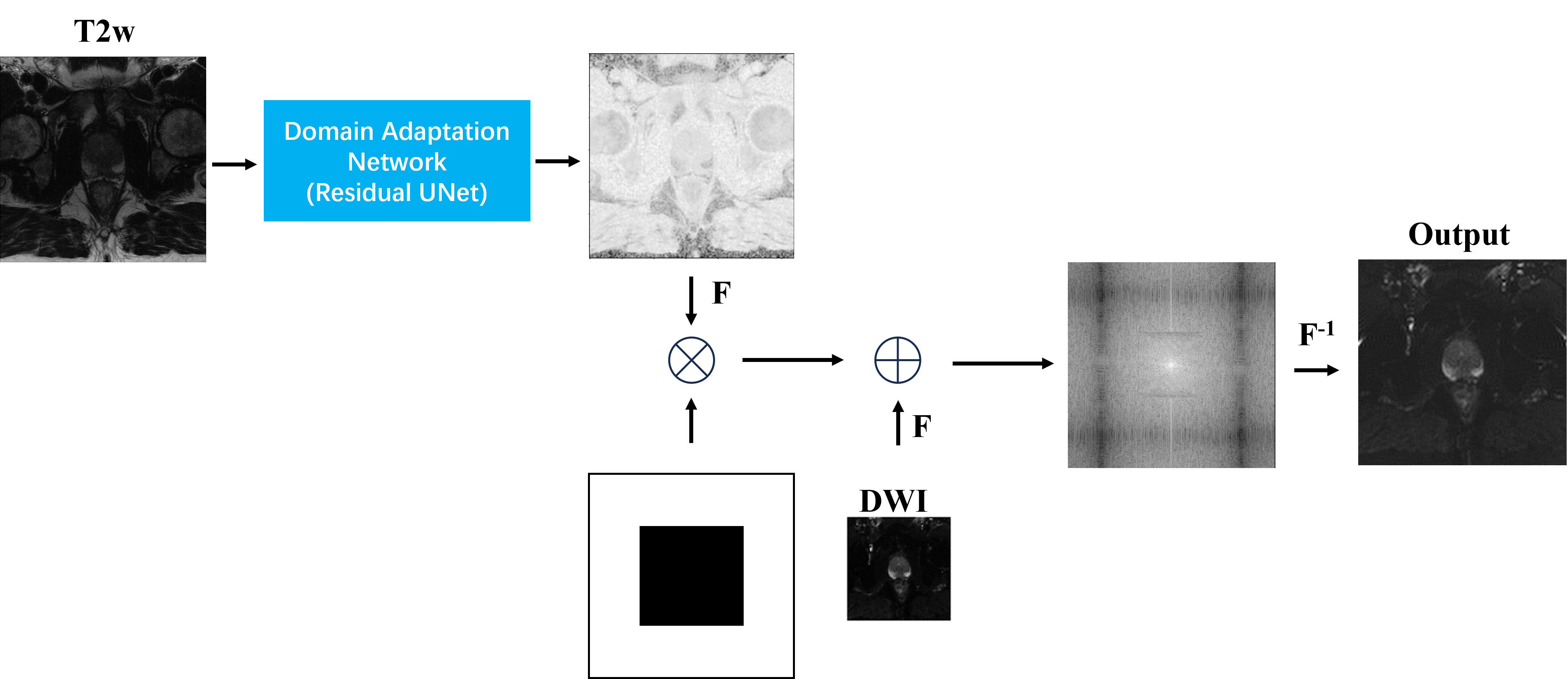
Approach: A UNet architecture is designed to perform same-resolution domain adaptation. During inference, the high frequency feature of the T2w images are used to fuse with the low frequency feature of original DWI in k-space to reconstruct high-resolution DWI.
Results: Our result shows improved SSIM score verified by paired student t-test. Our direct inference on HR DWI also exhibits improved sharpness.
Impact: This pilot work demonstrated that HR images (T2w) can be domain-adapted to provide high frequency prior to unsupervised super-resolution tasks using computationally efficient DL models.
Introduction
High-resolution (HR) Diffusion Weighted Imaging (DWI) is important for small tumor detection in prostate imaging1. However, HR DWI usually requires specialized sequences that are not widely available and longer scan time1,2. Previous studies on super-resolution DWI require high-resolution ground truth3-5, which is challenging in prostate due to noise, distortion, and motion-induced artifacts. The purpose of this work was to develop an unsupervised super-resolution approach that utilizes the domain-adapted HR T2-weighted imaging (T2w) prior to improve the in-plane resolution of standard DWI images by a factor of 12.75.Methods
In general, a super-resolution task aims to find a mapping function from LR (low resolution) image to HR image, where the field of view (FOV) stays unchanged. In the frequency domain, the super-resolution task can be defined as filling the missing information in the high-frequency region in k-space. The assumption of this work is that if a domain adaptation network is trained between LR images, the model will only learn to convert the LR contrast and preserve the HR information if HR images are used during inference. Therefore, the proposed process could be summarized as $$$I^{u \times v}_{\small HRDWI}(x,y) = F^{-1}(F(I^{m \times n}_{\small LRDWI})+F(f_{\small UNet}(\lambda I^{u \times v}_{\small HRT2})) \cdot H^{u \times v}(x,y))$$$, where $$$F$$$ denotes Fourier Transform, $$$\lambda$$$ denotes a hyperparameter, and $$$H(u,v)$$$ denotes high pass filter. Two three-layer residual UNets were adopted from Kerfoot et al6 for domain adaptation to b50 and b1000 images, and the ADC map was calculated from the transformed diffusion weighted images. We used Mean Absolute Error as the domain adaptation loss and correlation coefficient as the loss to preserve anatomical information between output and HR T2w images following Ge et al7. The detailed training process is illustrated in Figure 1. The generation of HR DWI is adapted from the Keyhole method in MRI8. We used the training set of the fastMRI prostate dataset9 for this study. A total of 218 cases were split into 6:2:2 for training, validation and testing. The DWI image with a resolution of 2mm was cropped to 90x90 pixels to ensure fixed FOV with T2w image with 320x320 pixels at 0.56mm resolution. Through-plane resolution remains the same.Results
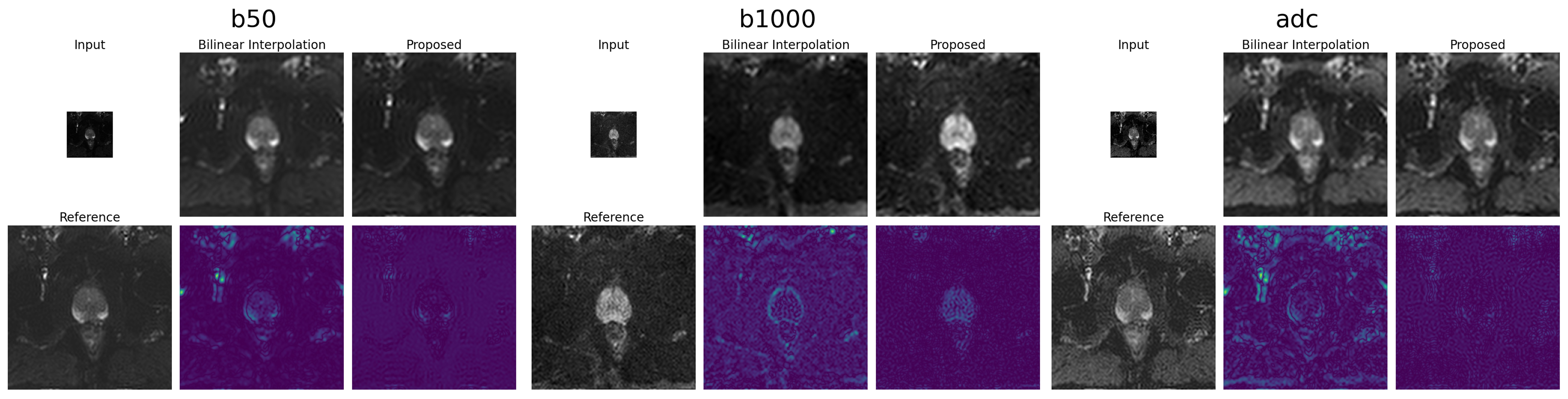
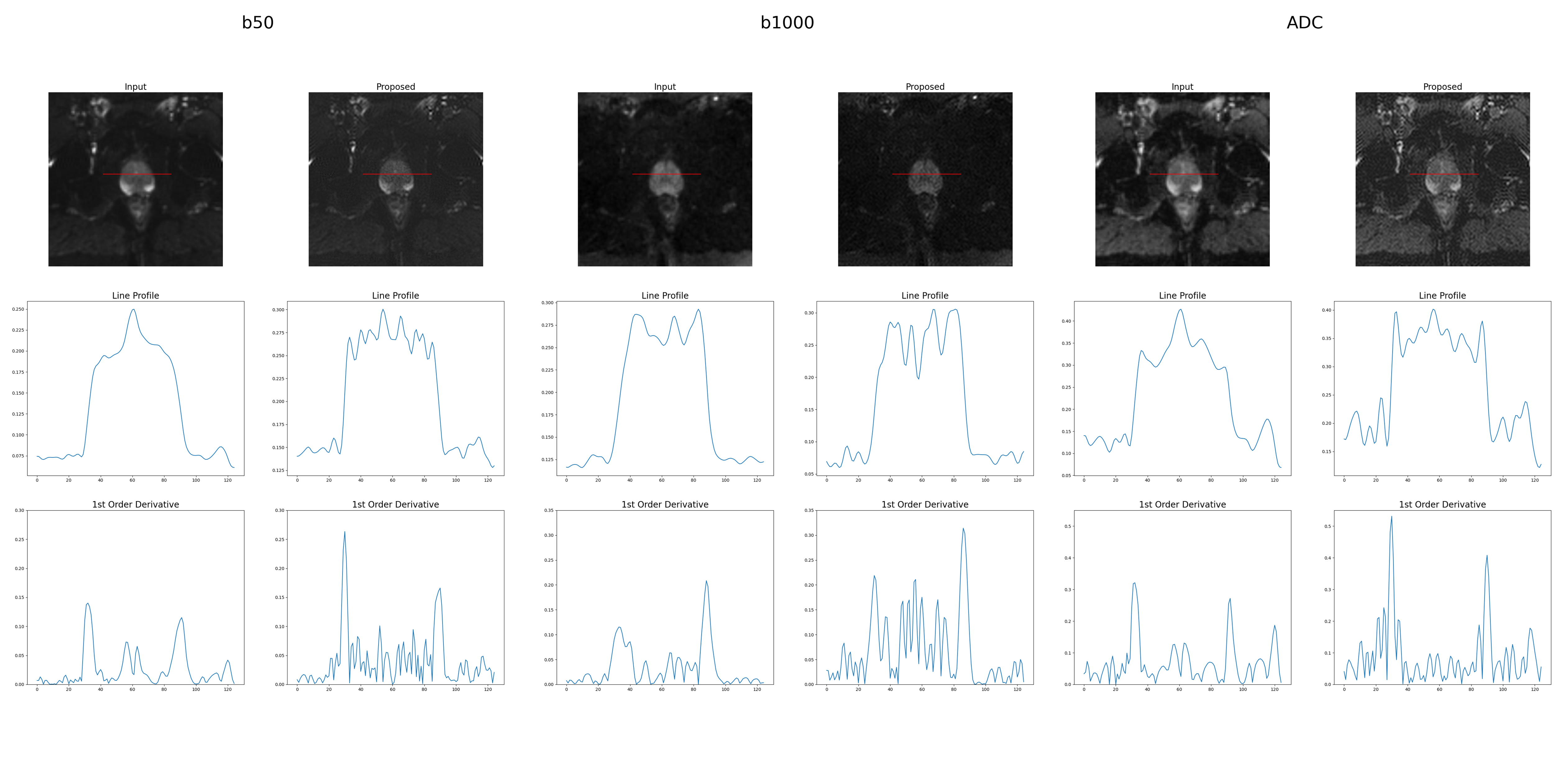
Two experiments were conducted to validate our approach. First, T2w and DWI images were downsampled to 50x50 (3.6-mm resolution) for training, and the standard DWI images served as the reference. As shown in Figure 2, Both b50 images and ADC images showed improved SSIM except for b1000 images when compared with bilinear interpolated 50x50 DWI images. Secondly, the standard resolution of DWI and down-sampled T2w images were used for training, and the sharpness of prostate boundary was measured from manually picked line profiles as the metric due to the lack of ground truth HR DWI images. Two line profiles were extracted from two separate slices for each case in the test set. Gaussian blurring and first-order derivative were applied in order, and the first two maximum value is extracted as the sharpness measure for the prostate boundary. During inference, we observed explicit improvement in image quality in all three contrasts. In addition, our proposed method showed better sharpness in all three contrasts. All comparisons in this study were proved statistically significant with the paired t-test with all p-values smaller than 0.05.Discussion
As shown in Figure 3, the model trained directly on 100x100 DWI and T2w images shows evident improvement in image fine details and sharpness. The lower SSIM of the proposed method in experiment 1, in which low-resolution DWI was used at b1000 could also be caused by scarce information in the original image and the high-frequency noise introduced by T2w prior. In addition, the adopted model is comparably smaller (6M parameters) than the contemporary SR models, and the performance could improve if more sophisticated models are employed. Therefore, future direction regarding this work could be employing other innovative ways to extract HR information of T2w images such as edge detection maps with first-order derivative. On the other hand, more sophisticated domain adaptation networks may further improve the super-resolution performance.Conclusion
This pilot work demonstrates the feasibility of improving the spatial resolution of standard DWI images in the prostate by a factor of 12 (from 2 by 2 to 0.56 by 0.56 mm2) using high frequency prior provided by HR T2w images for unsupervised super-resolution with computationally efficient DL models. Further evaluation is warranted to assess the potential impact of the proposed method on improving the diagnostic accuracy of prostate MRI.Acknowledgements
This study was partially supported by NIH R01CA217098.References
- Medved M et al. High-resolution diffusion-weighted imaging of the prostate. AJR Am J Roentgenol. 2014 Jul;203(1):85-90. doi: 10.2214/AJR.13.11098. PMID: 24951199.
- Baliyan V et al. Diffusion weighted imaging: Technique and applications. World J Radiol. 2016 Sep 28;8(9):785-798. doi: 10.4329/wjr.v8.i9.785. PMID: 27721941.
- Cao C et al. High-Frequency Space Diffusion Models for Accelerated MRI. 2020 Dec. 10.48550/arXiv.2208.05481.
- S. Chatterjee et al. ShuffleUNet: Super resolution of diffusion-weighted MRIs using deep learning. 29th European Signal Processing Conference (EUSIPCO), Dublin, Ireland. 2021, pp. 940-944. doi: 10.23919/EUSIPCO54536.2021.9615963.
- Ye X et al. Simultaneous superresolution reconstruction and distortion correction for single-shot EPI DWI using deep learning. Magn Reson Med. 2023 Jun;89(6):2456-2470. doi: 10.1002/mrm.29601. Epub 2023 Jan 27. PMID: 36705077.
- Kerfoot E et al. Left-Ventricle Quantification Using Residual U-Net. 9th International Workshop, STACOM 2018, Held in Conjunction with MICCAI 2018, Granada, Spain, September 16, 2018, Revised Selected Papers. 10.1007/978-3-030-12029-0_40.
-
Yunhao G et al. Unpaired whole-body MR to CT
synthesis with correlation coefficient constrained adversarial learning.
Proc. SPIE 10949, Medical Imaging 2019: Image Processing,
1094905 - van Vaals JJ et al. “Keyhole" method for accelerating
imaging of contrast agent uptake. J Magn Reson Imaging. 1993
Jul-Aug;3(4):671-5. doi: 10.1002/jmri.
1880030419 8347963 - Tibrewala R et al. FastMRI Prostate: A Publicly Available, Biparametric MRI Dataset to Advance Machine Learning for Prostate Cancer Imaging. ArXiv [Preprint]. 2023 Apr 18:arXiv:2304.09254v1. PMID: 37131871; PMCID: PMC10153282.
Figures